Techniques for identifying molecular structures and treating cell types lining a body lumen using fluorescence

a technology of molecular structure and cell type, applied in the field of fluorescence imaging of cell type in the walls of the body lumen, can solve the problems of inability to distinguish capsule systems, inability to inspect most of the small intestine, and inability to inspect some portions of the large intestine (colon), so as to enhance the local enhance the uptake of exogenous probes

Active Publication Date: 2009-04-07
THE JOHN HOPKINS UNIV SCHOOL OF MEDICINE
View PDF8 Cites 94 Cited by
  • Summary
  • Abstract
  • Description
  • Claims
  • Application Information

AI Technical Summary

Benefits of technology

[0018]According to another embodiment, to enhance local uptake of the exogenous probe, ultrasonic waves are emitted from a sound source on the solid support.
[0019]According to another embodiment, to enhance uptake of the exogenous probe, an electric field is generated from an electrode on the solid support.

Problems solved by technology

While suitable for inspection of the esophagus, stomach and portions of the large intestine, the endoscope is neither capable of inspecting some portions of the large intestine (colon) nor capable of inspecting most of the small intestine.
In the more advanced stages, when tumors are apparent by their morphology, the prior art capsule systems can not distinguish between a tumor mass containing dying cells that are responding to treatment, and a tumor mass with viable cells that are resisting treatment or are continuing to grow.
Therefore, existing capsule systems can not distinguish some significant disease-related types and functions of cells making up the structure of the walls.

Method used


Image

  • Techniques for identifying molecular structures and treating cell types lining a body lumen using fluorescence
  • Techniques for identifying molecular structures and treating cell types lining a body lumen using fluorescence
  • Techniques for identifying molecular structures and treating cell types lining a body lumen using fluorescence

Examples


Embodiment Construction

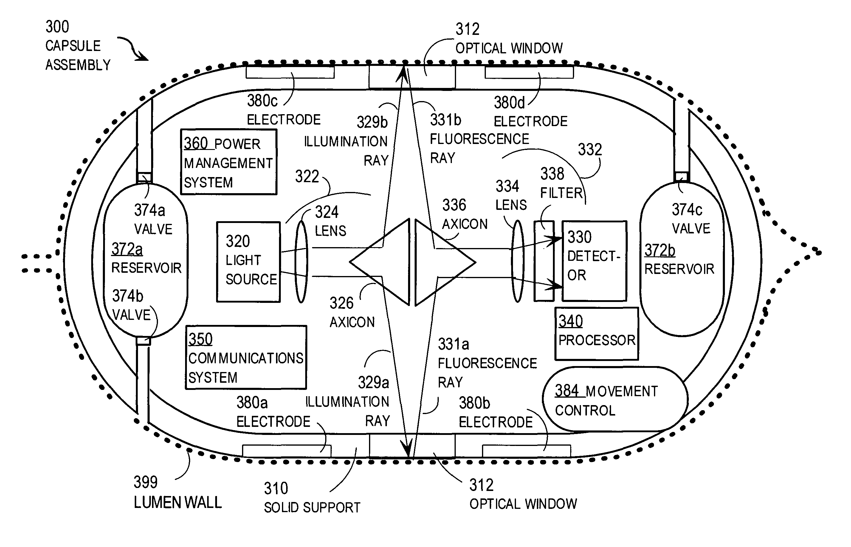
[0035]A method and apparatus are described for quantitative identification of specific molecular structures and tissue constituents as well as cell type and functions in the walls of a body lumen. In the following description, for the purposes of explanation, numerous specific details are set forth in order to provide a thorough understanding of the present invention. It will be apparent, however, to one skilled in the art that the present invention may be practiced without these specific details. In other instances, well-known structures and devices are shown in block diagram form in order to avoid unnecessarily obscuring the present invention.

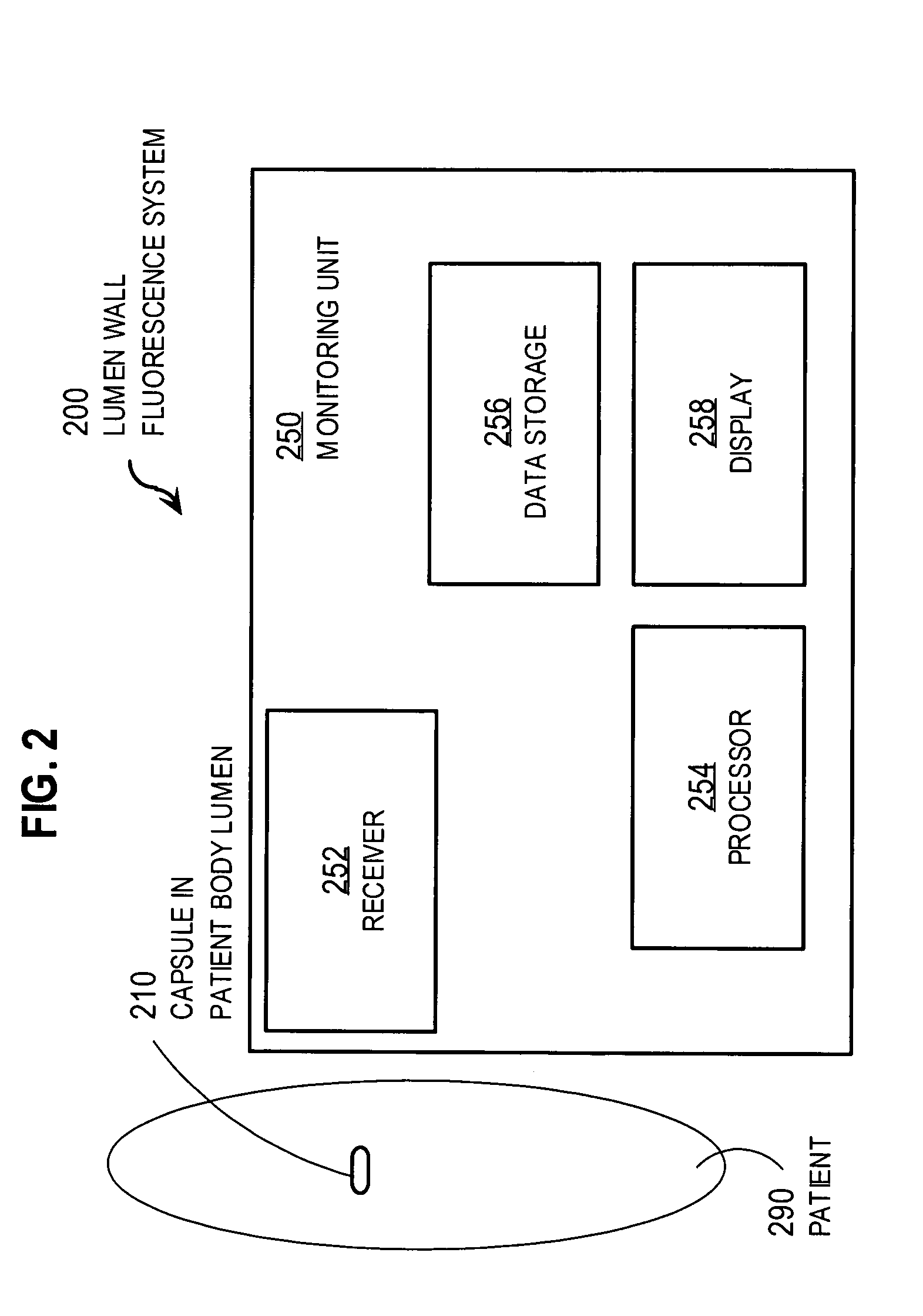
[0036]Embodiments of the invention are described primarily in the context of diagnosis and therapy for dysfunction of the human intestinal tract, but the invention is not limited to this context. For example, in other embodiments the techniques may be applied to non-human animals. Furthermore, in other embodiments, the techniques may be appli...


PUM


Abstract

Techniques for detecting fluorescence emitted by molecular constituents in a wall of a body lumen include introducing an autonomous solid support into the body lumen. Cells in a lumen wall of the body lumen are illuminated by a light source mounted to the solid support with a wavelength that excites a particular fluorescent signal. A detector mounted to the solid support detects whether illuminated cells emit the particular fluorescent signal. If the particular fluorescent signal is detected from the illuminated cells, then intensity or position in the lumen wall of the detected fluorescent signal, or both, is determined. These techniques allow the information collected by the capsule to support diagnosis and therapy of GI cancer and other intestinal pathologies and syndromes. For example, these techniques allow diagnostic imaging using endogenous and exogenous fluoroprobes, treating diseased sites by targeted release of drug with or without photoactivation, and determining therapeutic efficacy.

Description

CROSS-REFERENCE TO RELATED APPLICATIONS[0001]This application claims benefit of Provisional Appln. 60 / 400,325, filed Aug. 1, 2002, the entire contents of which are hereby incorporated by reference as if fully set forth herein, under 35 U.S.C. §119(e).BACKGROUND OF THE INVENTION[0002]1. Field of the Invention[0003]The present invention relates to identifying molecular structures and cell types in walls of a body lumen in animals; and in particular to fluorescent imaging of cell types in walls of a body lumen for diagnosis or therapy, such as in vivo therapy based on selective destruction of labeled tumor cells. The invention has application to the diagnosis and treatment of intestinal cancer and colon cancer, among other pathologies and syndromes.[0004]2. Description of the Related Art[0005]Cancer of the gastrointestinal (GI) tract is easily treated if detected early. Consequently a great deal of activity has been expended in developing systems to inspect the GI tract for early signs...

Claims


Application Information

Patent Timeline
Patent Type & Authority Patents(United States)
IPC IPC(8): A61B6/00A61K49/00
CPCA61B1/041A61K41/0057A61K49/0013A61K41/0071A61N5/062A61B2018/00982A61N1/327A61B5/0071A61B5/073A61B2560/0468A61B2562/0233A61B2562/162
Inventor MADAR, IGALMURPHY, JOHN C.
Owner THE JOHN HOPKINS UNIV SCHOOL OF MEDICINE
Why Eureka
Try Eureka