HPV E6, E7 mRNA assay and methods of use thereof

a technology of mrna assay and assay, which is applied in the field of hpv e6, e7 mrna assay, can solve the problems of pap smear sensitivity of only 58%, ineffective test to distinguish benign lesions from lesions with malignant potential, and pap smear

Active Publication Date: 2009-04-28
INCELLDX
View PDF6 Cites 58 Cited by
  • Summary
  • Abstract
  • Description
  • Claims
  • Application Information

AI Technical Summary

Problems solved by technology

The Pap smear, however, is not without its shortcomings; in particular, errors in cervical sampling and interpretation contribute to a Pap smear sensitivity of only 58%.
As the data in the foregoing table demonstrates, the majority of women diagnosed with ASCUS, LGSILS, and HGSILs do not progress to cancer; accordingly, the traditional Pap smear and LBC test, both of which merely identify the presence abnormal SILs are not effective tests to distinguish benign lesions from lesions with malignant potential.
Similarly, because the majority of women diagnosed with ASCUS, LGSILS, and HGSILs are usually infected with a high risk HPV, it follows that the identification of infection with a high risk HPV is also not a biologically relevant indicator for cervical cancer detection.

Method used


Image

  • HPV E6, E7 mRNA assay and methods of use thereof
  • HPV E6, E7 mRNA assay and methods of use thereof
  • HPV E6, E7 mRNA assay and methods of use thereof

Examples


example 1

Simultaneous Immunofluorescence and Ultrasensitive Fluorescence In Situ

Hybridization to Detect HPV E6, E7 (“In Cell HPV Assay”)

[0060]A 1 mL aliquot was removed from the liquid-based cervical cytology specimen isolated from the subjects. The cells were pelleted by centrifugation at 400× g and washed once in phosphate buffered saline (“PBS”), pH 7.4. Cells were resuspended in 100 μL of PBS, pH 7.4 and stained with a 1:10 dilution of phycoerythrin (“PE”)-conjugated anti-CAM 5.2 and PE / cy5-conjugated anti-CD16 (BDPharmingen, San Diego, Calif.). The cells were then incubated at 4° C. for 20 minutes in the dark. Following incubation, the cells were fixed, permeabilized, washed once in PBS, pH 7.4, pelleted by centrifugation at 400×g, washed again in 2×SSC, and pelleted by centrifugation. The cells were then resuspended in a hybridization mix consisting of 5×SSC, 30% formamide, and 100 μg / mL sheared salmon sperm DNA (“ssDNA”) and a cocktail of 5′- and 3′-fluorescein labeled HPV E6, E7 mRNA...

example 2

The Slide-Based In Cell HPV Assay

[0062]100 μL of cells (1×10⁶ cells / mL) were transferred from the liquid based cervical cytology specimen isolated from the subjects and put into a cytocentrifuge or liquid-based slide system. The slides were centrifuged at 800×g for 2 minutes at room temperature. Following a wash in 1×PBS, pH 7.4, the slides were incubated in 1× PermiFlow (Invirion, Inc., Frankfort, Mich.) fixation / permeabilization reagent in a Coplin jar at room temperature for 1 hour. The slides were then washed once in PBS and once in 2×SSC. The cells were hybridized to a cocktail of the HPV OncoTect probes a hybridization oven at 37° C. for 30-120 minutes. The slides were then washed for 5 minutes in a Coplin jar containing 50 mL pre-heated 2×SSC, 0.1% Triton X-100 and incubated for 15 minutes in a Coplin jar containing 50 mL pre-heated 0.1×SSC, 0.1% Triton X-100. Following a brief rinse in PBS, pH 7.4, the slides were coverslipped using Fluorsave mounting medium (CalBioChem, San...

example 3

Validation of the In Cell HPV Assay

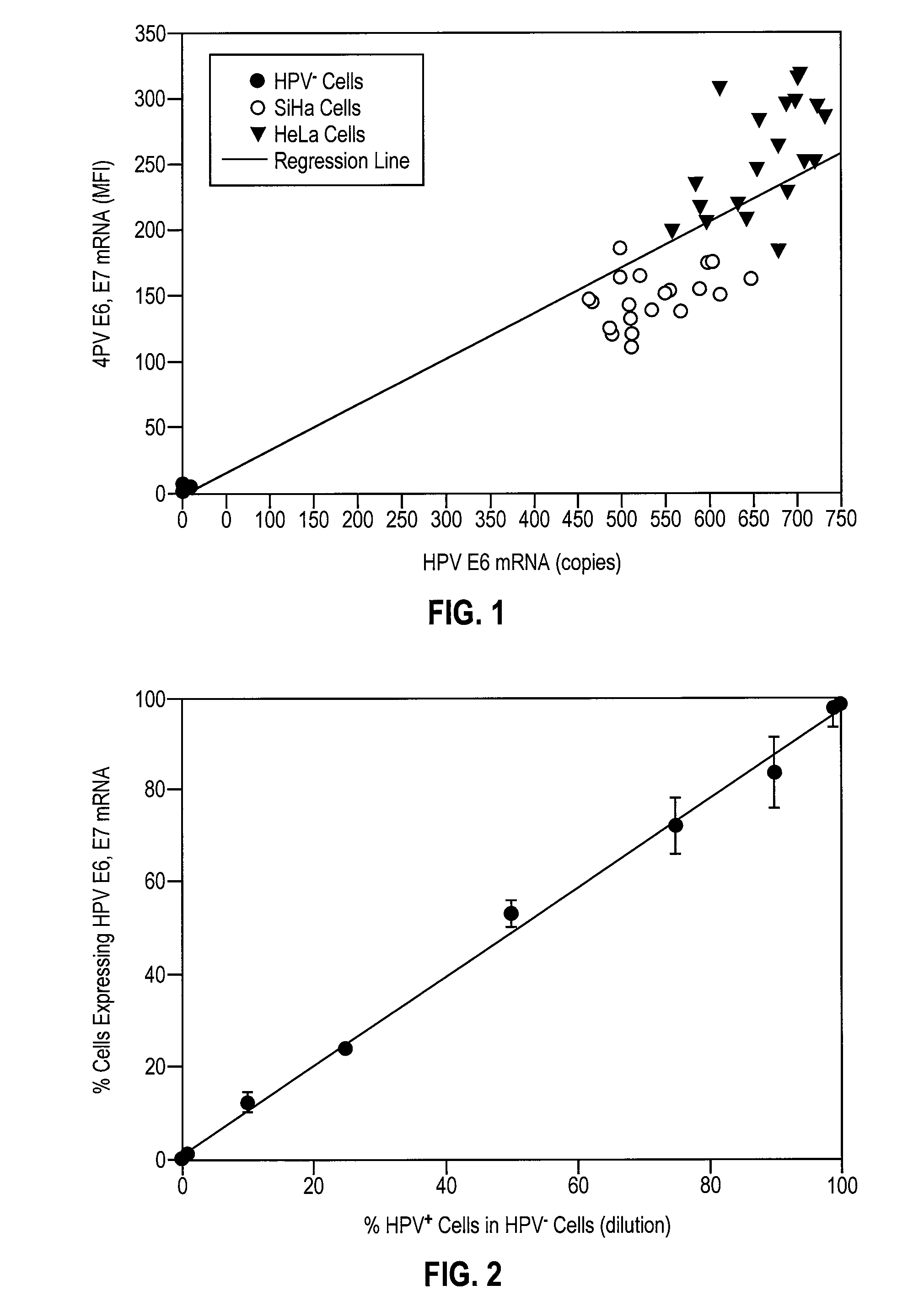
[0063]To validate the effectiveness of the In Cell HPV Assay, commercially available normal ectocervical cells, i.e., HPV− ectocervical cells, and HPV+ SiHa and HeLa cells were grown in culture and split into 20 samples and analyzed for HPV E6, E7 mRNA by the In Cell HPV Assay with flow cytometry and for HPV E6 mRNA by real-time RT-PCR with a detection limit of 10 copies per cell. FIG. 1 shows the relationship between the MFI of HPV E6, E7 mRNA and copies of HPV E6 mRNA in each of the cell populations. Linear regression analysis of the results of FIG. 1 resulted in a correlation of 0.88 with a p value of 0.001. Based upon the results of this experiment, the theoretical sensitivity of the In Cell HPV Assay was calculated to be 10 to 20 copies of HPV E6, E7 mRNA per cell. In all subsequent patient sample analyses, cells were considered positive for HPV E6, E7 mRNA if they exhibited an MFI greater than 200 copies per cell, 200 representing a copy numb...


PUM

PropertyMeasurementUnit
pHaaaaaaaaaa
molecular weightaaaaaaaaaa
fluorescence intensityaaaaaaaaaa

Abstract

Provided is an HPV E6, E7 mRNA assay, referenced herein as the “In Cell HPV Assay,” that is capable of sensitive and specific detection of normal cervical cells undergoing malignant transformation as well as abnormal cervical cells with pre-malignant or malignant lesions. The In Cell HPV Assay identifies HPV E6, E7 mRNA via in situ hybridization with oligonucleotides specific for HPV E6, E7 mRNA and quantitates the HPV E6, E7 mRNA via flow cytometry. The In Cell HPV Assay can be carried out in less than three hours directly from liquid-based cervical (“LBC”) cytology specimens. The In Cell HPV Assay provides an efficient and highly sensitive alternative to the Pap smear for determining abnormal cervical cytology.

Description

TECHNICAL FIELD[0001]The invention relates generally to an HPV mRNA assay for the detection of HPV-related disorders such as cervical cancer and cervical lesions. More specifically, the invention relates to an assay that identifies and quantitates HPV E6, E7 mRNA in cervical cells. The HPV E6, E7 mRNA Assay, referenced herein as the “In Cell HPV Assay,” is capable of sensitive and specific detection of cancerous and pre-cancerous abnormal cells as well as normal cells undergoing malignant transformation.BACKGROUND OF THE INVENTION[0002]Cervical cancer affects approximately 13,000 women per year in the United States and more than 400,000 women worldwide. Ninety percent of cervical cancers contain the high risk HPV DNA strains 16 and 18 (“HPV-16” and “HPV-18,” respectively). By contrast, low risk HPVs, such as HPV-6 and HPV-11, rarely develop into cancer. The presence of low risk and high risk HPVs are identified through the use of the polymerase chain reaction (“PCR”) or the Hybrid C...

Claims


Application Information

Patent Timeline
Patent Type & Authority Patents(United States)
IPC IPC(8): C12Q1/68G16Z99/00
CPCC12Q1/708C12Q1/6841G16Z99/00C12Q2600/158
Inventor PATTERSON, BRUCE K.
Owner INCELLDX
Why Eureka
Try Eureka