Integrated optical scanning image acquisition and display

a technology of optical scanning and image acquisition, applied in the field of microelectromechanical systems, can solve the problems of reducing the resolution and/or field of view of the device, limiting the possibility of pixels, and reducing the system's size using this approach

Inactive Publication Date: 2009-06-30
UNIV OF WASHINGTON +1
View PDF21 Cites 111 Cited by
  • Summary
  • Abstract
  • Description
  • Claims
  • Application Information

AI Technical Summary

Benefits of technology

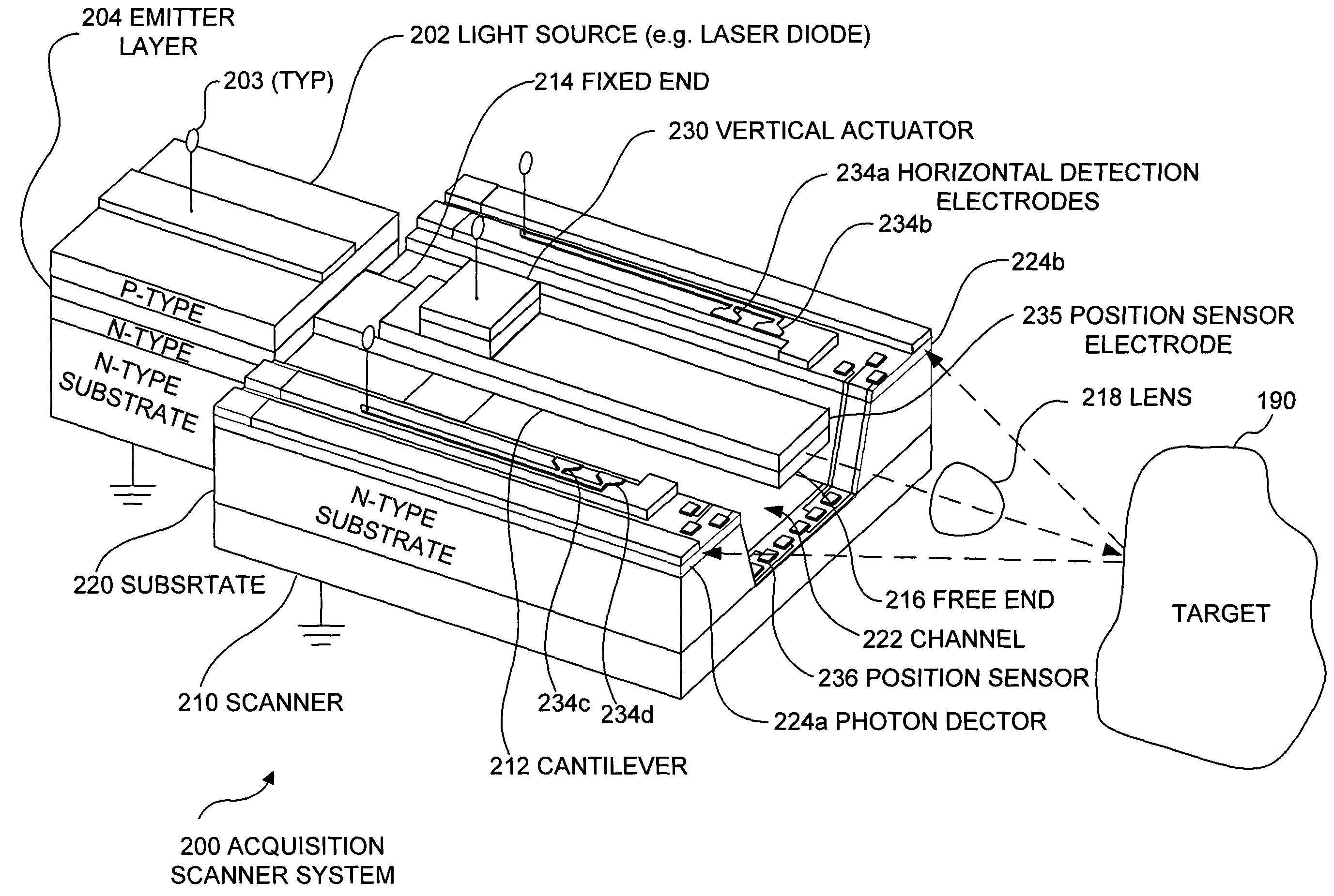
[0014]In accord with the present invention, an apparatus is defined for providing image acquisition and/or image display in a limited ROI. The apparatus comprises a micro-electro-mechanical system (MEMS) that integrates needed components into a miniature device that can be produced with conventional micro-fabrication techniques. The apparatus preferably includes one or more integrated light sources such as one or more laser diodes for illuminating an ROI, displaying an image, providing a therapy, and/or performing another function. Alternatively, the light source can include a generation component and delivery component, whereby only the delivery component is integrated into the apparatus. The light source can further include a modulator or filter, or modulation and filtering can be performed on the input or output of the light source. Also included is a cantilever having a fixed end and a free end. The fixed end is attached to a substrate that supports many or all other components of the apparatus. Preferably, the free end of the cantilever is released from the substrate during fabrication of the cantilever such that the cantilever can move in two orthogonal directions. The cantilever also preferably comprises a light-transmissive material such as an epoxy resin that acts as a waveguide to direct light from the light source toward the ROI. In that case, the one or more light sources are optically coupled to the fixed end of the cantilever waveguide, and the free end of the cantilever waveguide is adapted to be...

Problems solved by technology

Trying to minimize a system's size using this approach is limited by a number of factors, including the overall diameter of the fiber bundle, the number of pixel detectors on the camera detector, and diffractive properties of light beams.
Reducing the diameter of a conventional acquisition device reduces the possible number of pixels, and thus reduces the resolution and/or field of view (FOV) of the device.
This approach also has many size limitations.
For instance, light beams of less than 3 millimeter (mm) are impractical for displays using mirrors, because mirror scanners and grating deflectors must be significantly larger than the light beam diameter to avoid clipping the beam or adding diffraction.
Reducing the diameter of a conventional display device reduces the possible number of pixels, and thus reduces the resolution and/or field of view (FOV) of the device.
This technique requires a very complicated circuit to control the overall deflection of the reflectors and the overall size is quite large.
The current tools used for MIMPs cannot readily be integrated with an optical imaging system without increasing the size of the resultant instrument to an excessive degree.
As indicated above, currently available flexible scopes cannot be made smaller than this limit unless image field-of-view (FOV) or resolution is sacrificed.
Although imaging and some diagnostic capability can be integrated into existing scopes, such as standard tissue imaging in combination with fluorescence for early detection of cancers, the optical systems of current flexible scopes are not sufficiently small to provide integrated diagnoses and therapies at the required degrees of performance, size, and p...

Method used


Image

  • Integrated optical scanning image acquisition and display
  • Integrated optical scanning image acquisition and display
  • Integrated optical scanning image acquisition and display

Examples


Embodiment Construction

Example Applications of the Present Invention

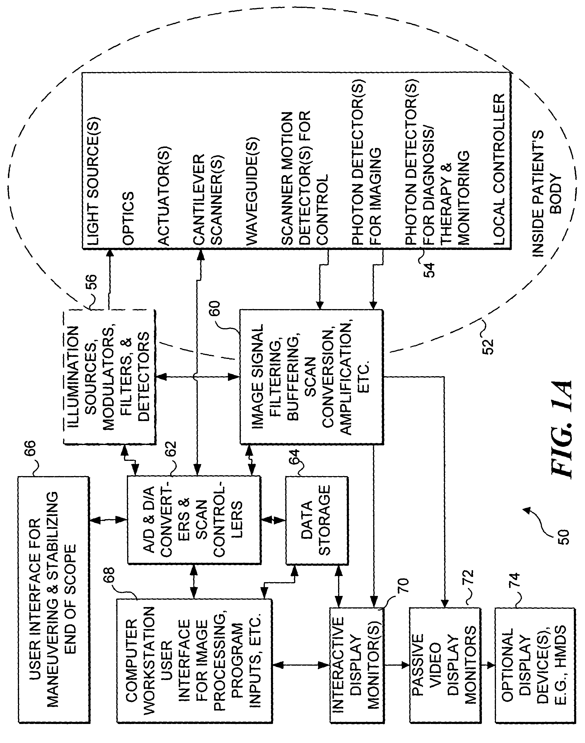
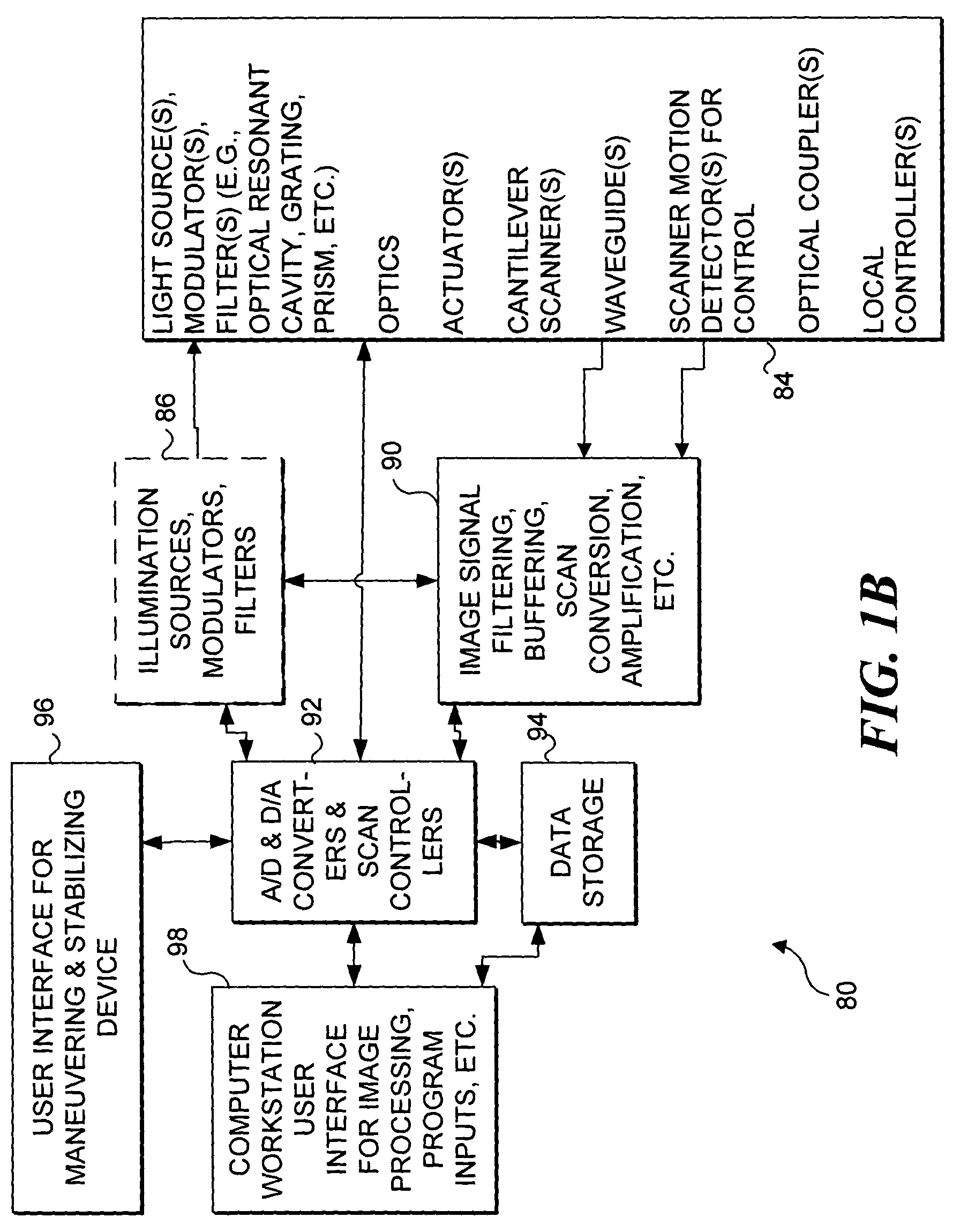
[0041]As indicated above, the present application is a continuation-in-part of copending utility patent application Ser. No. 09 / 850,594, filed on May 7, 2001, which is based on prior provisional patent application Ser. No. 60 / 212,411, filed on Jun. 19, 2000, both of which are hereby explicitly incorporated by reference. These applications describe a medical imaging, diagnostic, and therapy device. However, the present invention may be used for acquiring an image, for displaying an image, or for otherwise detecting or delivering light. Nevertheless, for exemplary purposes, the present invention is primarily described with regard to a preferred embodied as a miniature medical device such as an endoscope. Those skilled in the art will recognize that the present invention can also be embodied in a non-medical image acquisition device, a wearable display, a biological, chemical or mechanical sensor, or any other miniature high resolution, far-...


PUM


Abstract

An apparatus and method for providing image acquisition and/or image display in a limited region of interest (ROI). The apparatus comprises a micro electro-mechanical system (MEMS), preferably integrating a light source, a cantilever, a lens, an actuator, a light detector, and a position sensor. The light source provides light for illuminating the ROI, displaying an image, providing a therapy, and/or performing other functions. The cantilever comprises a resin waveguide with a fixed end attached to a substrate that supports many or all other components. A free end of the cantilever is released from the substrate during fabrication and includes the lens. The actuator scans the free end in orthogonal directions to illuminate the ROI or display an image. The position sensors detect the position of the free end for control. The light detector receives light backscattered from the ROI separate from, or at the fixed end the cantilever.

Description

RELATED APPLICATIONS[0001]This application is a continuation-in-part of patent application Ser. No. 09 / 850,594, filed on May 7, 2001, now U.S. Pat. No. 6,975,898 which is based on prior provisional patent application Ser. No. 60 / 212,411, filed on Jun. 19, 2000, the benefit of the filing dates of which is hereby claimed under 35 U.S.C. § 120 35 U.S.C. § 119(e), respectively, and both of which are hereby specifically incorporated by reference.GOVERNMENT RIGHTS[0002]This invention was made with government support under grant numbers CA094303 and CA096633 awarded by the National Institutes of Health. The government has certain rights in this invention.FIELD OF THE INVENTION[0003]The present invention generally relates to a micro-electro-mechanical system (MEMS) that conveys light to and from a region of interest (ROI), and more specifically, to a system that is selectively used for both imaging the ROI, and for displaying an image in the ROI.BACKGROUND OF THE INVENTION[0004]Conventional...

Claims


Application Information

Patent Timeline
IPC IPC(8): A61B6/00G01N21/64A61B1/00A61B1/005A61B1/04A61B5/00A61B5/107A61B18/20A61N5/06G01N21/27G01Q20/02G02B6/26G02B6/32G02B6/35G02B23/24G02B26/08G02B26/10H04N7/18
CPCA61B1/00048A61B1/0008A61B1/00165A61B1/00167A61B1/00172A61B1/00183A61B1/0051A61B1/0638A61B5/0062A61B5/0084G02B6/262G02B26/0875G02B26/10G02B26/101A61B1/00193A61B1/043A61B5/0066A61B5/0068A61B5/0071A61B5/0075A61B5/0086A61B5/1076A61N5/0601A61N5/062G02B6/32G02B6/3502G02B6/3566G02B6/357G02B6/3572G02B6/3578G02B23/2484A61B1/063A61B1/0655
Inventor WANG, WEI-CHISEIBEL, ERIC J.REINHALL, PER G.FAUVER, MARK E.BROWN, CHRIS M.
Owner UNIV OF WASHINGTON
Why Eureka
Try Eureka