Heparin binding motif and use thereof

a technology of eosinophil toxins and binding motif, which is applied in the direction of peptide/protein ingredients, peptide sources, drug compositions, etc., can solve the problems of not knowing much about the three-dimensional structure of hs, and the binding site of ecp has not been identified, so as to improve the ic50 value, less cytotoxic, and less interaction

Active Publication Date: 2009-09-29
NATIONAL TSING HUA UNIVERSITY
View PDF0 Cites 14 Cited by
  • Summary
  • Abstract
  • Description
  • Claims
  • Application Information

AI Technical Summary

Benefits of technology

[0051]The inhibition of lymphocyte and mammalian cell growth by ECP has been reported (Fan, T. C., et al. (2007) Traffic 8, 1778-1795; Maeda, T., et al. (2002) European Journal of Biochemistry 269, 307-316). As a physiological test, the cytotoxicity of ECP mt1 was monitored by MTT assay. The effect of ECP on the cell growth was determined by a colorimetric assay using 3-(4,5-dimethylthiazol-2-yl)-2,5-diphenyltetrazolium bromide (MTT) (US Biological, MA, USA). Cells were plated in a 96-well plate (5000 cells/well) and incubated at 37° C. overnight. Each sample was incubated with the indicated concentration (1-100 μM) of ECP. Forty-eight hours after treatment wi

Problems solved by technology

However, the heparin binding site in ECP has not been identified.
How

Method used


Image

  • Heparin binding motif and use thereof
  • Heparin binding motif and use thereof
  • Heparin binding motif and use thereof

Examples


example

Example 1

Materials

[0029]Mouse anti-MBP (maltose binding protein) was obtained from Santa Cruz Biotechnology (Santa Cruz, Calif.). RNase A was purchased from Promega (Madison, Wis.). Chemicals were purchased from Sigma-Aldrich (St. Louis, Mo.) unless otherwise specified. Human ECP peptide NYRWRCKNQNK-biotin (C1, SEQ ID NO: 3), EDN peptide NYQRRCKNQNK-biotin (D1, SEQ ID NO: 4), RNasel peptide MTQGRCKPVNK-biotin (R1, SEQ ID NO: 5), and HIV-TAT peptide YGRKKRRQRRRK-biotin (TAT, SEQ ID NO: 6) were purchased from Genemed Synthesis (South San Francisco, Calif.).

example 2

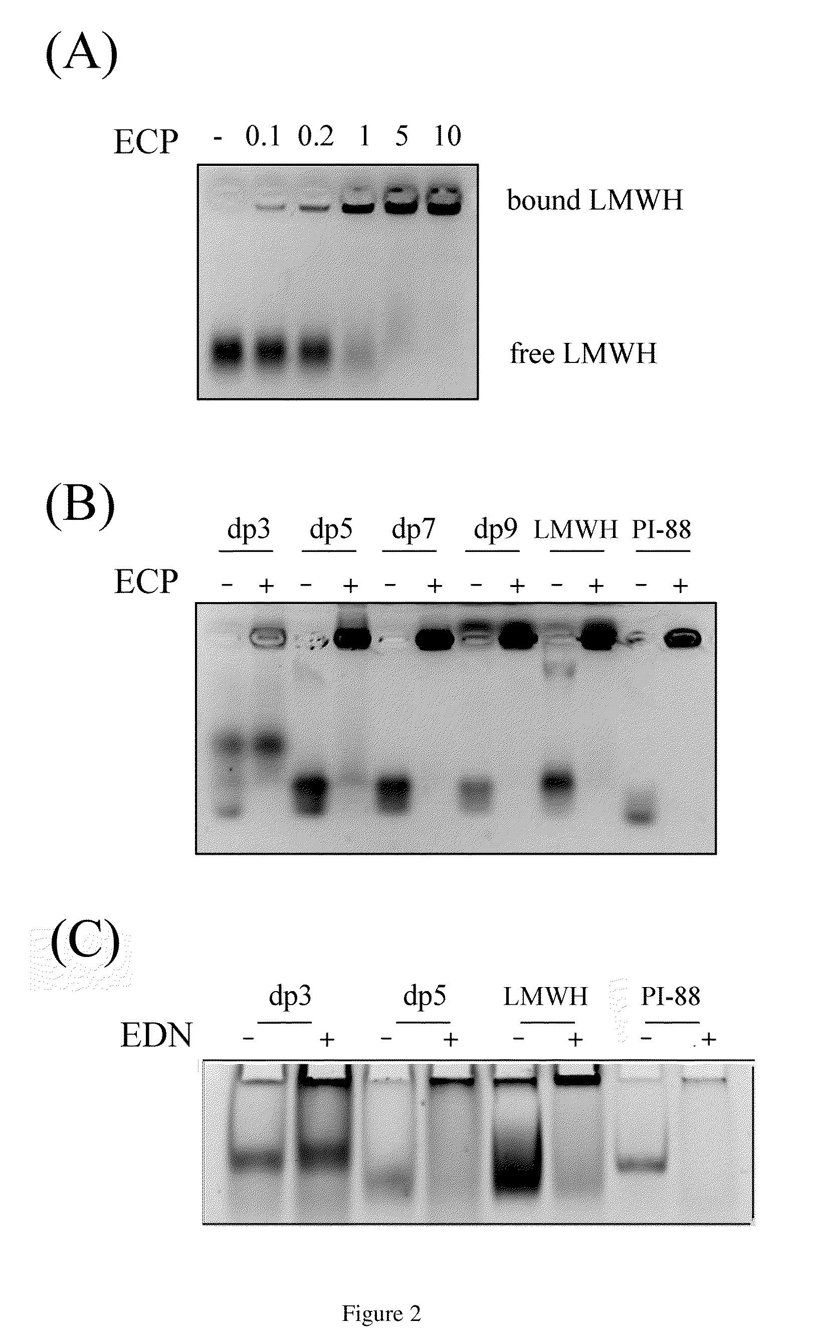
Oligosaccharide Length-Dependence of ECP-Heparin Interaction

[0030]Recombinant hECP containing a C-terminal His6 tag was expressed in E. coli BL21-CodonPlus(DE3) (Novagen, Madison, Wis.), purified by chromatography, and refolded as previously described (Boix, E., et al. (1999) Journal of Biological Chemistry 274, 15605-15614). MBP-ECP was purified using amylase affinity chromatography.

[0031]The prior art demonstrated that binding and endocytosis of ECP are HSPG dependent (Fan, T. C., et al. (2007) Traffic 8, 1778-1795). An early study demonstrated ECP purification using heparin-Sepharose chromatography (Gleich, G J., et al. (1986) Proc Natl Acad Sci USA 83, 3146-3150). Thus, the minimal heparin length (or disaccharide unit) required to interact with ECP was investigated by FACE (Fluorescence-assisted carbohydrate electrophoresis).

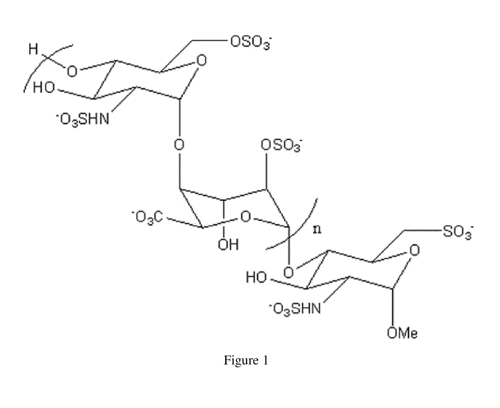
[0032]Synthetic heparin oligosaccharide (FIG. 1) (Lee, C. J., et al. (2004) J Am Chem Soc 126, 476-477), low molecular weight heparin (LMWH) and PI-88 (kind...

example 3

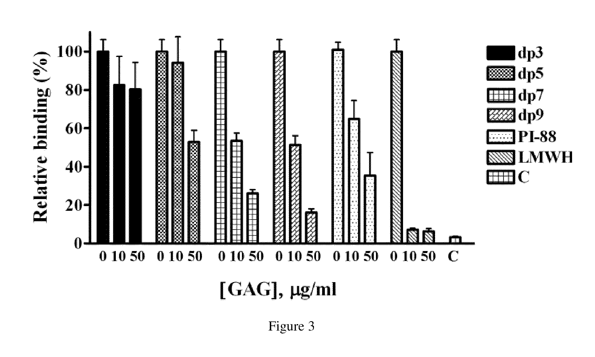
Competitive Inhibition of ECP Binding to Cells by Synthetic Oligosaccharides

[0034]Beas-2B, a human bronchial epithelial cell line, was cultured in RPMI 1640 medium (Sigma-Aldrich) supplemented with heat-inactivated 10% fetal bovine serum (Gibco / Invitrogen, Carlsbad, Calif.).

[0035]The ability of MBP-ECP to bind cells in the presence of serial dilutions of oligosaccharides or peptides was determined as described (Fan, T. C., et al. (2007) Traffic 8, 1778-1795). At the present invention, synthetic heparins (dp3-9) were tested for their ability to interfere with ECP binding to Beas-2B cells. Briefly, confluent monolayers of Beas-2B cells in 96-well plates were pretreated with various concentrations of oligosaccharides or peptides in serum-free RPMI 1640 medium at 4° C. for 30 min before incubation with 5 μg / ml MBP-ECP at 4° C. for 1 h. The cells were then washed with ice-cold PBS and fixed with 2% PFA at room temperature for 15 min prior to blocking with 2% BSA / PBS at room temperature f...


PUM


Abstract

A method for reducing cytotoxicity of eosinophil derived toxins comprising administering to a subject an effective amount of heparin, heparan sulfate, a potent heparanase inhibitor or a polypeptide which has sequence as follows: BZBXBX, XBBBXXBX, XBBXBX, BBXXBBBXXBB, BXBBXB, XBBBXXBBBXXBBX, or TXXBXXTBXXXTBB, wherein X represents any amino acid, Z represents an aromatic amino acid, and B represents a basic amino acid and T represents a turn.

Description

FIELD OF THE INVENTION[0001]The present invention relates to a heparin binding motif of eosinophil toxins and use thereof.BACKGROUND OF THE INVENTION[0002]Eosinophil cationic protein (ECP), a member of the ribonuclease A (RNase A) superfamily, is found in the specific granules of eosinophilic leukocytes. It is a single polypeptide with a molecular mass ranging from 16 to 21.4 kDa due to varying degrees of glycosylation. It shows a 67% amino acid sequence identity with eosinophil-derived neurotoxin (EDN), another eosinophil-secreted RNase. Although ECP shares the overall three-dimensional structure of RNase A, it has relatively lower RNase activity (Boix, E., et al. (1999) Journal of Biological Chemistry 274, 15605-15614). ECP released by activated eosinophils contributes to the toxicity against helminth parasites, bacteria, and single-strand RNA viruses (Lehrer, R., et al. (1989) Journal of Immunology 142, 4428-4434; Domachowske, J. B., et al. (1998) Nucleic acids research 26, 3358-...

Claims


Application Information

Patent Timeline
IPC IPC(8): C07K14/435C07K14/475
CPCC12N9/22C07K14/47A61P11/06
Inventor CHANG, MARGARET DAH-TSYRFAN, TAN-CHILIN, SHU-CHUAN
Owner NATIONAL TSING HUA UNIVERSITY
Why Eureka
Try Eureka