
Methods and devices for diagnosis of appendicitis

a technology for appendicitis and appendix, applied in chemical methods analysis, biochemistry apparatus and processes, instruments, etc., can solve the problems of insufficient identification and characterization of the appendix by imaging technology, limited ability to accurately recognize or diagnose the disease, and further handicapped by the expense of imaging technology, so as to improve the accuracy of method and increase the accuracy of appendicitis diagnosis. , the effect of increasing the accuracy of appendicitis

Inactive Publication Date: 2010-02-09
ASPENBIO PHARMA
View PDF26 Cites 5 Cited by
  • Summary
  • Abstract
  • Description
  • Claims
  • Application Information

AI Technical Summary

Benefits of technology

[0030]In one embodiment of this invention two or more molecules differentially associated with appendicitis are tested for. Identification of additional molecules provides greater accuracy to the method.
[0031]One molecule differentially associated with appendicitis is MRP8 / 14. Another is haptoglobin. Both these molecules can be tested for in diagnosing appendicitis. MRP8 / 14 levels in the range of about 1 to about 11 pg / ml are present in patients without appendicitis. Levels higher than this provide increased accuracy in diagnosing appendicitis. Levels higher than about 10, 11, 13, 15 or 20 μg / ml of MRP8 / 14 can be used to diagnose appendicitis. Haptoglobin levels in the range of about 27-139 mg / dL are found in patients without appendicitis. Levels higher than this, e.g., higher than 125, 130, 135, 139 and 150 provide increased accuracy in diagnosing appendicitis. Levels of 200 μg / ml indicate severe appendicitis.

Problems solved by technology

While medical science has an excellent understanding of appendicitis and its treatment, it is very limited in its ability to accurately recognize or diagnose the disease.
Imaging technology is inadequate in identifying and characterizing the appendix, especially in the early stages of the disease when treatment is likely to be most effective.
Imaging technology is further handicapped by its expense and its dependence upon the availability of highly trained and experienced people to interpret the studies.
This limitation affects thousands of people every year by inaccurately diagnosing their problem or by delaying the accurate diagnosis.
In cases of appendicitis, delays in diagnosis are the single most important factor leading to worsening of the condition and more complications related to the disease.
The misdiagnosis of appendicitis can lead not only to unnecessary surgery but also to delay of proper therapy for the actual underlying condition.
A dilemma for surgeons is how to minimize the negative appendectomy rate without increasing the incidence of perforation among patients referred for suspected appendicitis.
The secreted mucus of the appendix fills the closed lumen, causing an increase in intralumenal pressure and distension.
Currently, the diagnosis of appendicitis is difficult, and the difficulty persists during various stages in the progression of the condition.
If perforation occurs, a patient can present with severe pain and high fever.
At this very advanced stage, sepsis can be a serious risk with a potentially fatal outcome.
Also, this protein is not always associated with opportunistic infections (Froland, M. F., et al., 1994).
Without a reliable method for inexpensively testing the presence and severity of acute appendicitis, physicians must rely on costly imaging tests such as CT scans to tell them whether or not the patient truly has acute appendicitis and the urgency of appendectomy.
Furthermore, these imaging tests usually involve delays of several hours that could be used for appropriately treating the patient.

Method used


Image

  • Methods and devices for diagnosis of appendicitis
  • Methods and devices for diagnosis of appendicitis
  • Methods and devices for diagnosis of appendicitis

Examples


example 1

MRP8 / 14

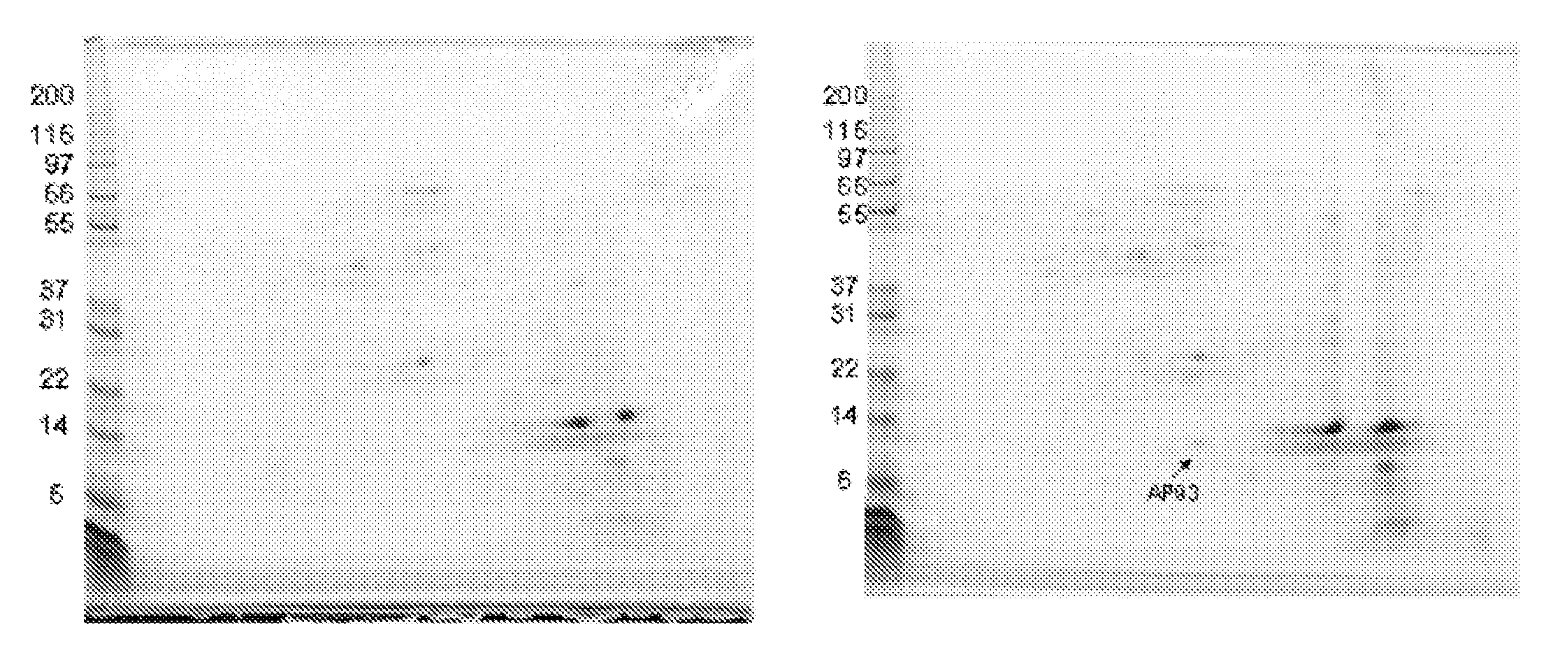
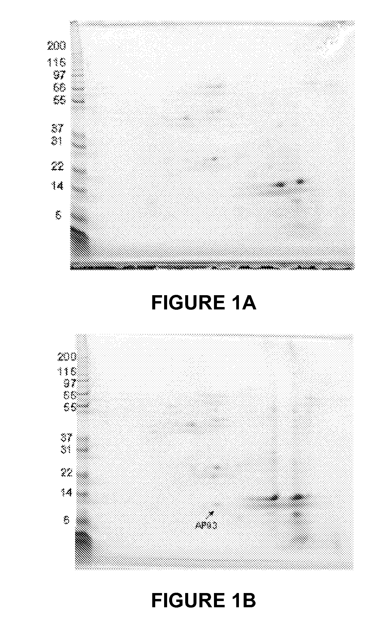
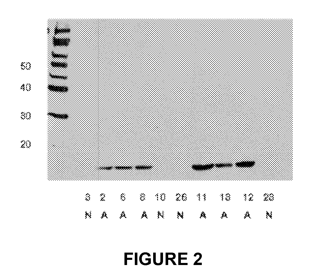
[0124]The objective of this study was to identify a tissue-specific marker that could contribute to the decision matrix for diagnosing early acute appendicitis. A proteomic screen was used to identify a protein in the appendix specifically upregulated in acute appendicitis. MRP8 / 14 was identified as present both in the diseased appendix and in serum of acute appendicitis patients.

[0125]MATERIALS AND METHODS. Specimen and Serum Collection. All patients enrolled in this study were treated according to accepted standards of care as defined by their treating physicians. Prior to being approached for inclusion in our study, all patients were evaluated by a surgeon and diagnosed by that surgeon as having appendicitis. The treating surgeon's plans for these appendicitis patients included an immediate appendectomy. The specifics of all treatments such as use of antibiotics, operative technique (either open or laparoscopic) were determined by the individual surgeon.

[0126]Exclusion Cri...

example 2

Haptoglobin

[0143]Using a proteomic screen of serum and appendix tissue, we determined that haptoglobin is upregulated in patients with acute appendicitis. The alpha subunit of haptoglobin is an especially useful marker in screening for the disease.

[0144]MATERIALS AND METHODS. Specimen and serum collection, appendicitis tissue processing, 2D gel analysis of extracted tissue samples, and western blot analysis of extracted appendix tissue samples were as described above in Example 1, except that for the western blot, affinity-purified anti-human haptoglobin (Rockland, 600-401-272) was used at a 1:5000 dilution in 0.5×uniblock for the primary antibody; and the secondary antibody was peroxidase anti-rabbit IgG (h+1), affinity purified (vector, pi-1000) in a 1:5000 dilution in uniblock.

[0145]RESULTS. Identification of proteins present in appendix tissue from appendicitis patients. A differential proteomic analysis was performed on depleted serum samples with the goal of identifying protei...

example 3

Method of Identifying Molecules using Fluid Samples

[0151]In variations of this example, fluid samples can include whole blood, serum, or plasma. The samples were whole blood collected from human patients immediately prior to an appendectomy. The specimens were placed on ice and transported to the lab. The blood was then processed by centrifugation at 3000 rpm for 15 minutes. Plasma was then separated by pouring into another container

[0152]Upon performing an appendectomy, a patient was classified as having appendicitis (AP) or non-appendicitis (NAP). The classification was based on clinical evaluation, pathology, or both as known in the art. For cases of appendicitis, the clinical condition was also characterized as either perforated or non-perforated.

[0153]The samples from AP patients were pooled and divided into aliquots. Pooled aliquots were treated so as to remove selected components such as antibodies and serum albumin. Similarly, the samples from NAP patients were pooled and di...


PUM

PropertyMeasurementUnit
temperatureaaaaaaaaaa
ionic strengthaaaaaaaaaa

Abstract

A method is provided for determining the severity of appendicitis in a patient that includes testing a blood, serum or plasma sample from the patient for the quantity of MRP8 / 14 in the sample and comparing it with the quantity of MRP8 / 14 present in standard samples correlated with an appendicitis severity scoring system. A histologically-based appendicitis severity scoring system is also provided. Immunoassays and kits for performing the appendicitis assays of this invention are also provided, as are standard samples and data correlating MRP8 / 14 quantities present in patient samples to histologically-based appendicitis severity grades. The methods and immunoassay devices and kits of this invention are useful for managing the treatment of patients presenting with appendicitis symptoms.

Description

CROSS-REFERENCE TO RELATED APPLICATIONS[0001]This application is a continuation-in-part application of U.S. application Ser. No. 11 / 189,120 filed Jul. 25, 2005, which claims priority to U.S. provisional application Ser. No. 60 / 590,631 filed Jul. 23, 2004, both of which are incorporated by reference to the extent not inconsistent herewith.BACKGROUND OF THE INVENTION[0002]Appendicitis is a common acute surgical problem affecting human beings of a wide age range. There are approximately 700,000 cases annually in the United States. A large proportion of cases occur in the 10 to 30 age group. An accurate diagnosis at a sufficiently early stage is a significant factor in achieving a successful outcome.[0003]Many people present to their physician with symptoms suggestive of appendicitis but caused by other ailments such as viral infections. Differentiating the appendicitis patients from those affected with other ailments is a daunting clinical task that physicians face daily. While medical...

Claims


Application Information

Patent Timeline
Patent Type & Authority Patents(United States)
IPC IPC(8): G01N33/00C12Q1/00G01N33/53
CPCC12Q1/6883G01N33/6893Y10T436/10C12Q2600/112C12Q2600/158G01N2800/06
Inventor COLGIN, MARK A.BEALER, JOHN F.DONNELLY, RICHARD G.
Owner ASPENBIO PHARMA
Learn More