Combination cancer immunotherapy with co-stimulatory molecules

a cancer immunotherapy and co-stimulatory technology, applied in the field of cancer immunotherapy, can solve the problems of incomplete tumor reduction, only a delay in the growth of well-established tumor implants, inadequate immune response, etc., and achieve the effect of reducing immunoregulatory t cell activity

Active Publication Date: 2010-04-13
UNIV OF SOUTHERN CALIFORNIA
View PDF13 Cites 85 Cited by
  • Summary
  • Abstract
  • Description
  • Claims
  • Application Information

AI Technical Summary

Benefits of technology

[0011]Provided are new cancer immunotherapeutic methods that combine administration of soluble co-stimulatory molecules with treatment to reduce immunoregulatory T cell activity in the individual.
[0022]The activity of immunoregulatory T cells in vivo may be reduced by administering a GITR ligand agonist.

Problems solved by technology

Most tumors express antigens that can be recognized to a variable extent by the host immune system, but in many cases, the immune response is inadequate.
Failure to elicit a strong activation of effector T-cells may result from the weak immunogenicity of tumor antigens or inappropriate or absent expression of co-stimulatory molecules by tumor cells.
However, in most of these studies, T-reg depletion resulted in either incomplete tumor reduction or only a delay in the growth of well-established tumor implants.

Method used


Image

  • Combination cancer immunotherapy with co-stimulatory molecules
  • Combination cancer immunotherapy with co-stimulatory molecules
  • Combination cancer immunotherapy with co-stimulatory molecules

Examples


example 1

Materials and Methods

[0102]Reagents and Mice

[0103]The Glutamine Synthetase Gene Amplification System, including the expression plasmid pEE12, was obtained from Lonza Biologics (Slough, U.K.). The plasimid pBJ, containing human B7.1 (huB7.1) cDNA (nucleotide sequence identical to GenBank accession number M27533), was obtained from the American Type Culture Collection (Manassas, Va.). Restriction endonucleases, T4 DNA ligase, Vent polymerase, and other molecular biology reagents were obtained from either New England Biolabs (Beverly, Mass.) or Boehringer Mannheim (Indianapolis, Ind.). Chloramine T, GSEM (50×), and 2,2′-azino-bis(3-ethylbenzthiazoline-6-sulfonic acid) diammonium salt (ABTS) were obtained from Sigma Chemical Co. (St. Louis, Mo.). Characterized and dialyzed fetal calf sera (FCS) were obtained from Hyclone Corp. (Logan, Utah), and RPMI 640 medium, Selective Hybridoma Medium (SFM) without L-glutamine, MEM non-essential amino acids solution (100×), and phosphate-buffered sa...

example 2

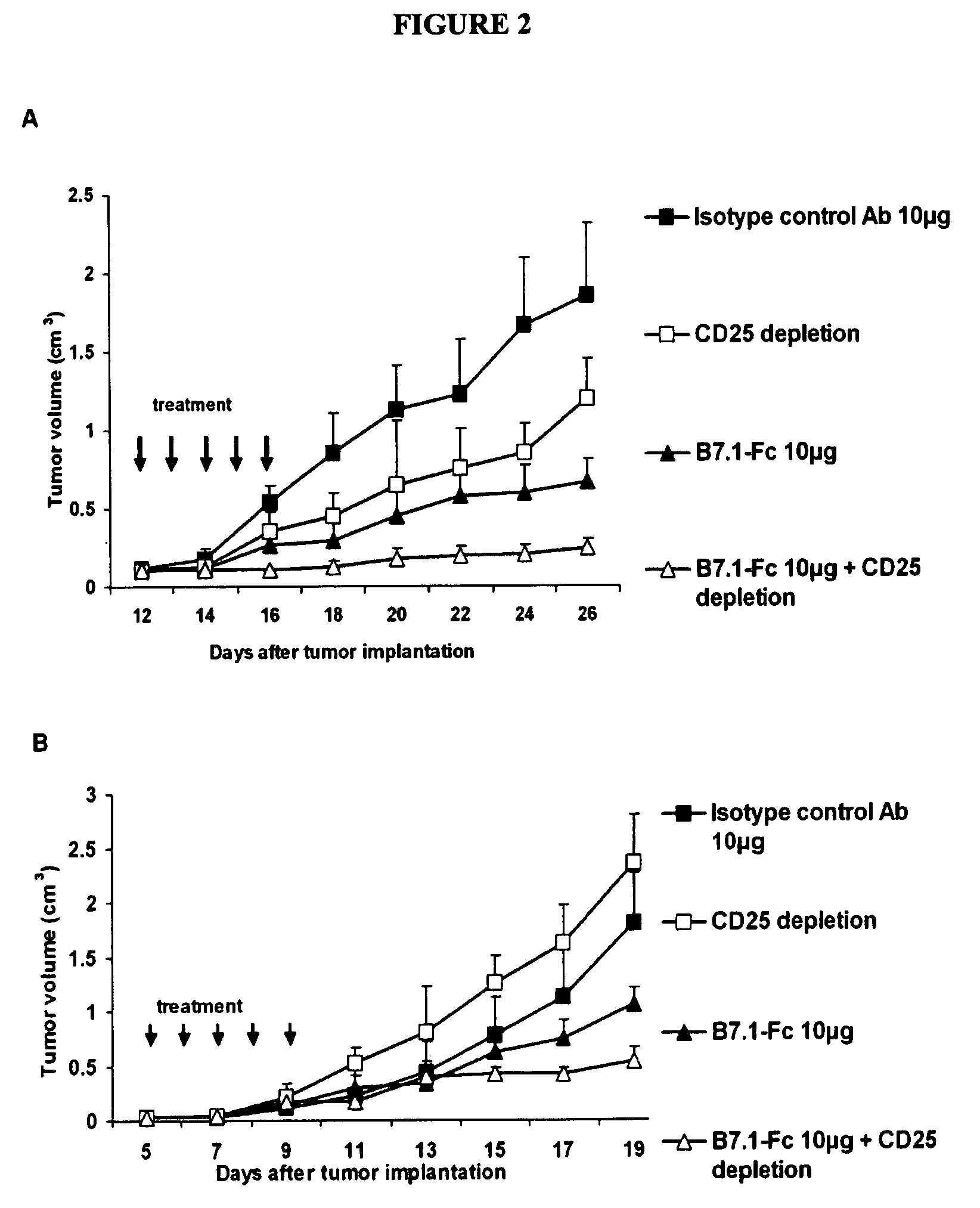
Results of B7.1-Fc and B7.2-Fc Cancer Therapy

[0133]Construction, Expression, and Purification of B7.1-Fc

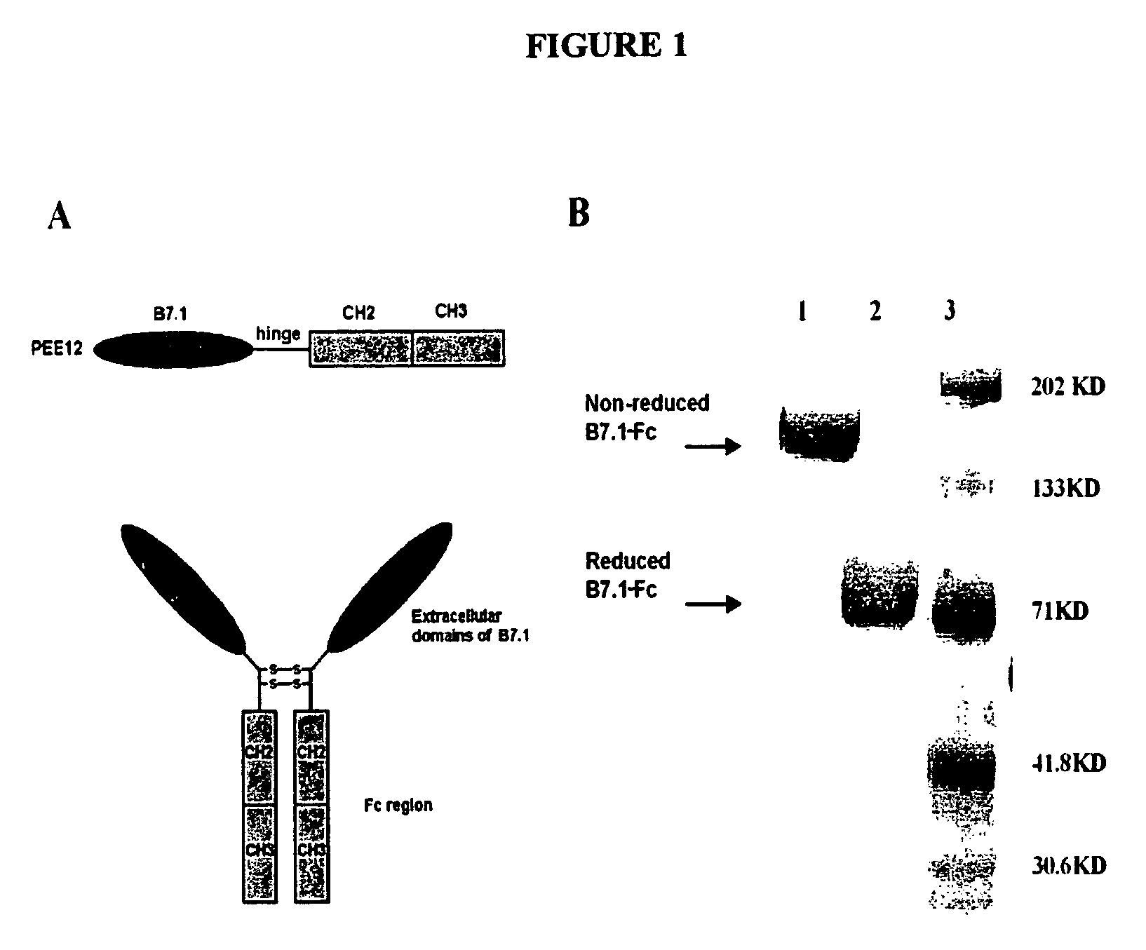
[0134]The C-terminus of B7.1 extracellular domain was fused onto the N-terminus of the hinge-CH2-CH3 molecule without any linker (FIG. 1). The fused B7.1-Fc was translated under human B7.1 signal sequence and the expressed fusion protein was a dimer as shown by SDS-PAGE. Cells expressing the highest leves of B7.1-Fc fusion protein were selected using a sandwich ELISA with monoclonal antibodies against human B7.1 or human IgG Fc. This subclone was found to produce over 100 μg / ml of fusion protein in aerated stir flask cultures.

[0135]The predicated molecular weight of the expressed fusion polypeptide was confirmed by SDS-PAGE under reducing conditions. The purity of the fusion protein was confirmed by HPLC which showed a main peak with a retention time of approximately 320 s. The activity of the fusion protein remains intact for one year when stored at −80° C.

[0136]Bioactivity of B7...

example 3

Cloning and Testing of B7.1 / NHS76 Fusion Protein

[0159]Construction, Expression, and Purification of B7.1 / NHS76 Fusion Protein

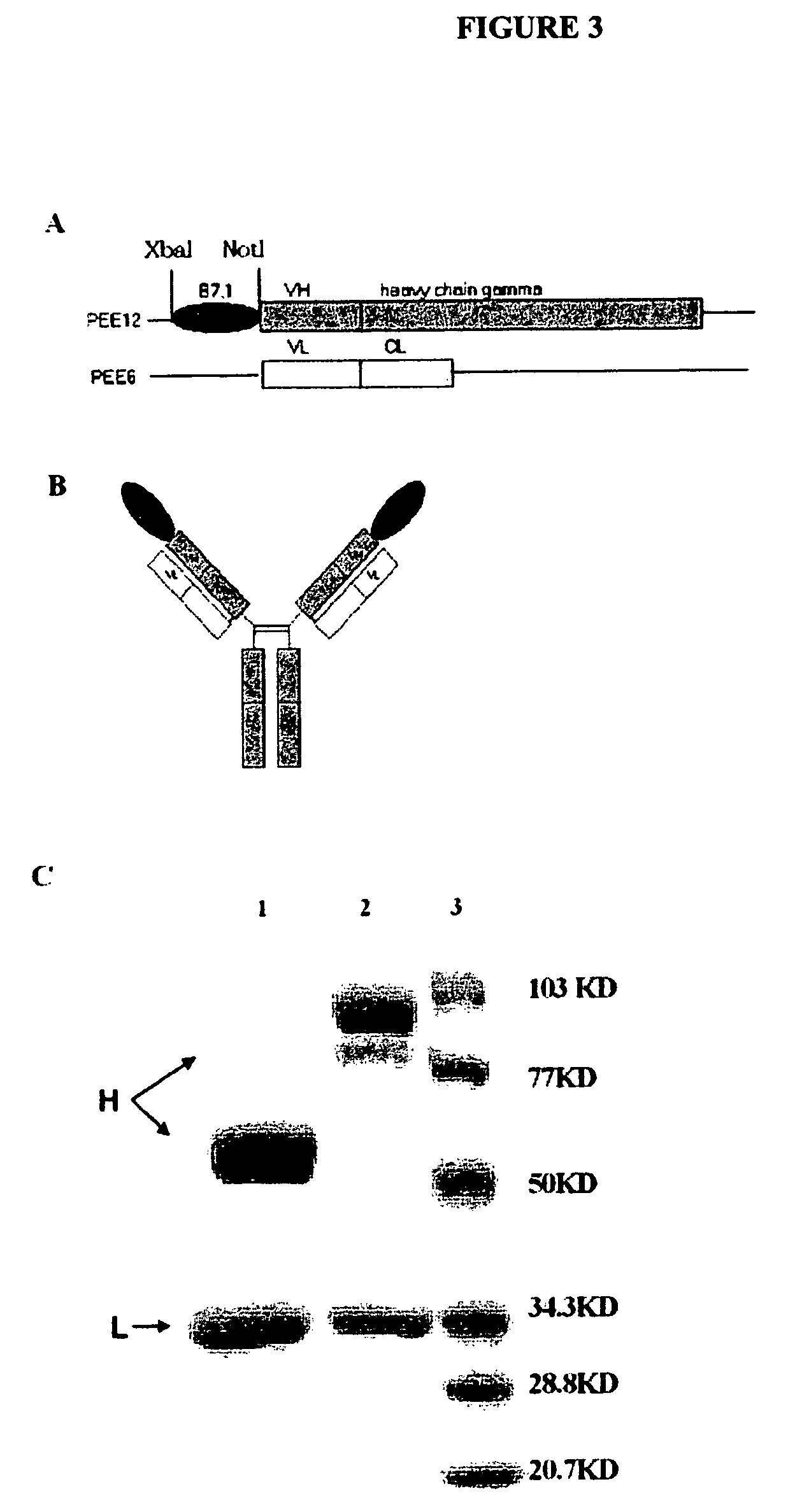
[0160]The plasmids carrying the light chain (pEE6 / NHS76λ) and heavy chain (pEE12 / NHS76 HC) sequences were constructed as described previously (see Sharifi, et al. 2001. Hybridoma Hybridomics 20:305-311). The extracellular domains of human B7.1 (Base 318-1040 Genbank: M27533) (Gordon, et al., 1989 J. Immunol. 143:2714-2722) were amplified by PCR from full-length cDNA of human B7.1 using primers 5′-TTC TCT AGA ATG GGC CAC ACA CGG-3′ (SEQ ID NO: 1) and 5′-AAT AGC GGC CGC ATC AGG AAA ATG CTC TTG-3′ (SEQ ID NO: 5). The PCR product was then inserted into the N-terminus of the NHS76 HC gene in pEE12 / NHS76 HC vector by XbaI and NotI under the translation of B7.1 leader sequence. B7.1 was linked at its C-terminus to the antibody variable region to preserve the activity of B7.1 which depends on its amino terminus. The resulting expression vector pEE12 / B7.1 / NHS76 HC was ...


PUM

PropertyMeasurementUnit
diameteraaaaaaaaaa
diameteraaaaaaaaaa
concentrationaaaaaaaaaa

Abstract

Provided are methods of reducing the size of a tumor or inhibiting the growth of cancer cells in an individual or inhibiting the development of metastatic cancer by administering an effective amount of a soluble form of a co-stimulatory molecule from an antigen presenting cell and by reducing the activity of immunoregulatory T cells in the individual. Methods of reduction in the activity of immunoregulatory T cells involve removing them ex vivo or depleting or inactivating them in vivo. Also provided are cancer therapeutic compositions comprising a soluble form of a co-stimulatory molecule from an antigen presenting cell and an antibody specific for an intracellular antigen.

Description

CROSS REFERENCE TO RELATED PATENT APPLICATIONS[0001]This application claims priority under 35 U.S.C. §119(e) to U.S. Application Ser. No. 60 / 623,849, filed Oct. 29, 2004, incorporated herein by reference in its entirety.FIELD OF THE INVENTION[0002]The present invention relates to cancer immunotherapeutic methods.BACKGROUND OF THE INVENTION[0003]The following discussion of the background of the invention is merely provided to aid the reader in understanding the invention and is not admitted to describe or constitute prior art to the present invention.[0004]Surgery, radiation therapy, and chemotherapy have been the standard accepted approaches for treatment of cancers including leukemia, solid tumors, and metastases. Immunotherapy (sometimes called biological therapy, biotherapy, or biological response modifier therapy), which uses the body's immune system, either directly or indirectly, to shrink or eradicate cancer has been studied for many years as an adjunct to conventional cancer...

Claims


Application Information

Patent Timeline
Patent Type & Authority Patents(United States)
IPC IPC(8): A61K38/17A61K38/16A61K39/395A61K39/00
CPCC07K14/70532C07K16/2866C07K16/30C07K16/2878A61K2039/505C07K2319/75C07K2319/30A61P35/00A61P35/04
Inventor EPSTEIN, ALANHU, PEISHING
Owner UNIV OF SOUTHERN CALIFORNIA
Why Eureka
Try Eureka