

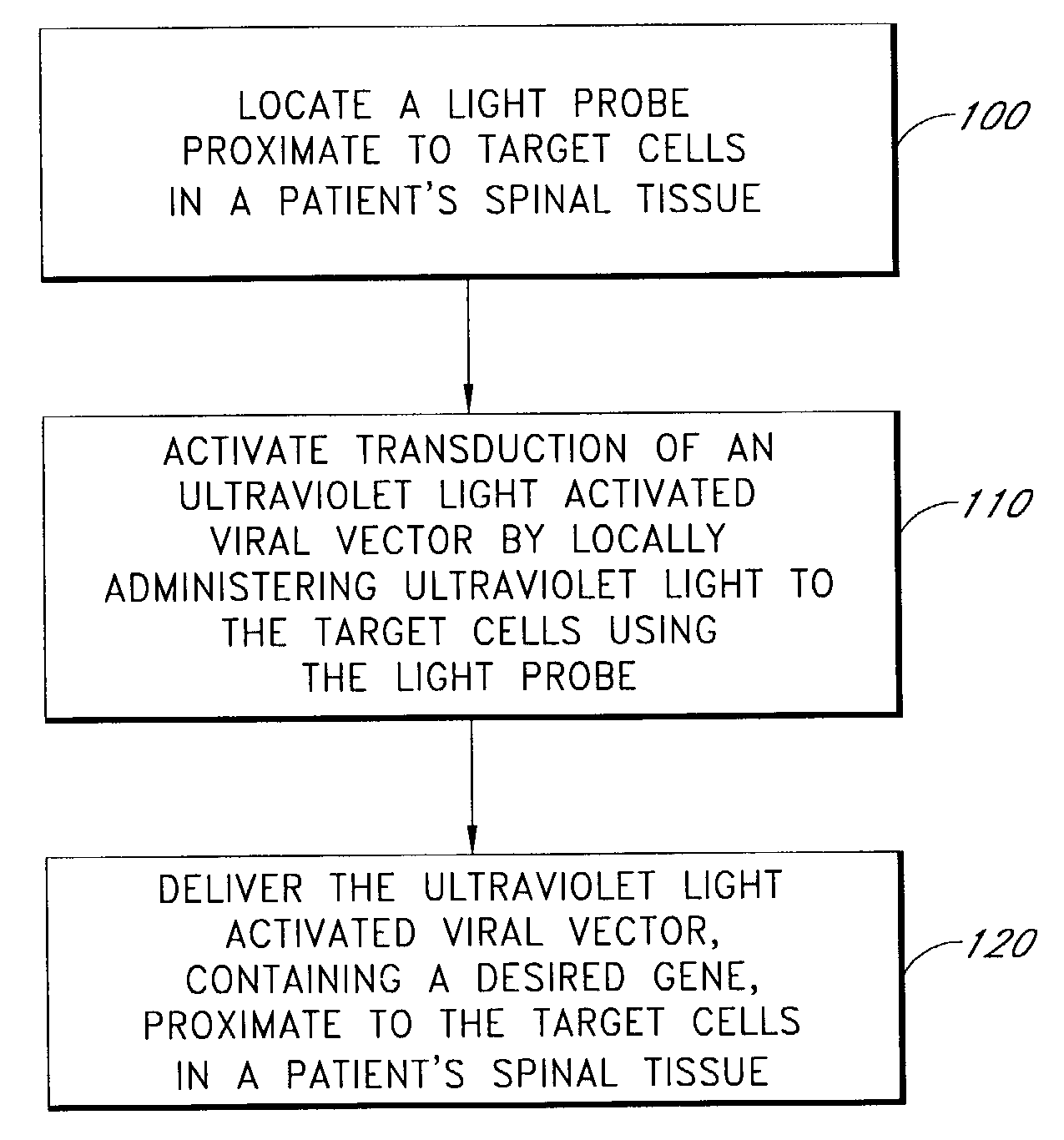
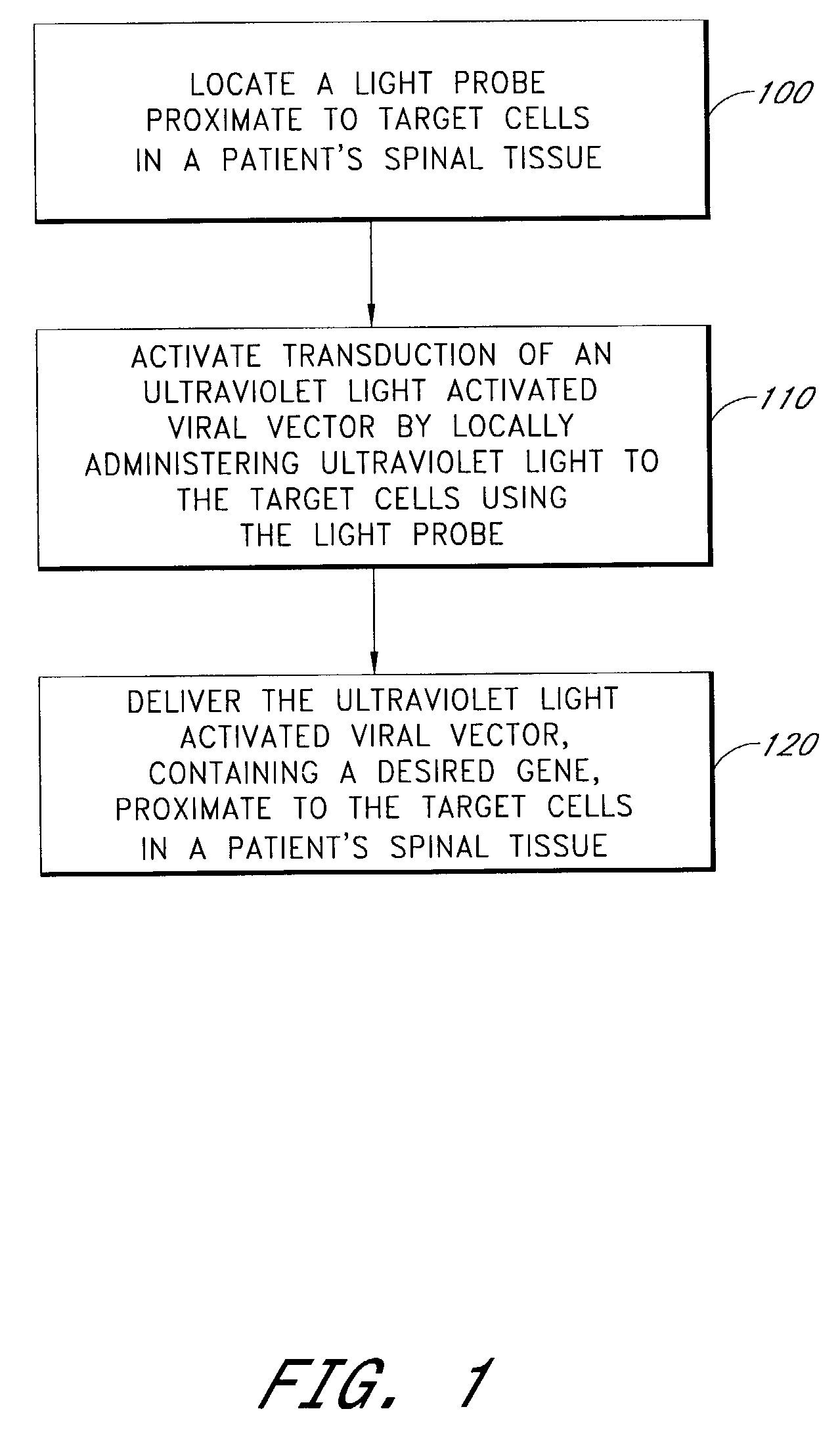
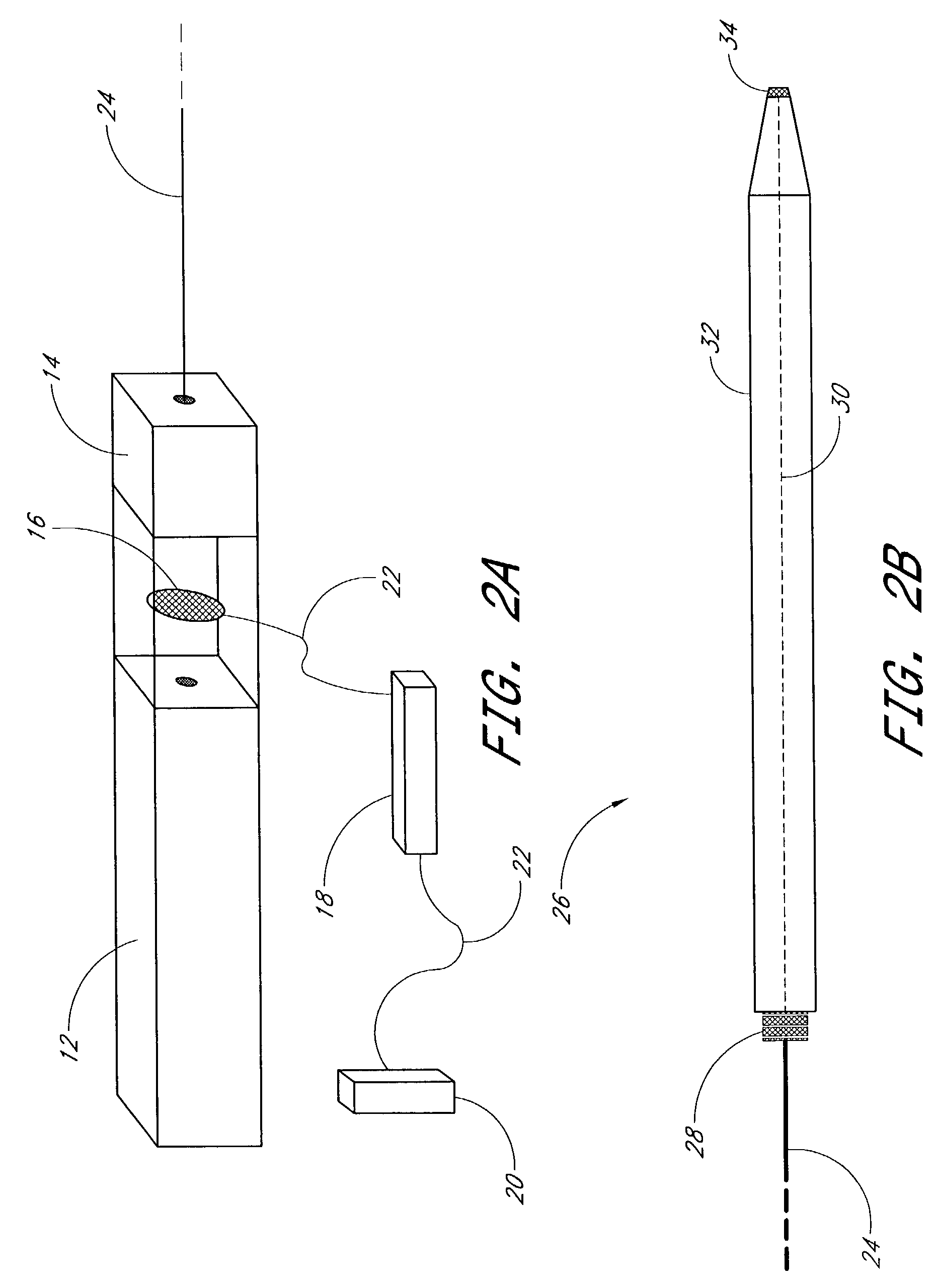
Method for introducing an ultraviolet light activated viral vector into the spinal column
a technology of ultraviolet light and activated viral vector, which is applied in the field of gene therapy, can solve the problems of reducing the range of motion and pain and/or disability, increasing the rate of second strand synthesis, and reducing the absorption of biomechanical shock, so as to facilitate the rebuilding of important components and effectively stimulate the transduction of uv activated viral vector.
- Summary
- Abstract
- Description
- Claims
- Application Information
AI Technical Summary
Benefits of technology
Problems solved by technology
Method used
Image

Examples
example 1
I. Methods
[0058]A. Isolation of Human Mesenchymal Stem Cells
[0059]Human Mesenchymal Stem Cells (hMSC) were isolated from patient blood samples harvested from the iliac crest. The blood samples were diluted in an equal volume of sterile Phosphate Buffered Saline (PBS). The diluted sample was then gently layered over 10 ml of Lymphoprep (Media Prep) in a 50 ml conical tube (Corning). The samples were then centrifuged at 1800 rpm for 30 minutes. This isolation protocol is a standard laboratory technique, and the resulting gradient that formed enabled the isolation of the hMSCs from the layer immediately above the Lymphoprep. The isolated fraction was placed into a new 50 ml conical tube, along with an additional 20 ml of sterile PBS. The sample was centrifuged at 1400 rpm for 8 minutes. The supernatant was removed the cell pellet was resuspended in 20 ml for fresh PBS, and centrifuged again for 8 minutes at 1400 rpm. Afterwards the supernatant was removed, the cell pellet was resuspend...
PUM
| Property | Measurement | Unit |
|---|---|---|
| wavelength | aaaaa | aaaaa |
| wavelength | aaaaa | aaaaa |
| wavelength | aaaaa | aaaaa |
Abstract
Description
Claims
Application Information