Method and system for digital image based tissue independent simultaneous nucleus cytoplasm and membrane quantitation

a tissue independent, cytoplasm and membrane quantitation technology, applied in the field of digital image processing, can solve the problems of affecting the work of many investigators, the enormous amount of image data generated in such cases, and the need for efficient software analysis tools

Active Publication Date: 2010-07-20
VENTANA MEDICAL SYST INC
View PDF10 Cites 46 Cited by
  • Summary
  • Abstract
  • Description
  • Claims
  • Application Information

AI Technical Summary

Problems solved by technology

Converting large amounts raw data including raw data on digital images generated in these experiments into meaningful information that can be used by an analyst to formulate an opinion remains a challenge that hinders many investigators.
The image data generated in such cases is tremendous and require efficient software analysis tools.
Since, cancer is life threatening when it becomes invasive, it carries a potential for spreading and metastasis.
Clinical studies in patients with breast cancer over the last decade have convincingly demonstrated that amplification / over expression of HER-2 / neu is associated with a poor medical prognosis.
Gene amplification of HER-2 / neu is associated with aggressive cell behavior and poor prognosis.
Prostate cancer strikes as many men (and causes almost as many deaths annually) as breast cancer does in women, but lacks the national awareness and research funding breast cancer currently receives.
However, many images do not have pixel values that make effective use of the full dynamic range of pixel values available on an output device.
The result in either case is that the output is relatively dull in appearance.
There are several problems associated with using existing digital image analysis techniques for analyzing digital images for determining know medical conditions.
One problem is that existing digital image analysis techniques are typically used only for analyzing measurements of chemical compounds applied to biological samples such as groups of cells from a tissue sample.
Another problem is the manual method used by pathologists is time consuming and prone to error including missing areas of the slide including tumor or cancer cells.
There is a considerable gap between the pace at which drug discovery is progressing and automated tools available to assist the drug researcher.
Often researchers are forced to resort to manual methods which are subjective, time consuming and could be inconsistent.
Difficulties are reported with the performance and interpretation of IHC and FISH in the medical oncology community over the last several years.
In particular the detection of estrogen and progesterone receptors and the detection of HER2 / neu targets is not consistent.
Problems encountered include, but are not limited to, identification of cutoff points in assay scores to define positive vs. negative results, broad interlaboratory variability in performance of the assays, discrepancies between laboratories with high volume vs low volume throughput, use of “home brew” antibodies for IHC, deviations from recommended methods in the package insert leading to altered performance characteristics of the assays, conflicting data in the published literature, and lack of data from prospectively conducted studies.
There are several problems associated with using existing digital image analysis techniques for analyzing FISH images.
Once there are a large number of image sections to be processed together, issues like seamless composition of tiles becomes an issue.
3) Another problem with FISH based analysis is the need to stack planes in three dimensions to get a focused image.
Failure to obtain interpretable results after two test runs occurred in 5.0% of samples tested by FISH and 0.08% of those tested by immunohistochemistry.
On the website (www.ihcworld.com / _protocols / general_IHC / immunoenzyme_double.htm), it is stated that this enzyme based double staining method is limited for the demonstration of two proteins at different locations.
Often, the tissue origin of a tumor cannot be determined with routine histology.
Early metastasis can be difficult to detect using conventional histology.
Limitation of the techniques available in prior art is the ability to identify and quantify individual cell components.
Each of the three components can get stained or unstained or counterstained.
Observing a three dimensional cell in two dimensions has its own issues.
Simultaneous identification of components becomes complex if more than one component behaves similar.
That is, if nucleus and cytoplasm take stain and appear brown red, detecting the boundary between cytoplasm and membrane is much more difficult than the case where only membrane gets stained.
Similarly, it will be very difficult to locate nucleus in a cell if both nucleus and cytoplasm are stained similar.
However, cocktail staining is found to be unsuitable for some applications.
None of the existing products are capable of providing simultaneous quantitation of nucleus, cytoplasm and membrane, which is a key factor for studies of translocation.
However, these attempts still do not solve all of the problems with automated biological analysis systems that have been developed to improve the speed and accuracy of the testing process.

Method used


Image

  • Method and system for digital image based tissue independent simultaneous nucleus cytoplasm and membrane quantitation
  • Method and system for digital image based tissue independent simultaneous nucleus cytoplasm and membrane quantitation
  • Method and system for digital image based tissue independent simultaneous nucleus cytoplasm and membrane quantitation

Examples


Embodiment Construction

Exemplary Biological Sample Analysis System

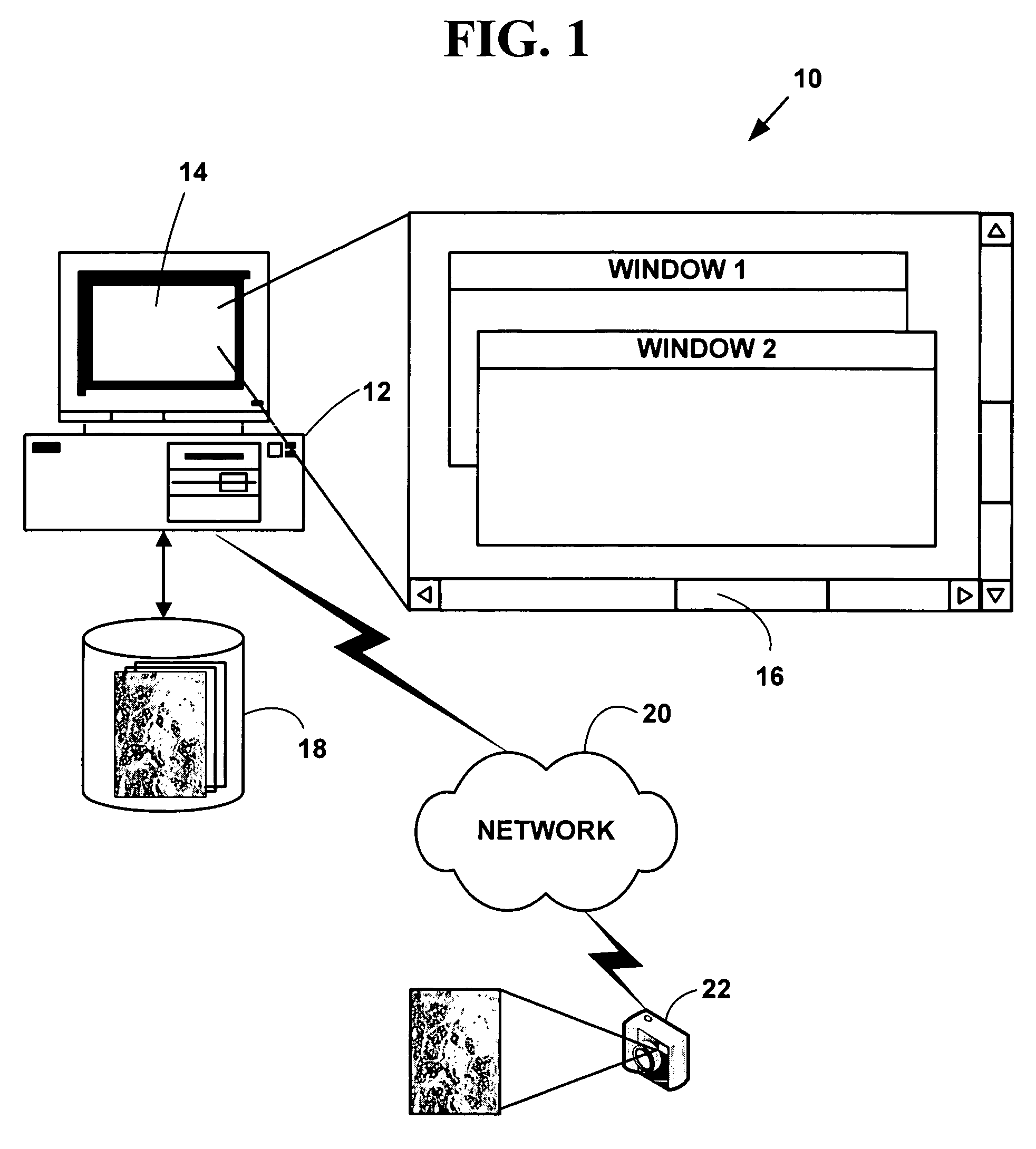
[0101]FIG. 1 is a block diagram illustrating an exemplary biological sample analysis processing system 10. The exemplary biological sample analysis processing system 10 includes one or more computers 12 with a computer display 14 (one of which is illustrated). The computer display 14 presents a windowed graphical user interface (“GUI”) 16 with multiple windows to a user. The present invention may optionally include a microscope or other magnifying device (not illustrated in FIG. 1) and / or a digital camera 18 or analog camera. One or more databases 20 (one or which is illustrated) include biological sample information in various digital images or digital data formats. The databases 20 may be integral to a memory system on the computer 12 or in secondary storage such as a hard disk, floppy disk, optical disk, or other non-volatile mass storage devices. The computer 12 and the databases 20 may also be connected to an accessible via one or more...


PUM


Abstract

A method and system for automatic digital image based tissue independent simultaneous nucleus, cytoplasm and membrane quantitation. Plural types of pixels comprising cell components including at least cell cytoplasm and cell membranes from a biological tissue sample to which a chemical compound has been applied and has been processed to remove background pixels and pixels including counterstained components are simultaneously identified. The identified cell components pixels are automatically classified to determine a medical conclusion such as a human breast cancer, a human prostrate cancer or an animal cancer.

Description

CROSS REFERENCES TO RELATED APPLICATIONS[0001]This application claims priority to U.S. Provisional Patent Application No. 60 / 655,465, filed Feb. 23, 2005, the contents of which are incorporated by reference. This application is a CIP of U.S. patent application Ser. No. 10 / 938,314, filed Sep. 10, 2004, which claims priority to U.S. Provisional Patent Application No. 60 / 501,412, filed Sep. 10, 2003, and U.S. Provisional Patent Application No. 60 / 515,582 filed Oct. 30, 2003, and this application is a alp of U.S. patent application Ser. No. 10 / 966,071, filed Oct. 15, 2004 which claims priority to U.S. Provisional Patent Application No. 60 / 530,714, filed Dec. 18, 2003, the contents of all of which are incorporated by reference.COPYRIGHT NOTICE[0002]Pursuant to 37 C.F.R. 1.71(e), applicants note that a portion of this disclosure contains material that is subject to and for which is claimed copyright protection, such as, but not limited to, digital photographs, screen shots, user interface...

Claims


Application Information

Patent Timeline
Patent Type & Authority Patents(United States)
IPC IPC(8): G06K9/00
CPCG06K9/00127Y10S128/922G06V20/69
Inventor GHOLAP, ABHIJEET S.GHOLAP, GAURI A.JOSHI, APARNABASU, AMITABHARAO, CHIRUVOLU V. K.JADHAV, PRITHVIRAJ
Owner VENTANA MEDICAL SYST INC
Why Eureka
Try Eureka