Time-lapse cell cycle analysis of unstained nuclei

a cell cycle analysis and time-lapse technology, applied in image analysis, image enhancement, instruments, etc., can solve the problems of inability to distinguish between the various phases of the cell cycle, toxic side effects of fluorescent dyes, and barriers to long-term cellular imaging

Inactive Publication Date: 2010-10-19
GENERAL ELECTRIC CO
View PDF29 Cites 9 Cited by
  • Summary
  • Abstract
  • Description
  • Claims
  • Application Information

AI Technical Summary

Problems solved by technology

Further, it is also useful to determine if an uncharacterized compound has the effect of increasing progression through the cell cycle, because such a compound may be potentially carcinogenic.
The common use of fluorescent dyes presents barriers for long-term cellular imaging.
While such dyes may be suitable for fixed cell assays or for live assays of short duration (e.g. several hours), fluorescent dyes have toxic side effects that prevent long-term studies.
If cells are stained with a nuclear dye and cultured for extended periods, they die, either because they cannot replicate their DNA when it is intercalated with dye, or because the dye interferes with chromosome segregation during mitosis.
This approach also does not distinguish among the various phases of the cell cycle.

Method used


Image

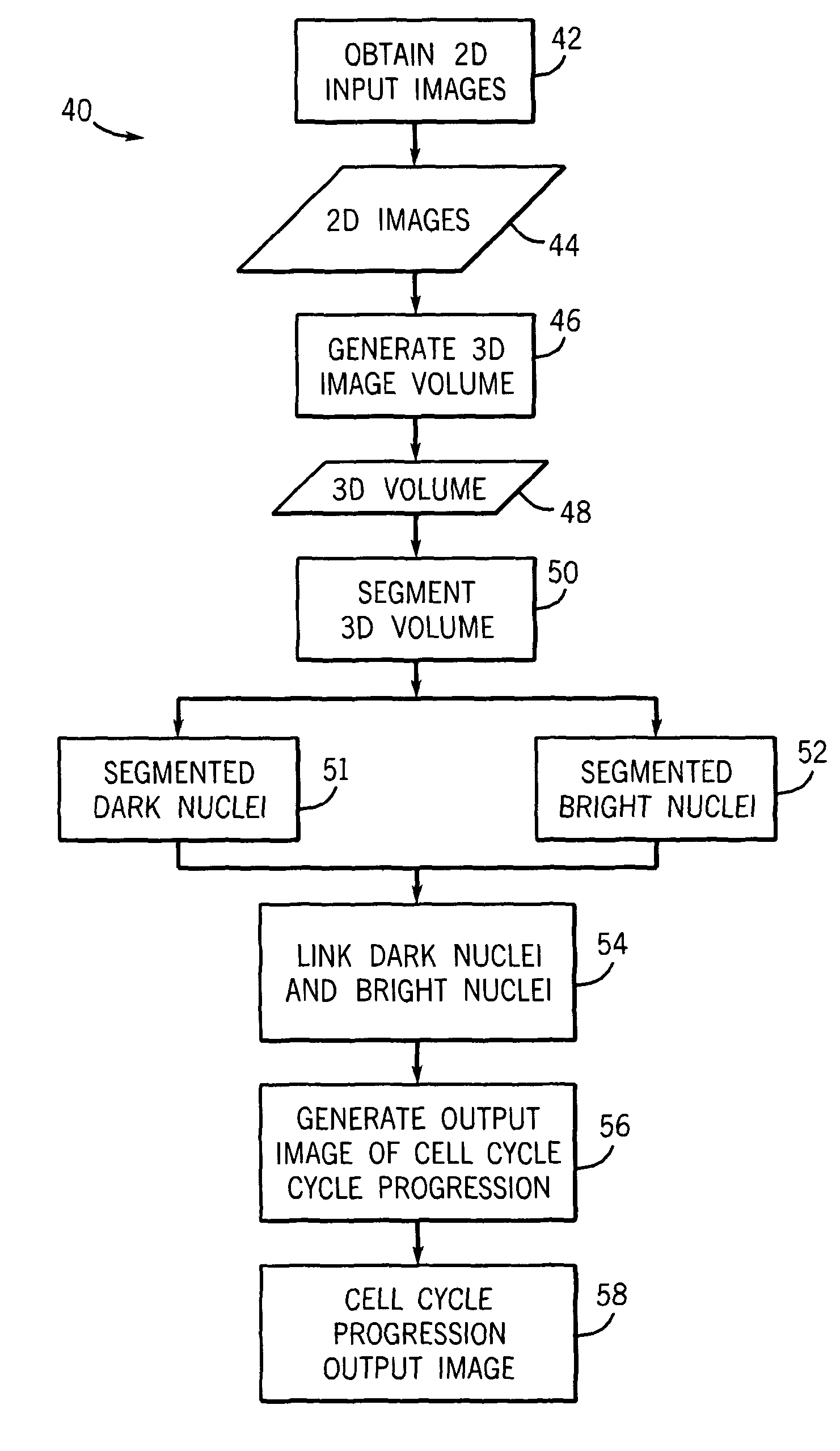
  • Time-lapse cell cycle analysis of unstained nuclei
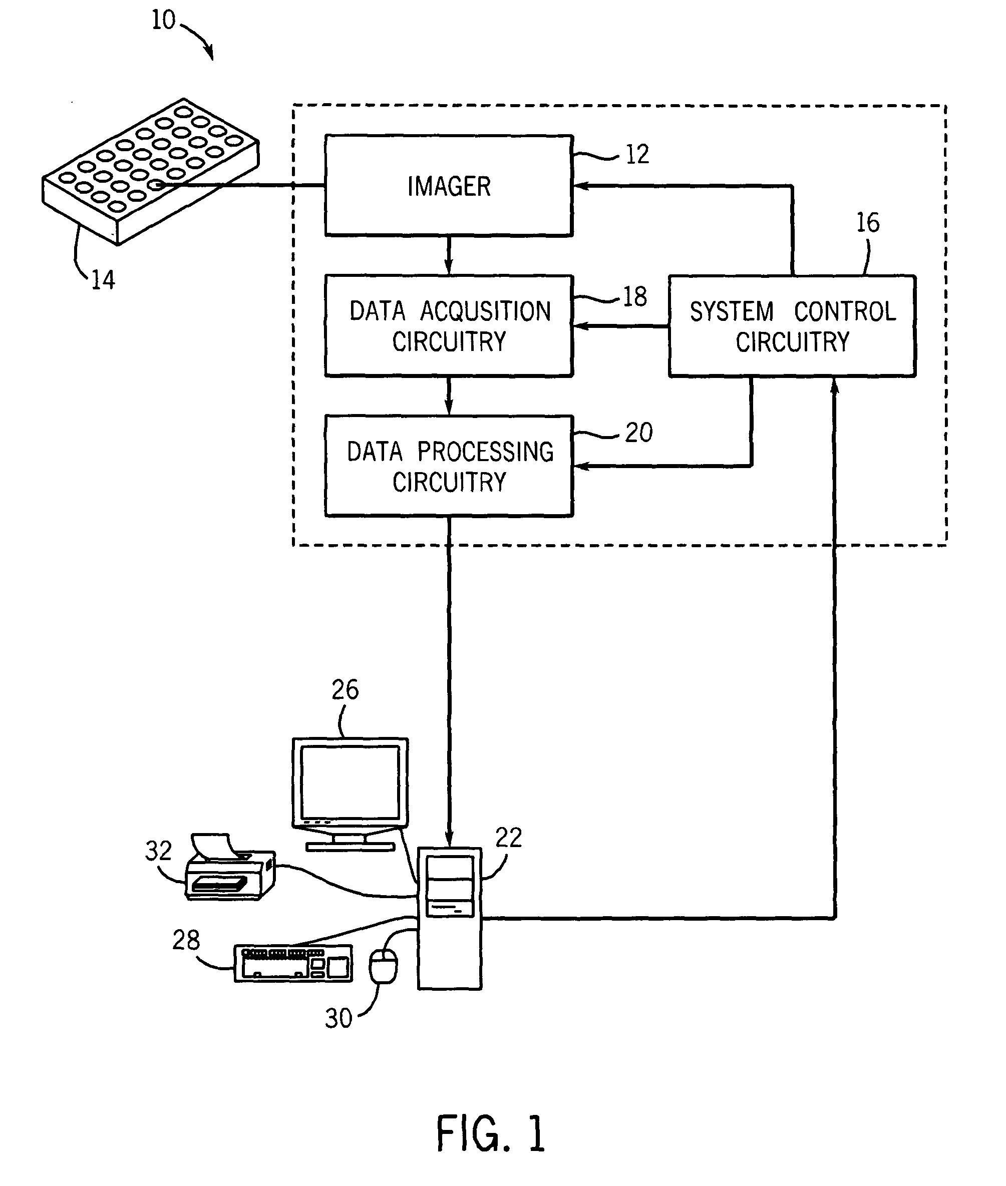
  • Time-lapse cell cycle analysis of unstained nuclei
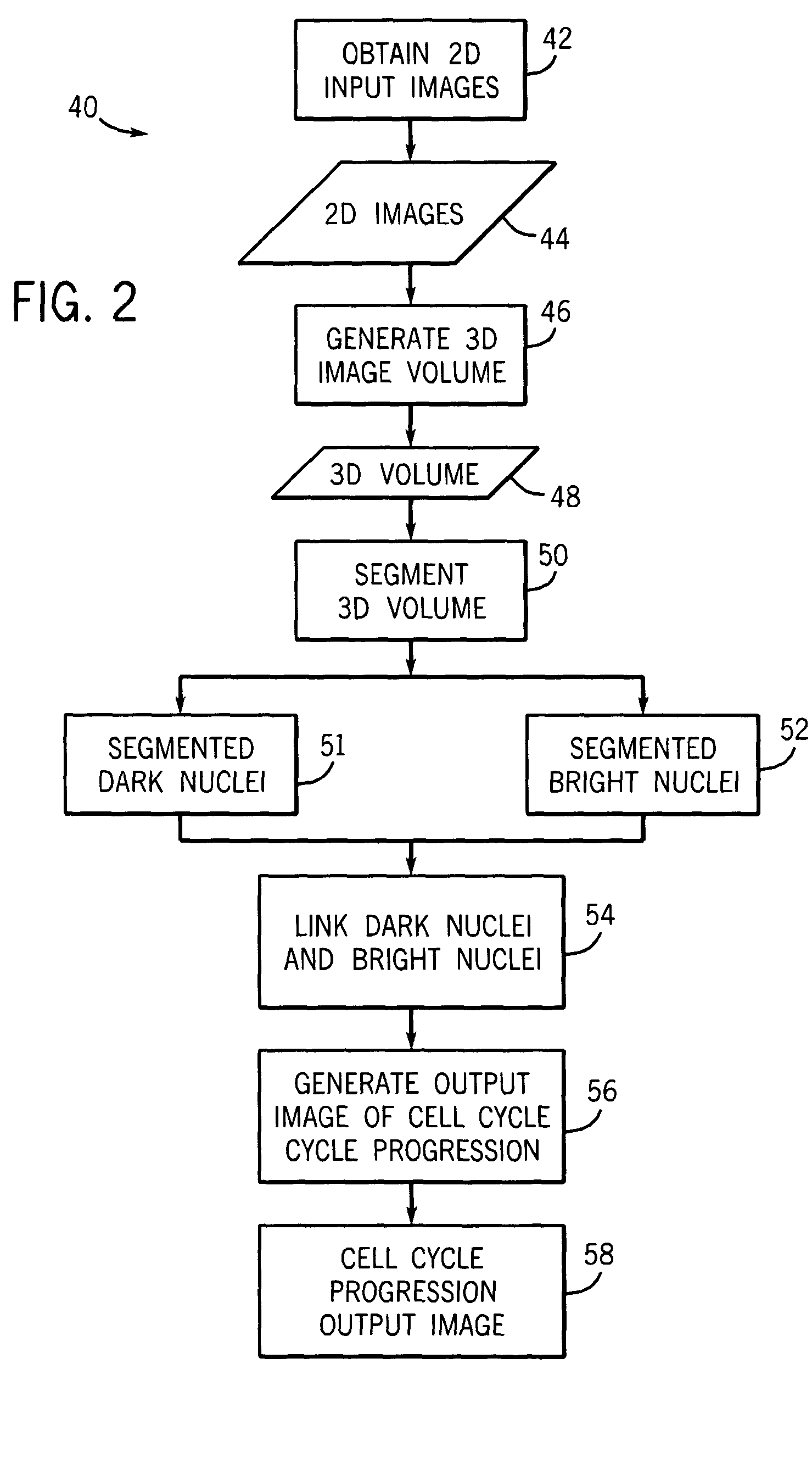
  • Time-lapse cell cycle analysis of unstained nuclei

Examples


Embodiment Construction

[0024]In the field of medicine it is often desirable to be able to track the progress of a cell through different phases of the cell cycle. Generally, the cell cycle may be divided into four distinct phases. DNA in the cell nucleus is replicated during the “S” phase of the cell cycle (for “synthesis”). The entire cell-division phase is denoted as the “M” phase (for “mitosis”). This leaves the period between the M phase and the start of DNA synthesis (the S phase), which is called the “G1” phase (first gap phase), and the period between the completion of DNA synthesis and the next M phase, which is called the “G2 ” phase. Interphase is a term for the non-mitotic phases of the cell cycle, and includes G1, S, and G2 phases. In a typical cell, interphase may comprise 90% or more of the total cell cycle time. However, in cells that have been treated with one or more cell cycle altering compounds or agents and / or a cell-cycle altering stimulus (e.g. irradiation), normal progress through t...


PUM


Abstract

The present invention provides a cell imaging technique for automatically tracking the progression of a cell through the cell cycle over time through segmentation of a volume of two-dimensional time-lapse images. The technique allows long-term tracking of the cell cycle progression of an individual cell or multiple cells. Further, the invention provides a unique display of cell cycle progression, allowing an end user to easily determine changes to cell cycle progression for a cell of interest.

Description

CROSS REFERENCE TO RELATED APPLICATIONS[0001]This application is a Non-Provisional Application of Provisional Patent Application No. 60 / 736,345, entitled “TIME-LAPSE CELL CYCLE ANALYSIS USING THREE-DIMENSIONAL SEGMENTATION OF UNSTAINED NUCLEI IN LINE SCAN CONFOCAL FLUORESCENCE IMAGES”, filed Nov. 12, 2005, which is herein incorporated by reference.BACKGROUND[0002]The present technique relates generally to image analysis designed to determine the cell cycle phase of a particular cell. More specifically, the present technique relates to automatically segmenting a cell into a cell cycle phase using three-dimensional segmentation of two-dimensional time-lapse images.[0003]When eukaryotic cells replicate, they pass through a tightly regulated series of events known as the cell cycle. The cell cycle generally includes four phases: G1, S, G2, and M. Each phase of the cell cycle is marked by distinctive characteristics in cell morphology and total DNA content as the DNA is replicated and th...

Claims


Application Information

Patent Timeline
Patent Type & Authority Patents(United States)
IPC IPC(8): G06K9/00G06T17/00
CPCG06K9/0014G06T7/0012G06T7/0081G06T7/0083G06T7/0089G06T7/0097G06T7/20G06T2207/10016G06T2207/10056G06T2207/10064G06T2207/20156G06T2207/20161G06T2207/30024G06T7/11G06T7/12G06T7/149G06T7/174G06V20/695
Inventor PADFIELD, DIRK RYANSEBASTIAN, THOMAS BABYRITTSCHER, JENSTHOMAS, NICHOLAS
Owner GENERAL ELECTRIC CO
Why Eureka
Try Eureka