
Systems, devices, methods, apparatus and computer-accessible media for providing optical imaging of structures and compositions

a technology of optical imaging and optical imaging catheter, which is applied in the direction of catheter, fluorescence/phosphorescence, instruments, etc., can solve the problems of multimodality imaging of luminal organs, difficult to place the acquired signature in the appropriate morphologic context, and the inability to ascertain information such as molecular expression and tissue composition from the ofdi signal, etc., to facilitate simultaneous multimodal imaging of biological tissues.

Active Publication Date: 2012-04-26
THE GENERAL HOSPITAL CORP +1
View PDF5 Cites 74 Cited by
  • Summary
  • Abstract
  • Description
  • Claims
  • Application Information

AI Technical Summary

Benefits of technology

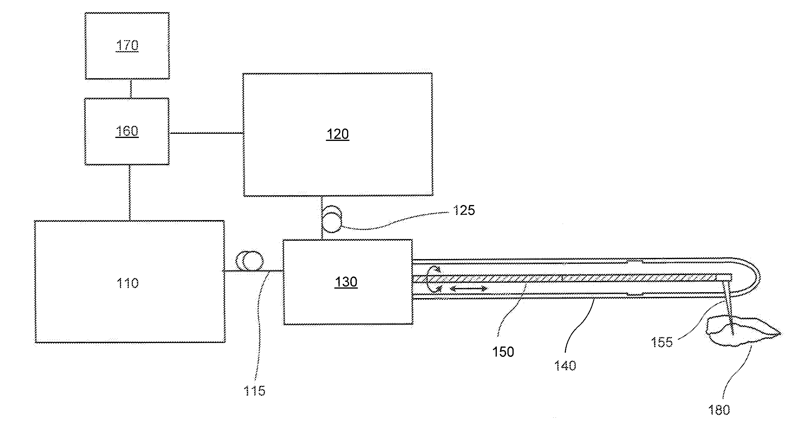
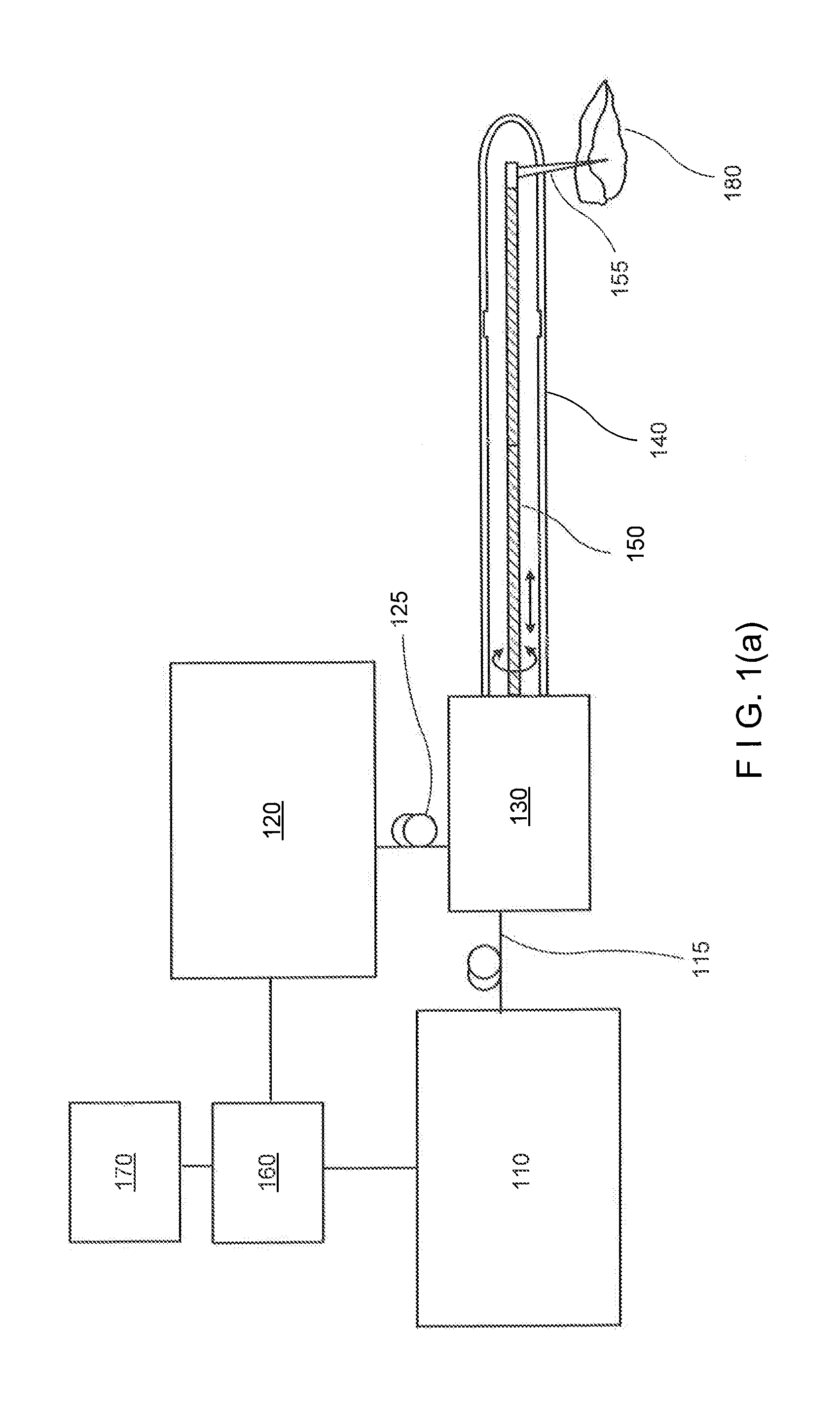
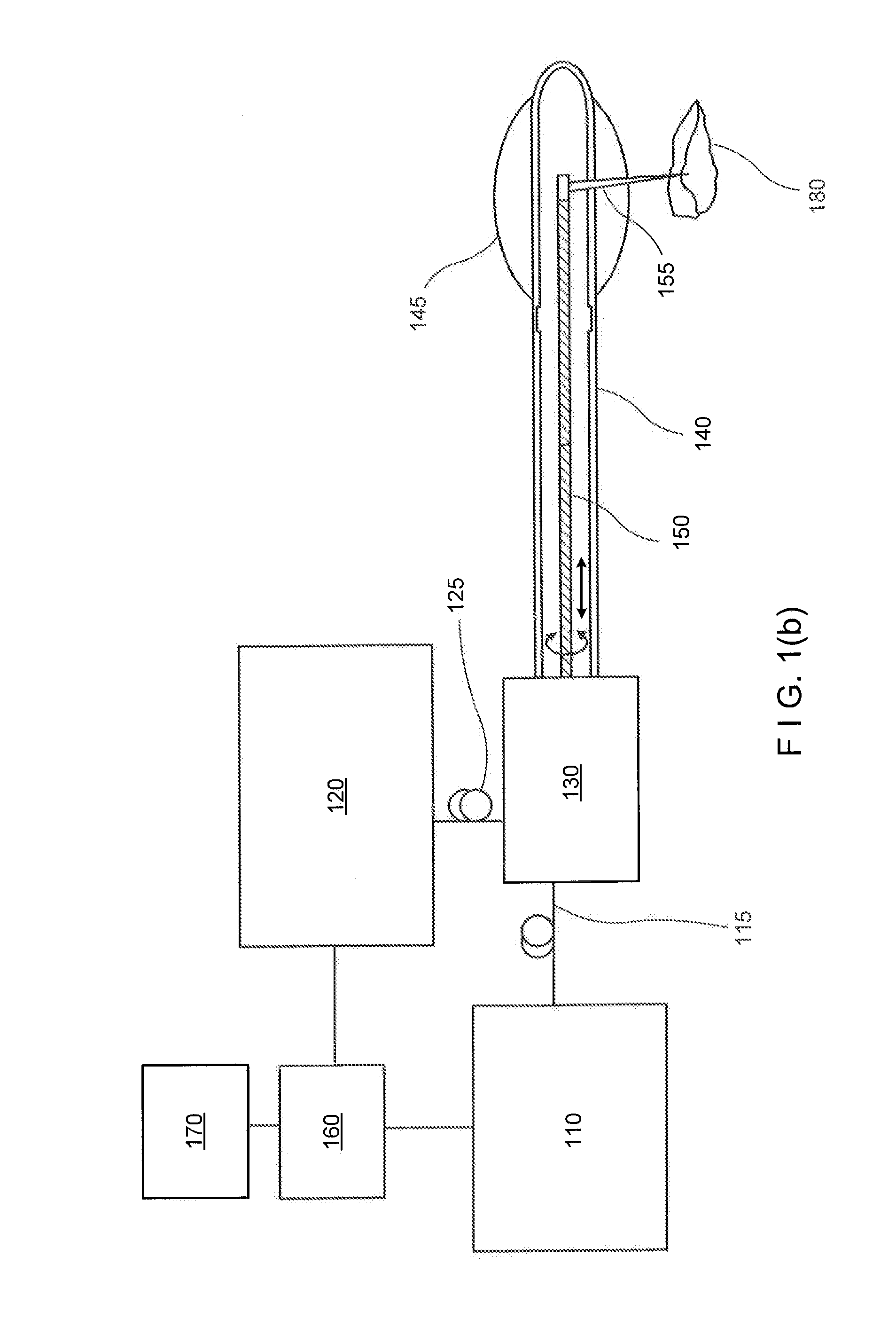
The present invention provides a system and method for simultaneously imaging biological tissues using optical techniques. The system includes a multimodality catheter that illuminates the tissues and collects signals from the inside of the lumen, a multimodality rotary junction that connects the catheter to a stationary system, and a device / apparatus that can transmit electro-magnetic radiation between waveguides in an optical fiber. The system can obtain information about the anatomical structure and molecular / chemical / biological compositions of the tissues using optical coherence tomography, optical frequency domain interferometry, optical florescence, optical acoustics, and other modalities. The invention can be used in optical coherence tomography, probe, catheter, eye box, endoscope, and other applications. The method and computer-accessible medium can provide a way to determine characteristics of structures or compositions in real-time.

Problems solved by technology

However, information such as molecular expression and tissue composition may not be ascertained from the OFDI signal.
However, these exemplary techniques operate without knowledge of the underlying microscopic structure, making it difficult to place the acquired signatures in the appropriate morphologic context.
However, in vivo multimodality imaging of luminal organs, such as coronary arteries, gastrointestinal tract, and respiratory system has not been appropriately described.
Although multimodality imaging techniques in a catheter form have been utilized, such techniques could not provide comprehensive three-dimensional (3D) information from the luminal organs yet, due to the lack of fast rotation, high speed acquisition, and / or small catheter for in vivo data collection.
A sequential acquisition of such diverse information using multiple independent catheters and systems has, however, likely been untenable due to the impracticality of conducting multiple procedures, subject motion, and difficulties registering datasets.

Method used


Image

  • Systems, devices, methods, apparatus and computer-accessible media for providing optical imaging of structures and compositions
  • Systems, devices, methods, apparatus and computer-accessible media for providing optical imaging of structures and compositions
  • Systems, devices, methods, apparatus and computer-accessible media for providing optical imaging of structures and compositions

Examples


Embodiment Construction

[0007]It is therefore one of the objects of the present invention to reduce or address the deficiencies and / or limitations of such prior art approaches, procedures and systems. In accordance with certain exemplary embodiments of the present disclosure, exemplary systems, devices, methods, apparatus and computer-accessible media can be provided which facilitate a simultaneous multimodality imaging of biological tissues, such as, e.g., luminal organs in vivo, using optical techniques.

[0008]According to one exemplary embodiment of the present disclosure, a device / apparatus can be provided which can include a multimodality catheter that illuminates the tissues and collects signals from the inside of the lumen, a multimodality system which generates light sources, detects returning lights, and processes signals, and a multimodality rotary junction which rotates and pulls back the catheter and connects the moving catheter to the stationary system. In another exemplary embodiment, a dual-m...


PUM


Abstract

Exemplary systems, devices, methods, apparatus and computer-accessible media for providing and / or utilizing optical frequency domain imaging (OFDI) and fluorescence of structures and, e.g., multimodality imaging using OFDI techniques and fluorescence imaging techniques are described. For example, an arrangement can provide at least one electro-magnetic radiation to an anatomical structure. Such exemplary arrangement can include at least one optical core and at least one cladding at least partially surrounding the fiber(s). A region between the optical core(s) and the cladding(s) can have an index that is different from indexes of the optical core(s) and the cladding(s). The arrangement can also include at least one apparatus which is configured to transmit the radiation(s) via the optical core(s) and the cladding(s) to the anatomical structure.

Description

CROSS-REFERENCE TO RELATED APPLICATION(S)[0001]The present application is based on and claims priority from U.S. Patent Application Ser. No. 61 / 348,071, filed on May 25, 2010, and U.S. Patent Application Ser. No. 61 / 353,424, filed on Jun. 10, 2010, the entire disclosures of which are incorporated herein by reference.FIELD OF THE DISCLOSURE[0002]The present disclosure relates generally to exemplary systems, devices, methods, apparatus and computer-accessible media for providing optical imaging catheter of structures and compositions, and more particularly to systems, devices, methods, apparatus and computer-accessible media for providing and / or utilizing optical frequency domain imaging (OFDI) and fluorescence of structures and compositions and, e.g., multimodality imaging using OFDI techniques and fluorescence imaging techniques.BACKGROUND INFORMATION[0003]A majority of diseases arise within luminal organs such as the gastrointestinal tract and coronary arteries. Understanding and d...

Claims


Application Information

Patent Timeline
Patent Type & Authority Applications(United States)
IPC IPC(8): A61B6/00
CPCA61B5/0066A61B5/6852G01N21/4795G01B9/02091G01N21/65G01B9/02029G01B9/0205G01N21/64A61B5/0071A61B5/0084G01N21/6456G01N2201/0866
Inventor TEARNEY, GUILLERMO J.BOUMA, BRETT EUGENEYOO, HONGKISHISHKOV, MILENGARDECKI, JOSEPHSHUBOCHKIN, ROMANMORSE, THEODOREJAFFER, FAROUC AMIN
Owner THE GENERAL HOSPITAL CORP
Learn More