

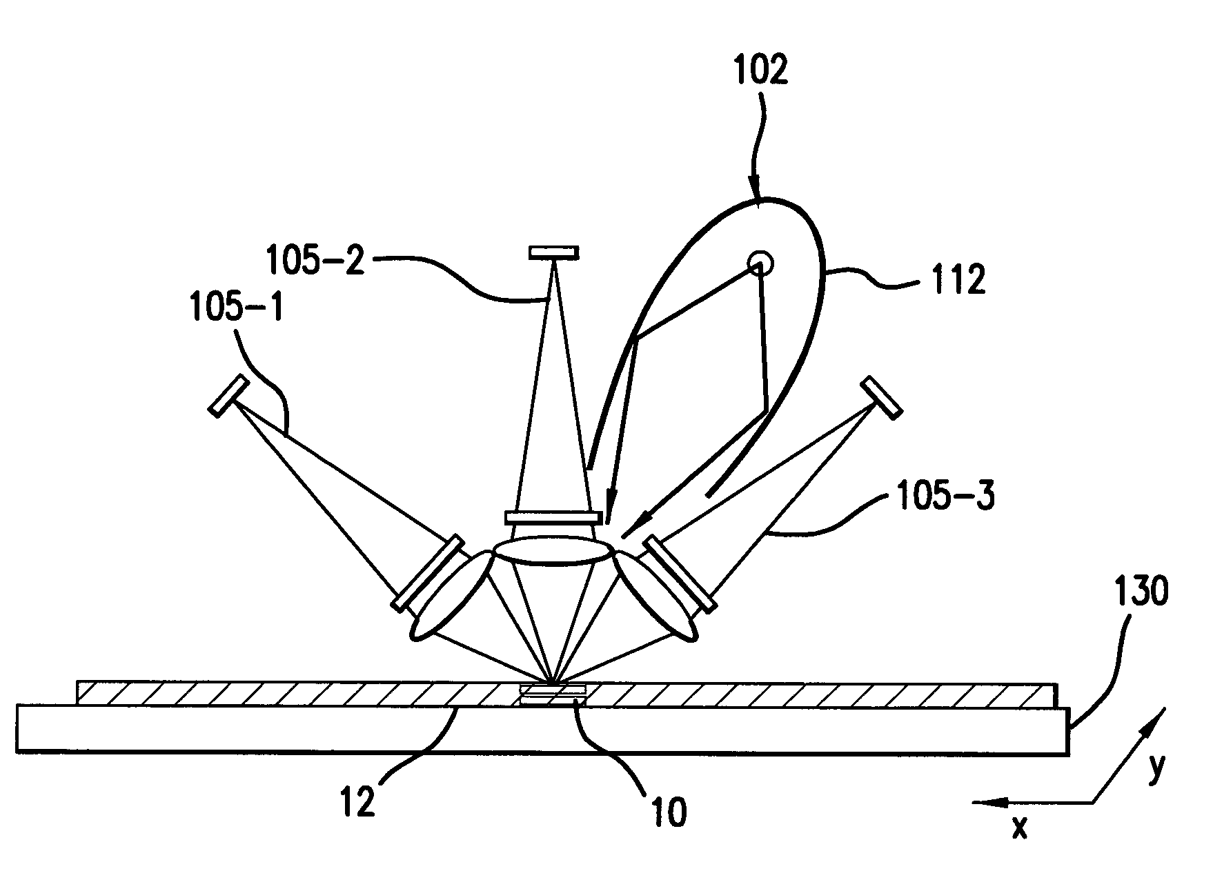
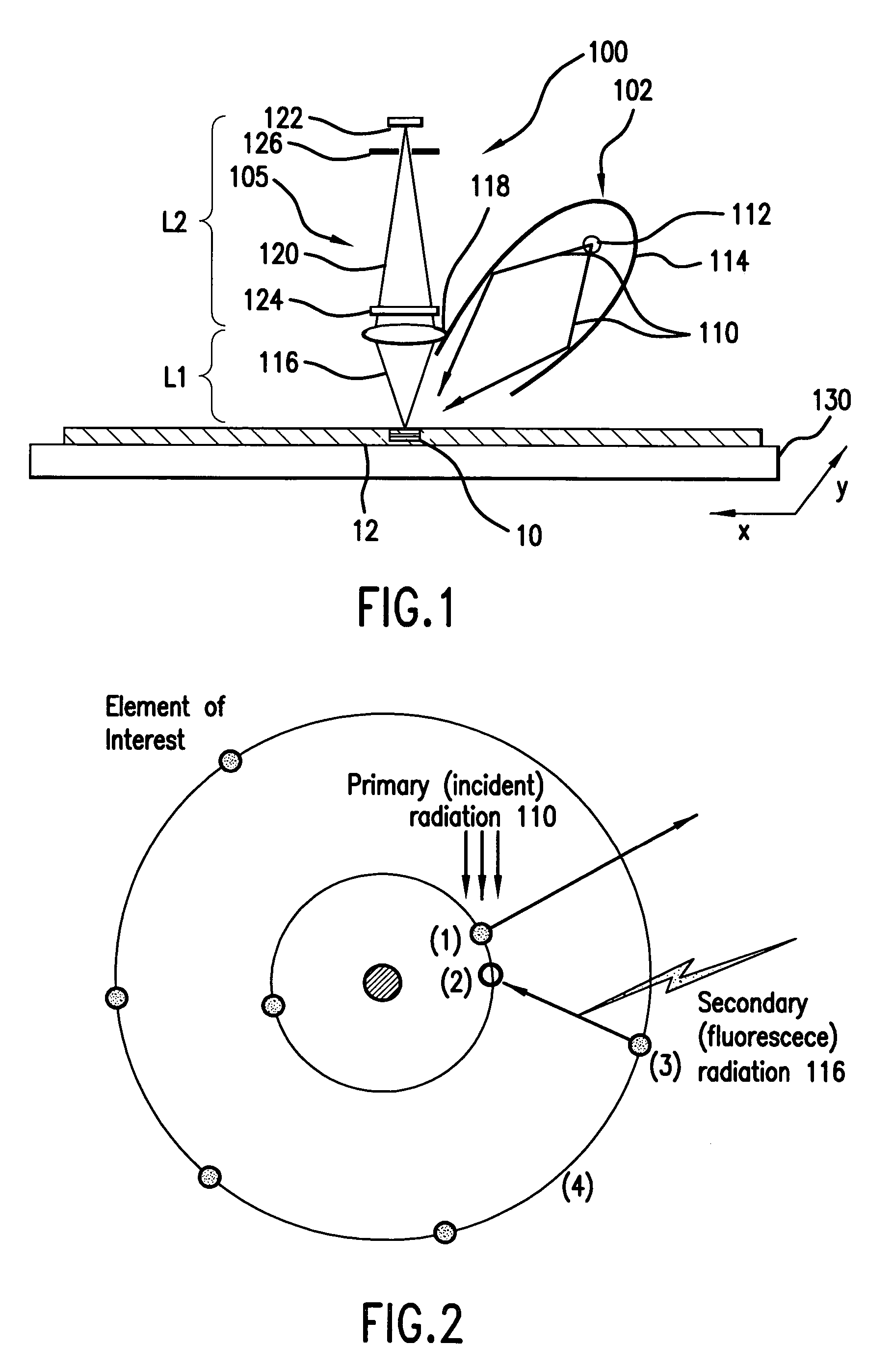
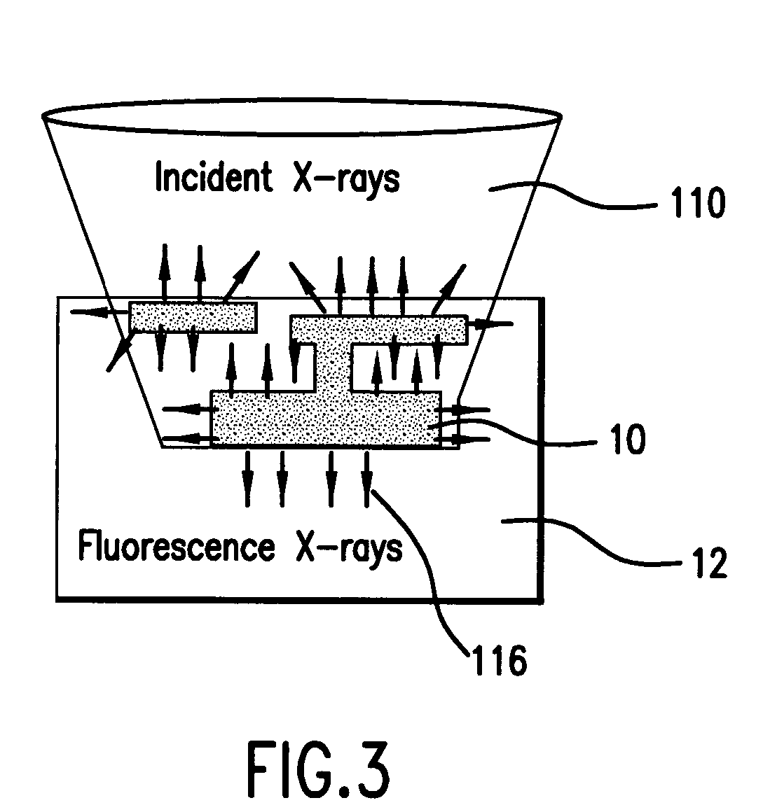
Element-specific X-ray fluorescence microscope and method of operation
a fluorescence microscope and element-specific technology, applied in the direction of instruments, material analysis using wave/particle radiation, nuclear engineering, etc., can solve the problems of limiting throughput and unpractical element-specific imaging systems using laboratory x-ray sources, and achieves high signal to noise and better elemental specificity
- Summary
- Abstract
- Description
- Claims
- Application Information
AI Technical Summary
Benefits of technology
Problems solved by technology
Method used
Image

Examples
examples
[0077]The X-ray fluorescence from important materials of an IC are induced with radiation energy in the approximately 100 to 8000 eV range. The materials that produce the x-ray fluorescence are copper at 930 eV (La) and 8046 eV (Ka), silicon at 99 eV (La) and 1740 eV (Ka), germanium at 1186 eV (La), tantalum at 1710 eV (Ma), titanium at 452 eV (La) and 4510 eV (Ka), cobalt at 776 eV (La) and 6929 eV (Ka), phosphorus at 2013 eV (Ka), arsenic at 1282 eV (La), and aluminum at 1486 eV (Ka), for example. Boron at 183 eV (Ka) is another alterative.
[0078]Considering copper as an example, FIG. 4 shows the efficiency of a 500 nanometer (nm) copper zone plate in the 900 to 950 eV range, where copper has a fluorescence line. Note that the efficiency peaks to about 30% around 932 eV, which is close to the energy of a Cu La fluorescence line at 930 eV.
[0079]FIG. 5 shows the transmission of copper over this 900 to 950 eV range. For example, 200 nm of copper has a transmission above 933 eV of only...
PUM
| Property | Measurement | Unit |
|---|---|---|
| grazing incidence angle | aaaaa | aaaaa |
| solid angle | aaaaa | aaaaa |
| atomic numbers | aaaaa | aaaaa |
Abstract
Description
Claims
Application Information