Method and a dual-array transducer probe for real time mechanical imaging of prostate

a technology of mechanical imaging and transducer probe, applied in the field of medical devices, can solve the problems of small improvement in prostate cancer detection, lack of operator's skills, and disappointing experience with trus as a means of prostate cancer screening and staging, and achieve the effect of removing the influence of operator's skills

Inactive Publication Date: 2010-10-26
ARTANN LAB
View PDF24 Cites 16 Cited by
  • Summary
  • Abstract
  • Description
  • Claims
  • Application Information

AI Technical Summary

Benefits of technology

[0017]Accordingly, it is an object of the present invention to overcome these and other drawbacks of the prior art by providing a novel method and device for objective and consistent mechanical imaging of a palpable organ eliminating the influence of operator's skills on the results of such examination.
[0018]It is another object of the present invention to provide a mechanical imaging device for examination of prostate shaped in such a way as to allow proper compression of the prostate organ only and minimizing distortions caused by pressing the device against other surrounding tissues and organs.
[0020]It is another object of the present invention to provide a probe and a method of its use allowing teaching the medical personnel the proper technique of prostate examination.
[0027]Importantly, the processing of data is preferably conducted by using data from both the probe head pressure sensor array (used as a primary or first source of pressure data) and from shaft pressure sensor array (used as a secondary source of pressure data). This allows moving of the probe relative to the prostate while maintaining the common identified features of each obtained mechanical image. In other words, every time the probe is moved from one position to the next, the processing means of the device are adapted to follow certain identifiable features and the distance from the sphincter from the previous mechanical image to the next one such that a complete 2D or 3D image may be constructed. That way, there is less need for knowing the absolute position in space of both the prostate and the probe in order to accurately relate each successive pressure pattern to a certain part of the prostate.
[0029]Importantly, the shape and size of the head pressure sensor array is selected such that it provides for uniform compression of the area of interest about the prostate gland and not other areas of the rectum. Further facilitating this aspect of the invention is the S-shaped design and an angular offset between the head of the probe and the shaft of the probe. Such advantageous shape increases the accuracy of obtained pressure data and reduces the artifacts caused by inadvertent tilting of the probe to avoid sphincter trauma.

Problems solved by technology

Unfortunately, to date the experience with TRUS as a means of prostate cancer screening and staging has been disappointing.
It adds little to screening by DRE and PSA, and the small improvement in prostate cancer detection does not justify its cost.
As a screening test, TRUS has a low specificity and a high false positive rate.
However, its positive predictive value is limited.
The DRE alone is even less useful.
However, combining the two modalities nearly doubles the cancer detection rate.
The main disadvantage of DRE is its high degree of subjectivity.
There may not be a sufficient number of skilled users available for large-scale mass prostate screenings.
Another limitation of DRE is that a physician performing the examination cannot objectively record the state of the examined prostate.
Therefore, it is difficult to objectively compare the results of consecutive examinations of the same prostate.
The effectiveness and reliability of DRE are highly dependent on the skill of the user, since the finger does not provide a quantitative or objectively verifiable assessment.
While prior art mechanical imaging devices provided for data collection, the ability to recreate the 2D and 3D images of the prostate were limited by the insufficiently accurate information obtained from the transrectal probe with regard to the examined prostate in the course of examination.
Upon compressing the area about the prostate, it is difficult to obtain uniform compression of that area alone and not load surrounding tissues and organs, especially the sphincter.
Such compression of surrounding tissues and organs would distort data collection away from the desired area and introduce errors associated with tilting the probe and stretching the sphincter or other tissues of the rectum.
Another reason for reduced sensitivity is because the prostate can be hard to find initially and it can also shift from its original place during the examination procedure.
Therefore, the prior art methods have a fundamental disadvantage in that as the examination progresses, no means are available to properly locate the prostate and then compensate for the probe position and orientation shift relative for the moving prostate.

Method used


Image

  • Method and a dual-array transducer probe for real time mechanical imaging of prostate
  • Method and a dual-array transducer probe for real time mechanical imaging of prostate
  • Method and a dual-array transducer probe for real time mechanical imaging of prostate

Examples


Embodiment Construction

[0052]A detailed description of the present invention follows with reference to accompanying drawings in which like elements are indicated by like reference letters and numerals.

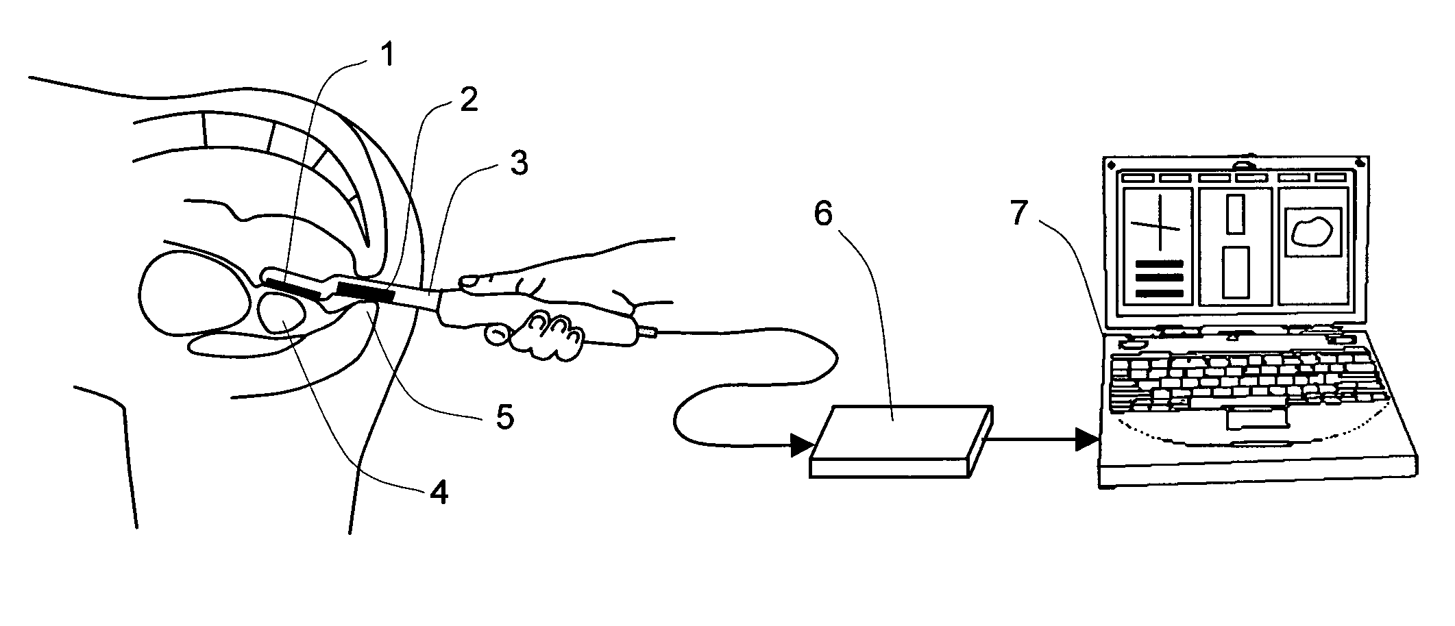
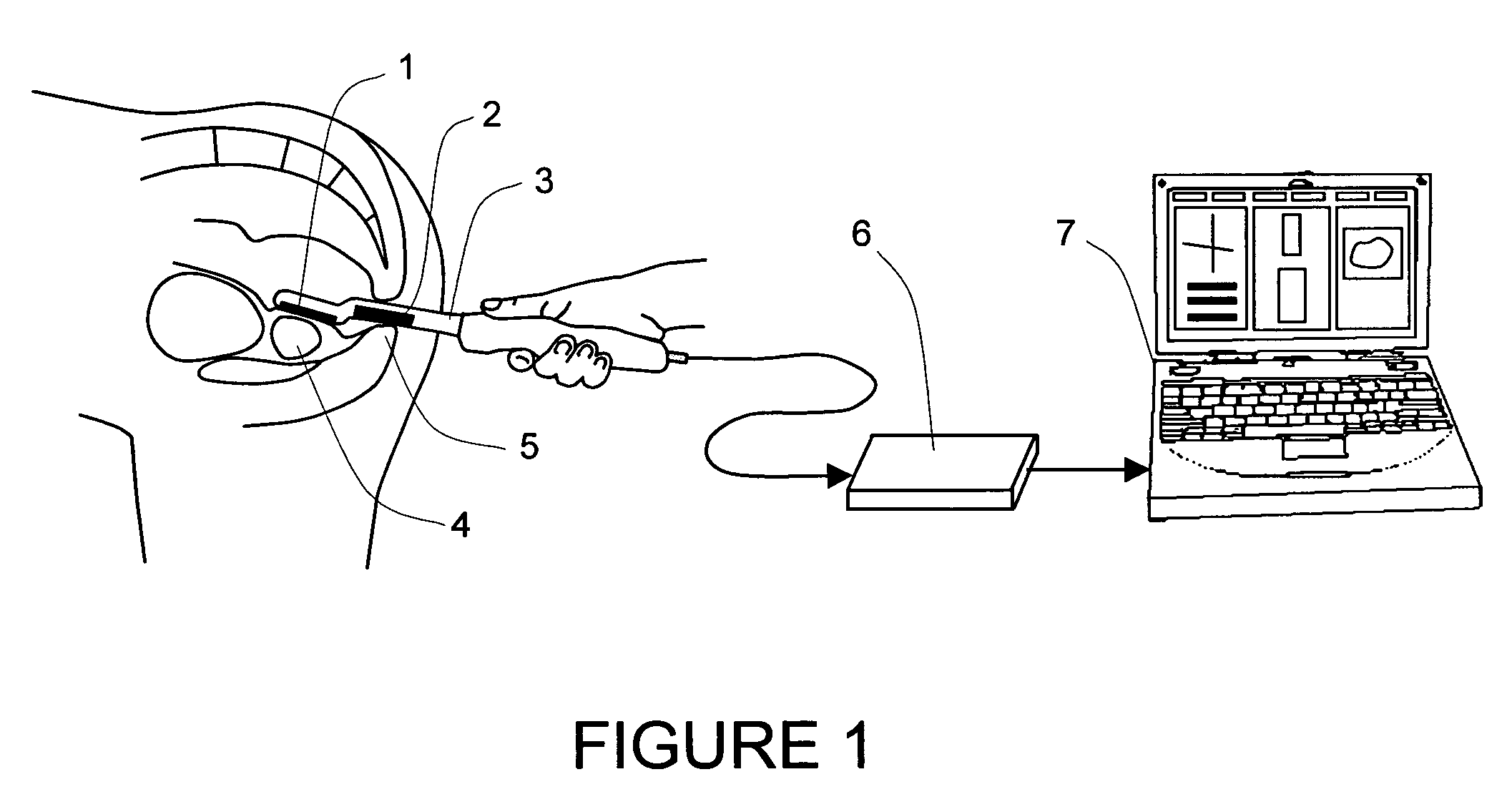
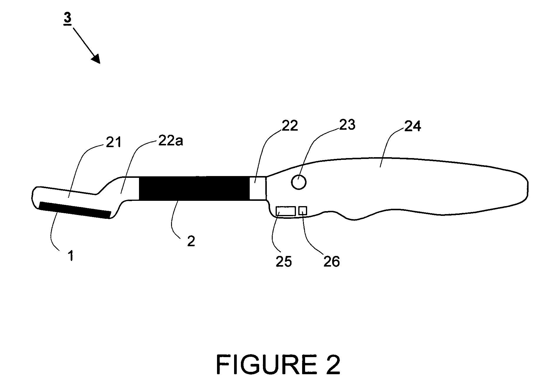
[0053]Referring now to the drawings, FIG. 1 is a schematic view of a preferred embodiment of a device for generating a mechanical image of a three-dimensional prostate volume from a plurality of data frames corresponding to a scan of the prostate. The device comprises the following major elements:[0054]a dual-array transrectal probe 3 with incorporated head pressure sensor array 1 for receiving pressure response data for the prostate 4 and shaft pressure sensor array 2 for receiving supplemental pressure response data for a sphincter area 5,[0055]electronic unit 6, and[0056]a processing and displaying means 7, which may be for example incorporated into a compact personal computer.

[0057]The prostate examination is performed using the following general steps. The patient is instructed to take off all clothes b...


PUM


Abstract

The present invention relates to a transrectal probe and method for real time mechanical imaging of a prostate. The probe is equipped with dual-array pressure sensors—one on the probe head and another on the shaft of the probe spaced away from the head with an angular and linear offset forming an S-shaped transition between the shaft and the head of the probe. The addition of the shaft pressure sensor array together with orientation tracking sensors allows precise calculation of the current head position throughout the examination of the prostate. Display means are used to guide the user in the proper manipulation of the probe in order to reduce the forces on surrounding tissues and organs and to minimize patient's discomfort.

Description

CROSS-REFERENCE DATA[0001]This application is a continuation-in-part of a co-pending U.S. patent application Ser. No. 11 / 123,999 filed May 6, 2005 and entitled “Method and device for real time mechanical imaging of prostate”.STATEMENT REGARDING FEDERALLY SPONSORED RESEARCH OR DEVELOPMENT[0002]This invention was made with government support under SBIR Grant 2 R44 CA82620-02A1 awarded by the National Institutes of Health, National Cancer Institute. The government has certain rights in this invention.BACKGROUND OF THE INVENTION[0003]The present invention relates generally to medical devices. More specifically, it relates to a mechanical imaging system and process for examining, mapping, and diagnosing diseases of a palpable organ such as a prostate gland in a male patient, especially the prostate cancer. It is also applicable more generally to mechanical imaging of palpable tissues, including but not limited to, through natural body openings in a human being, i.e. mouth, ear(s), rectum...

Claims


Application Information

Patent Timeline
Patent Type & Authority Patents(United States)
IPC IPC(8): A61B5/103
CPCA61B5/103A61B5/4381A61B5/0053A61B5/6885A61B2562/0247A61B2562/046A61B8/4254
Inventor SARVAZYAN, ARMEN P.EGOROV, VLADIMIRAYRAPETYAN, SUREN
Owner ARTANN LAB
Why Eureka
Try Eureka