Method for the processing of radiological images in tomosynthesis for a detection of radiological signs

a radiological sign and tomosynthesis technology, applied in the field of radiological sign detection detection methods, can solve the problems of large quantity of information to be managed, time-consuming and laborious, and the new tomosynthesis mammography device has drawbacks, so as to reduce the time spent on the detection of radiological signs, the effect of reducing the time and reducing the time spent on identification

Active Publication Date: 2010-12-14
GENERAL ELECTRIC CO
View PDF4 Cites 17 Cited by
  • Summary
  • Abstract
  • Description
  • Claims
  • Application Information

AI Technical Summary

Benefits of technology

[0008]Embodiments of the invention are aimed precisely at overcoming the above-mentioned drawbacks of the prior art. To this end, the invention proposes an image-processing method enabling the automatic detection of the radiological signs in series of 3D data. The time spent to locate these radiological signs is greatly reduced. Hence, the invention can prevent the radiologist from possibly overlooking lesions during the examination. With this image-processing method, the radiological signs are easier to identify and thus enable a more efficient examination.
[0009]Embodiments of the invention propose a novel approach to mammography that advantageously exploits the techniques of digital processing of the radiology image to improve the readability of the information.
[0010]This image-processing method can be used to cope with a large quantity of the data by developing novel strategies capable of reducing time and simplifying access to information of clinical interest.

Problems solved by technology

In particular, since standard mammography images are the result of projections, they represent superimposed structures that disturb the visibility of the structures of the breast, sometimes leading to a falsely positive interpretation when a superimposition of these structures resembles a lesion or to a falsely negative interpretation when the structures obscure the visibility of a lesion.
However, this new tomosynthesis mammography device has drawbacks.
Consequently, the quantity of information to be managed is very great.
Similarly, access to a piece of information having clinical interest takes much more time since this information is sought sequentially in the volume, slice by slice.
However, the new tomosynthesis mammography devices cannot be subjected to high frequency of use because the time of access to information of clinical interest is very great.
Another problem, which is more specific to mammography but can, however, occur in other fields, is related to the necessity of being able to analyze certain radiological signs, which become clinically interesting between 100 μm and 1 mm.

Method used


Image

  • Method for the processing of radiological images in tomosynthesis for a detection of radiological signs
  • Method for the processing of radiological images in tomosynthesis for a detection of radiological signs
  • Method for the processing of radiological images in tomosynthesis for a detection of radiological signs

Examples


first embodiment

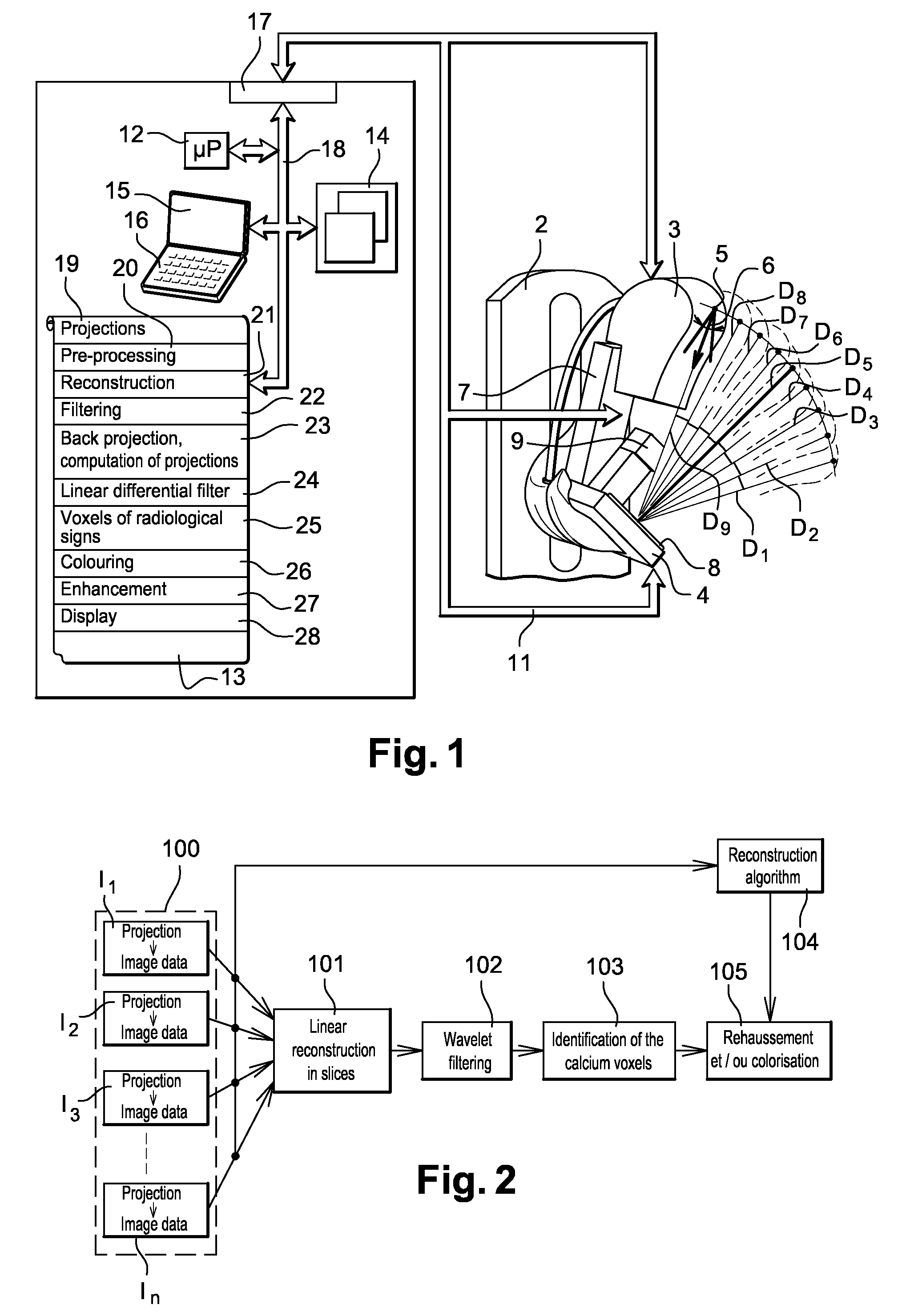
[0043]FIG. 2 shows the invention. In FIG. 2, the X-ray tube 3, in the step 100, emits x-ray intensities going through the patient's breast for a multiplicity of projections P1 to Pn, in a pre-determined path. The detector 4 acquires the pieces of x-ray image data I1 to In respectively representing the projection images P1 to Pn. The control logic unit processes the pieces of x-ray image data I1 to In.

[0044]At the step 101, the control logic unit applies a simple back-projection reconstruction algorithm. This algorithm is used to rebuild the volume in different slice planes parallel to the detector. The term used here is tomosynthesis of the breast. All the pieces of image data I1 to In are used during this tomosynthesis reconstruction to provide a digital volume of the breast. This tomosynthesis technique enables the reconstruction of the 3D volume of the breast being studied from a small number of 2D projections or pieces of image data, distributed on a restricted angular domain an...

second embodiment

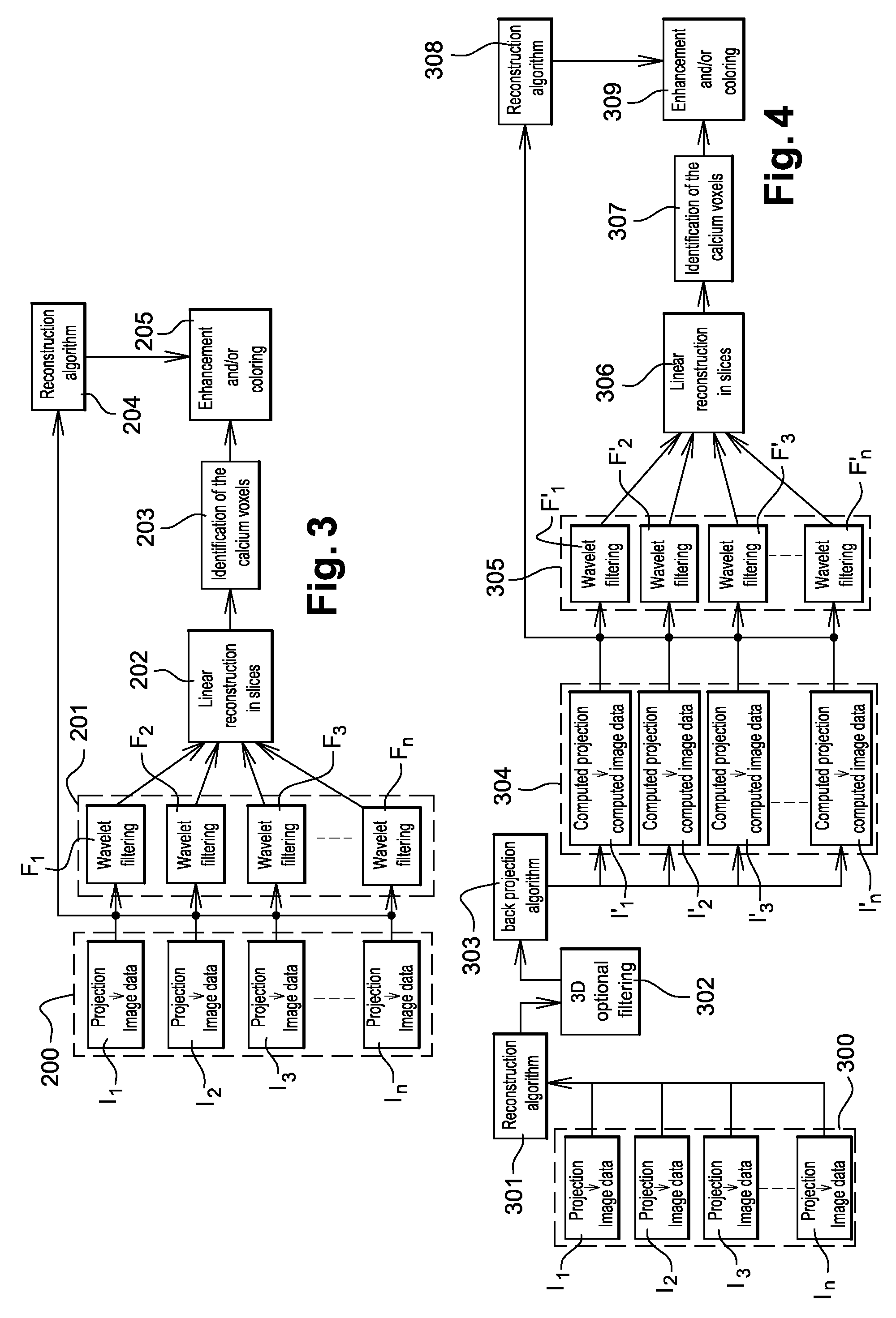
[0090]The invention has thus implemented a second embodiment comprising a faster algorithm that depends only on the number of projections and not on the number of slices in assuming a parallel geometry, as illustrated in FIG. 3.

[0091]FIG. 3 shows another illustration of means implementing the method according to an embodiment of the invention. In the implementation of the invention, the image-processing method is applied to each piece of image data I1 to In, respectively representing each projection P1 to Pn obtained at the step 200. These pieces of image data are given directly by the digital detector to the control logic unit. It is on the basis of these pieces of image data that the processing method enables the location of elements likely to form radiological signs. These radiological signs may be microcalcifications or opacities.

[0092]At the step 201, the control logic unit respectively applies a binary mask F1 to Fn to each piece of image data I1 to In in order to separate the...


PUM


Abstract

In an image-processing method for the detection of radiological signs in series of 3D data, an algorithm is used to detect radiological signs in a digital volume according to their contrasts. This algorithm is applied to reconstructed slices or directly to the series of projections. This algorithm is made by means of linear differential filters for signal analysis. It is used to color or enhance the intensity of the detected radiological signals according to the degree of malignancy.

Description

FIELD OF THE INVENTION[0001]An embodiment of the present invention provides a method for the processing of radiology images for the detection of radiological signs. Embodiments of the present invention can be applied to particular advantage but not exclusively in the field of medical imaging and, more particularly, that of mammography. Embodiments of the present invention also relate to a medical image reviewing station comprising an image-processing method of this kind.DESCRIPTION OF THE PRIOR ART[0002]Today, mammography is widely used for the detection of lesions and the prevention of breast cancer. The signs to be detected in mammography images are radiological signs associated with lesions. These signs may be either calcium deposits or cases of opacity. Calcium deposits are called microcalcifications and individually form small-sized elements (ranging from 100 μm to 1 mm in diameter) that are more opaque to X-rays than the surrounding tissues. Opacities are dense regions where t...

Claims


Application Information

Patent Timeline
Patent Type & Authority Patents(United States)
IPC IPC(8): G06K9/00A61B6/04A61B6/00A61B6/02A61B6/03G06T1/00
CPCG06T7/0012G06T11/008G06T2207/10072G06T2207/10116G06T2207/20064G06T2207/30068G06T2211/436
Inventor BERNARD, SYLVAINMULLER, SERGESOUCHAY, HENRIPETERS, GEROIORDACHE, RAZVAN
Owner GENERAL ELECTRIC CO
Why Eureka
Try Eureka