Fluorescent proteins and chromoproteins from non-Aequorea hydrozoa species and methods for using same

a technology of fluorescence proteins and chromoproteins, which is applied in the field of fluorescence proteins, can solve the problems of hammering the use of these proteins in many applications, and achieve the effect of high stringency

Inactive Publication Date: 2011-05-31
ZAKRYTOE AKTSIONERNOE OBSCHESTVO EVROGEN
View PDF7 Cites 17 Cited by
  • Summary
  • Abstract
  • Description
  • Claims
  • Application Information

AI Technical Summary

Problems solved by technology

The major drawback of the Anthozoa-derived GFP-like is strong oligomerization that hampers the use of these proteins in many applications (Lauf et al., FEBS Lett.

Method used


Image

  • Fluorescent proteins and chromoproteins from non-Aequorea hydrozoa species and methods for using same
  • Fluorescent proteins and chromoproteins from non-Aequorea hydrozoa species and methods for using same
  • Fluorescent proteins and chromoproteins from non-Aequorea hydrozoa species and methods for using same

Examples


example 1

[0135]phiYFP Cloning, Sequencing and Recombinant Protein Production

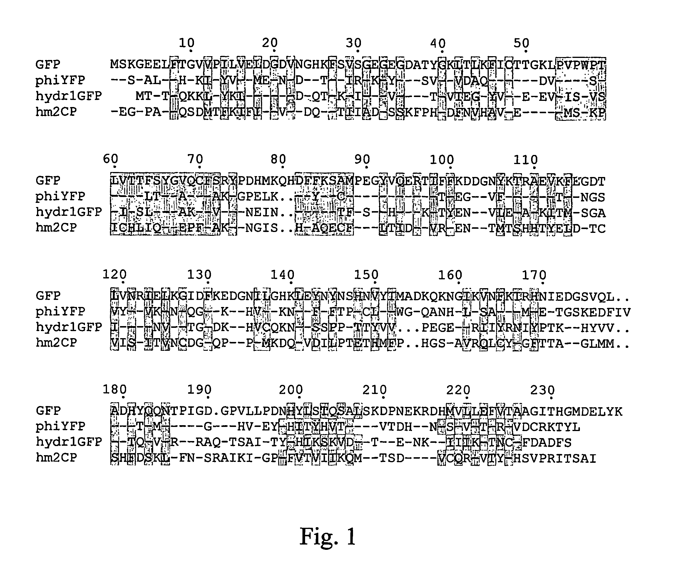
[0136]Bright yellow fluorescence was detected using a fluorescent microscope in Hydromedusa Phialidium sp. (Cnidaria; Hydrozoa; Hydroida; Leptomedusae; Campanulariidae). To find the protein responsible for fluorescence in this jellyfish, a strategy based on the screening of an expression cDNA library in E. coli was chosen. Amplified cDNA samples were prepared using a SMART cDNA amplification kit (Clontech) and cloned into PCR-Script vector (Stratagene). About 105 recombinant clones were screened visually using a fluorescent stereomicroscope. Two fluorescent clones encoding the same yellow fluorescent proteins were found and were named phiYFP. The nucleic acid and amino acid sequences for phiYFP are shown in SEQ NOs: 01, 02 and 23. Comparison of phiYFP with A. victoria GFP is shown in FIG. 1. phiYFP appears to be more similar to GFP (50% identity) than to coral-derived fluorescent proteins.

[0137]To facilitate protein ...

example 2

[0138]PhiYFP Mutagenesis

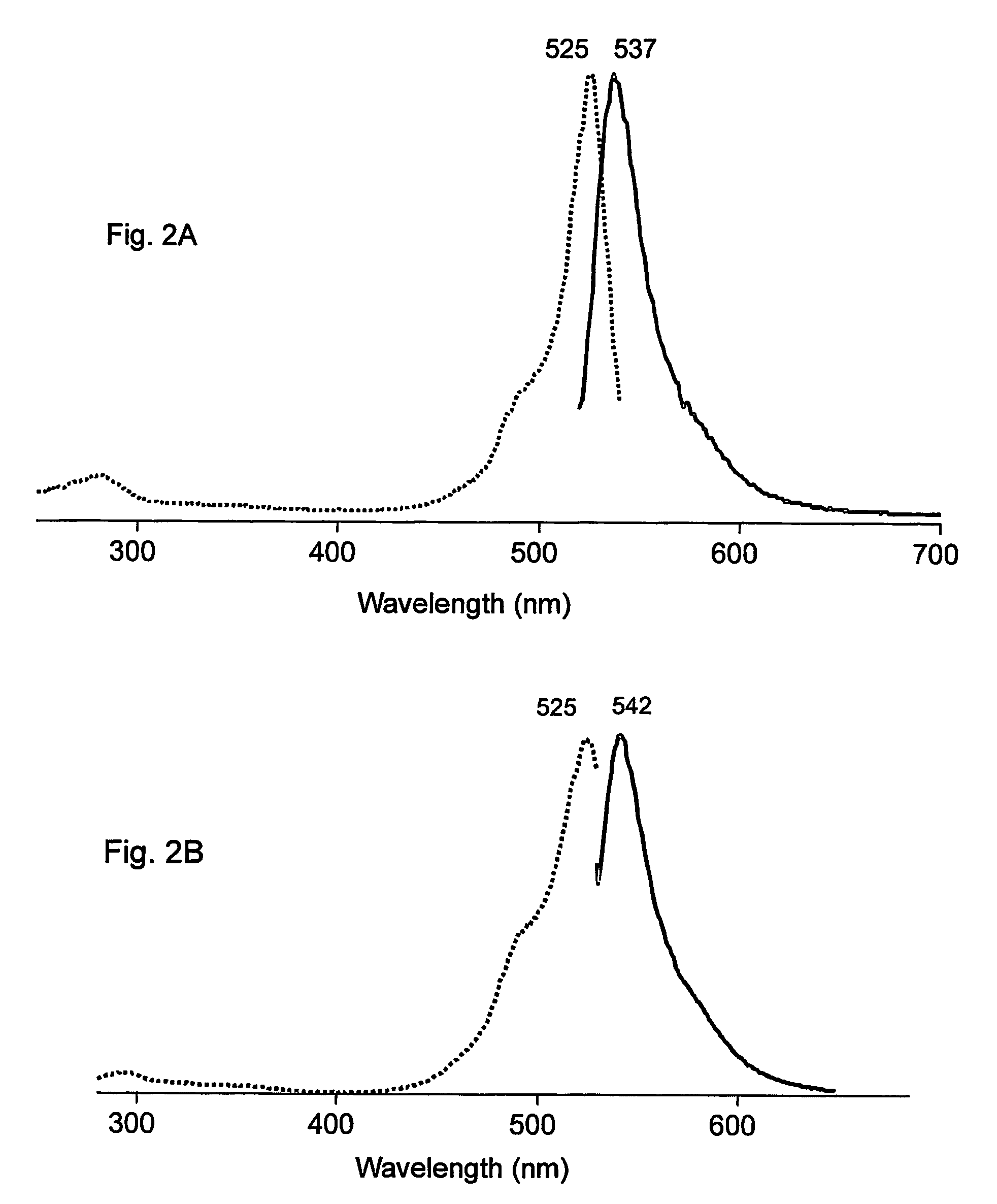
[0139]The PhiYFP nucleic acid coding sequence was prepared as described above in the Example 1. We have modified the encoded wild-type protein by random mutagenesis. Random mutagenesis of phiYFP resulted in the generation of a brighter mutant named phiYFP-Y1 with a slightly altered excitation-emission spectra. This mutant contained three amino acid substitutions, specifically S2P, E174G, I201M (SEQ ID NOs: 03, 04, and 24). phiYFP-Y1 exhibited a brightness 1.5 to 2 fold higher than the wild type phiYFP in a side-by-side visual comparison of E. coli colonies expressing these fluorescent proteins. In addition, phiYFP-Y1 demonstrates a slightly red-shifted emission spectrum that peaked at 542 nm (see FIG. 2B).

[0140]Both phiYFP and phiYFP-Y1 proteins were found to be dimeric. It was demonstrated by protein gel-electrophoresis of non-heated protein samples (see Baird et al., supra, 2000). Under these conditions these FPs migrated as yellow fluorescent band at about...

example 3

[0144]hydr1GFP Cloning, Sequencing and Recombinant Protein Production

[0145]Bright green fluorescence was detected using a fluorescent microscope in a hydromedusa 1 (about 1 mm in length, FIG. 4) of sub-order Anthomedusae (Cnidaria, Hydrozoa, Anthomedusae). To search for the gene responsible for the fluorescence in this jellyfish, a strategy based on screening of an expression cDNA library in E. coli was implemented. Amplified cDNA samples were prepared using a SMART cDNA amplification kit (Clontech) and cloned into the PCR-Script vector (Stratagene). About 105 recombinant clones were screened visually using a fluorescent stereomicroscope. Three fluorescent clones were identified, each encoding the same green fluorescent protein, which was named hydr1GFP. The nucleotide and amino acid sequences for this protein are shown in SEQ ID NOS: 11, 12, and 28. A comparison of hydr1GFP with A. victoria GFP is shown in FIG. 1. hydr1GFP appears to be more similar to GFP (37% identity) than to fl...


PUM

PropertyMeasurementUnit
wavelengthaaaaaaaaaa
timeaaaaaaaaaa
timeaaaaaaaaaa

Abstract

The present invention provides nucleic acid molecules encoding a fluorescent and chromo-proteins and mutants, variants and derivatives thereof, as well as proteins and peptides encoded by these nucleic acids. The nucleic acid molecules and proteins of interest are isolated from non-Aequorea Hydrozoa species. The proteins of interest include yellow fluorescent protein, phiYFP, from Phialidium sp., green fluorescent protein hydr1GFP and purple chromoprotein, hm2CP from hydroid medusae of sub-order Anthomedusae. Also of interest are proteins that are substantially similar to, or derivatives, or homologues, or mutants of, the above-referenced specific proteins. Also provided are fragments of the nucleic acids and the peptides encoded thereby, as well as antibodies specific to the proteins and peptides of the invention. In addition, host-cells, stable cell lines and transgenic organisms comprising above-referenced nucleic acid molecules are provided. The subject protein and nucleic acid compositions find use in a variety of different applications and methods, particularly for labeling of biomolecules, cell or cell organelles. Finally, kits for use in such methods and applications are provided.

Description

[0001]This application claims the benefit of U.S. Provisional Application(s) Nos. 60,425,570 filed 12 Nov. 2002, 60 / 429,795 filed 27 Nov. 2002, 60 / 464,258 filed 21 Apr. 2003 and 60 / 480,080 filed 20 June 2003.FIELD OF THE INVENTION[0002]This invention relates generally to the field of biology and chemistry. More particularly, the invention is directed to fluorescent proteins.BACKGROUND OF THE INVENTION[0003]Labeling of a protein, cell, or organism of interest plays a prominent role in many biochemical, molecular biological and medical diagnostic applications. A variety of different labels have been developed and used in the art, including radiolabels, chromolabels, fluorescent labels, chemiluminescent labels, and the like, with varying properties and optimal uses. However, there is continued interest in the development of new labels. Of particular interest is the development of new protein labels, including fluorescent protein labels.[0004]Green Fluorescent Protein (GFP), its mutants...

Claims


Application Information

Patent Timeline
Patent Type & Authority Patents(United States)
IPC IPC(8): C07H21/02C07H21/04C12P21/06C07K14/435C07K16/18C12Q1/68G01N33/533
CPCC07K14/43595G01N33/533C12Q1/6897C07K16/18A01K2217/05
Inventor LUKYANOV, SERGEI ANATOLIEVICHSHAGIN, DMITRY ALEXEEVICHYANUSHEVICH, YURY GRIGORIEVICH
Owner ZAKRYTOE AKTSIONERNOE OBSCHESTVO EVROGEN
Why Eureka
Try Eureka