
Methods and devices for measuring tear film and diagnosing tear disorders

a technology of tear film and tear disorder, applied in the field of methods and devices for measuring tear film and diagnosing tear disorders, can solve the problems of unsuitable contact lens wear for individuals with moderate to severe dry eye, patients with autoimmune diseases, and computer users who are particularly susceptible to developing dry ey

Active Publication Date: 2011-06-21
JOHNSON & JOHNSON SURGICAL VISION INC
View PDF5 Cites 7 Cited by
  • Summary
  • Abstract
  • Description
  • Claims
  • Application Information

AI Technical Summary

Benefits of technology

The patent is about methods and devices for evaluating the effects of ophthalmic formulae on the tear film and diagnosing dry eye conditions. The technical effects of the patent include improved methods for diagnosing dry eye using objective measures such as tear film break-up time and non-invasive tear film break-up time, as well as improved devices for performing these evaluations. These methods and devices can help to better diagnose and treat dry eye conditions, which can affect millions of people worldwide.

Problems solved by technology

Contact lens wearers, computer users, patients who live and / or work in dry environments, and patients with autoimmune disease are all particularly susceptible to developing dry eye.
Individuals with moderate to severe dry eye are unsuitable for contact lens wear and must wear eyeglasses or undergo refractive surgery for their vision correction needs.
For various reasons, all of these methods are imperfect and lacking in precision.
However, it is unstable and breaks up after a short period of time.
However, the instillation of fluorescein often stimulates reflex tearing, obviating the measurement of TBUT.
Also, the presence of fluorescein in the tear film changes the properties of tears, which means that the measurements may not be truly physiological.
Furthermore, tear breakup time is abnormal in many different dry eye states and thus cannot easily differentiate between dry eye types.
Most clinicians regard the Schirmer test as unduly invasive and of little value for the diagnosis of mild to moderate dry eyes.
The test cannot properly diagnose lipid deficient dry eyes.
The Schirmer test cannot properly diagnose dry eye states wherein sufficient or even excess aqueous tears are produced.
The test, with or without prior use of an anesthetic eyedrop, is also considered to lack precision and accuracy.
This test is also nonetheless still invasive and lacks sufficient precision and utility for mild to moderate dry eye diagnosis.
Since redness occurs in ocular conditions other than dry eye (e.g., during infection), this test is unsuitable as an independent diagnostic for dry eye.
Osmolarity, lactoferrin, impression cytology and brush cytology diagnostic methods all involve substantial chemical laboratory work and are thus not suited for general clinical use.
Osmolarity cannot independently diagnose dry eye conditions wherein sufficient or even excess aqueous tears are produced.
Since this method does not evaluate the tear film lipid layer, it is not accurately diagnostic for tear lipid deficiencies, which account for 60% or more of dry eye cases.
Tearscope-based diagnoses exclude an assessment of the aqueous fluid volume and thus are limited.
However, both blink frequency and maximum interblink interval measurements have not been routinely used to diagnose dry eye due to the inherent complexity of their measurement, involving video recording and video frame analysis.
This method suffers from at least two problems: first, the fluorescein washes out of the eye at a rate different from that of the formulation components of interest and secondly it diffuses into the ocular tissue.
The latter creates a source of error in formulation retention time measurements as it is difficult to distinguish between fluorescence of the thin film from fluorescence from the tissue.
However, these methods involve the use of radioisotopes and therefore necessitate expensive equipment and a laboratory suited for the handling of isotopes.
Also, the radioactive compounds typically have low molecular weights so they too may freely diffuse out of the viscous vehicle and into ocular tissue or be deposited on the lid margins that will result in erroneous retention measurements.
While this method tracks the fluorescence of the high molecular weight fluorescein to obtain a more reliable retention time, it does not measure tear film aqueous or aqueous+lipid layer thickness.
However, it is reported in the January 2003 IOVS reference that the interferometer in that reference, the best of the three systems in the aforementioned three references, cannot measure mean thicknesses of less than 1 micron, meaning it cannot measure the tear lipid layer.
3000 nm), this only limits the lipid layer diagnostic capability of these instruments.
Despite the optical interferometer instrument capabilities disclosed in the prior art, the effects of an ophthalmic formula topically applied directly onto the ocular surface on aqueous or aqueous+lipid or lipid tear film thickness have not been fully determined.
Thus, this method and instrument also suffers from limited observation capabilities.

Method used


Image

  • Methods and devices for measuring tear film and diagnosing tear disorders
  • Methods and devices for measuring tear film and diagnosing tear disorders
  • Methods and devices for measuring tear film and diagnosing tear disorders

Examples


example 1

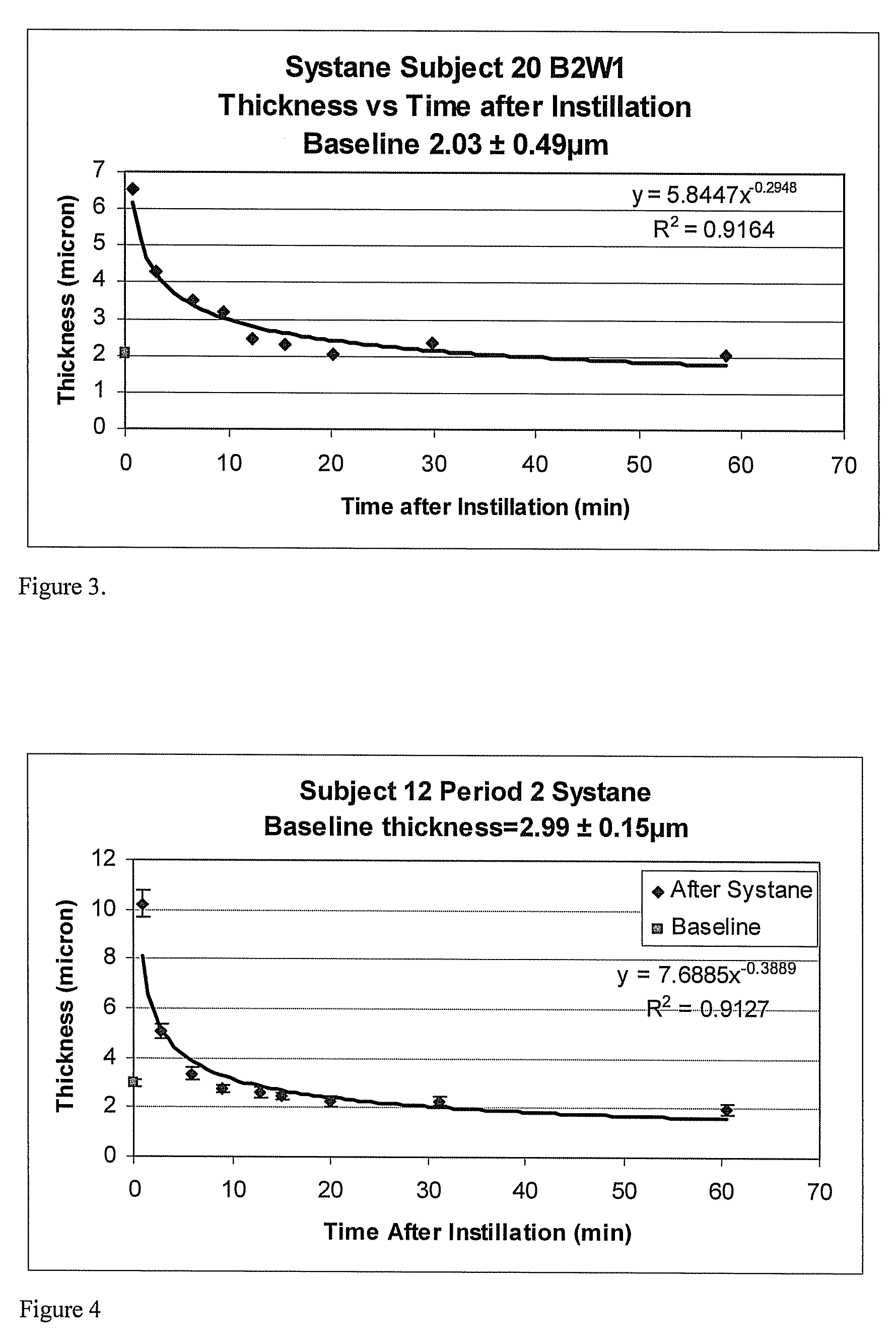
[0078]In this example, the combined aqueous+lipid layers tear film thickness was measured in the right eye of a subject (Subj 20, B2W1) prior to the application of Systane drops. The aqueous+lipid layers combined thickness was measured with a wavelength-dependent optical interferometer of the type disclosed in King-Smith, P E et al. The Thickness of the Human Precorneal Tear Film: Evidence from Reflection Spectra. Invest. Opthalmol. Vis. Sci. 2000 October; 41(11): 3348-3359, which is incorporated herein by reference in its entirety. 50 measurements were taken of the tear film at a 12.5×133 um spot at the apex of the cornea, each 504 msec, over a 25.2 second interval, to yield a baseline pre-eye drop combined aqueous+lipid layers tear film thickness of 2.03±0.49 microns. Thereafter, a single 40 uL drop of Systane drops, lot 62314F, exp November 2007 (Alcon Laboratories, Fort Worth, Tex.), was instilled into the right eye of the same subject and the combined aqueous+lipid layers thick...

example 2

[0079]In this example, the combined aqueous+lipid layers tear film thickness was measured in the right eye of a subject (Subj 12, B2W1) prior to the application of Systane drops. The aqueous+lipid layers combined thickness was measured using the instrument of example 1, to yield a baseline pre-eye drop combined aqueous+lipid layers tear film thickness of 2.99±0.15 microns. Thereafter, a single 40 uL drop of Alcon® Systane drops, lot 62314F, exp November 2007 (Fort Worth, Tex.), was instilled into the right eye of the same subject and the combined aqueous+lipid layers thickness was measured several times over a period of 1 hour, as indicated in Table 1.

[0080]

TABLE 1time, min00.882.755.929.0712.8715.0519.9331.1260.55thickness, microns2.9910.215.033.352.752.592.482.252.271.96std dev, microns0.150.530.290.240.170.190.160.230.170.22rtbaselinep p p p p p

[0081]T-test statistical comparisons were calculated between the baseline thickness and the thickness values at 9.07 minutes and thereaf...

example 3

[0083]In this example, the combined aqueous+lipid layers tear film thickness was measured in the right eye of a subject (Subj 20, B1W1) prior to the application of a commercially available eye drop, blinks tears lubricating eye drops (Advanced Medical Optics, Inc., Santa Ana, Calif.). The aqueous+lipid layers combined thickness was measured using the instrument of example 1, to yield a baseline pre-eye drop combined aqueous+lipid layers tear film thickness of 1.89±0.24 microns. Thereafter, a single 40 uL drop of Blink® tears, was instilled into the right eye of the same subject and the combined aqueous+lipid layers thickness was measured several times over a period of 1 hour. It can be seen in FIG. 5 that the instillation of Blink® Tears thickened the tear film over a period of time. Again, although general thickening of the tear film following topical application of an ophthalmic formula is conventionally expected from the prior art, it is surprising that measurement of tear film t...


PUM


Abstract

Methods and devices measure eye blinks and tear film lipid and aqueous layer thickness before and following ophthalmic formula application onto the ocular surface, especially wherein the ophthalmic formula is an artificial tear. The methods and devices are suitable for dry eye diagnosis. The methods and devices are suitable for use to evaluate ophthalmic formula effects on the tear film and to use such information to diagnose ophthalmic formula treatment of ocular disease conditions such as dry eye in the absence of contact lens wear or post-surgical eye drop treatment and diagnosis. The methods and devices are also suitable for use in the optimization of ophthalmic drug dosage forms and sustained drug release.

Description

BACKGROUND OF THE INVENTION[0001]1. Field of the Invention[0002]Embodiments of the invention relate to methods and devices for use in evaluating ophthalmic formula effects on the tear film and to use such information to diagnose ophthalmic formula treatment of ocular disease conditions such as dry eye or post-surgical ophthalmic formula treatment and diagnosis.[0003]2. Description of the Related Art[0004]Dry eye syndrome is a prevalent condition among both men and women for which there is no cure, although symptoms may be relieved with proper diagnosis and treatment. The condition affects more than 3.2 million American women middle-aged and older alone (Schaumberg D A, Sullivan D A, Buring J E, Dana M R. Prevalence of dry eye syndrome among US women. Am J Ophthalmol 2003 August; 136(2):318-26). Most dry eye patients are prescribed artificial tears to treat their dry eye conditions. Contact lens wearers, computer users, patients who live and / or work in dry environments, and patients ...

Claims


Application Information

Patent Timeline
Patent Type & Authority Patents(United States)
IPC IPC(8): A61B3/00A61B3/10
CPCA61B3/101
Inventor HUTH, STANTRAN, DENISE
Owner JOHNSON & JOHNSON SURGICAL VISION INC
Learn More