
Method for improving magnetic resonance imaging of the breast

a magnetic resonance imaging and breast technology, applied in the field of breast enhancement, can solve the problems of non-uniformity of the magnetic resonance system, the 2-dimensional coil pad is not provided with image enhancement, and the presentation of variable specificity that represents a major limitation, so as to enhance the magnetic resonance image of breast tissue, high density, and high resilien

Inactive Publication Date: 2011-12-13
ORGAMEND
View PDF18 Cites 4 Cited by
  • Summary
  • Abstract
  • Description
  • Claims
  • Application Information

AI Technical Summary

Benefits of technology

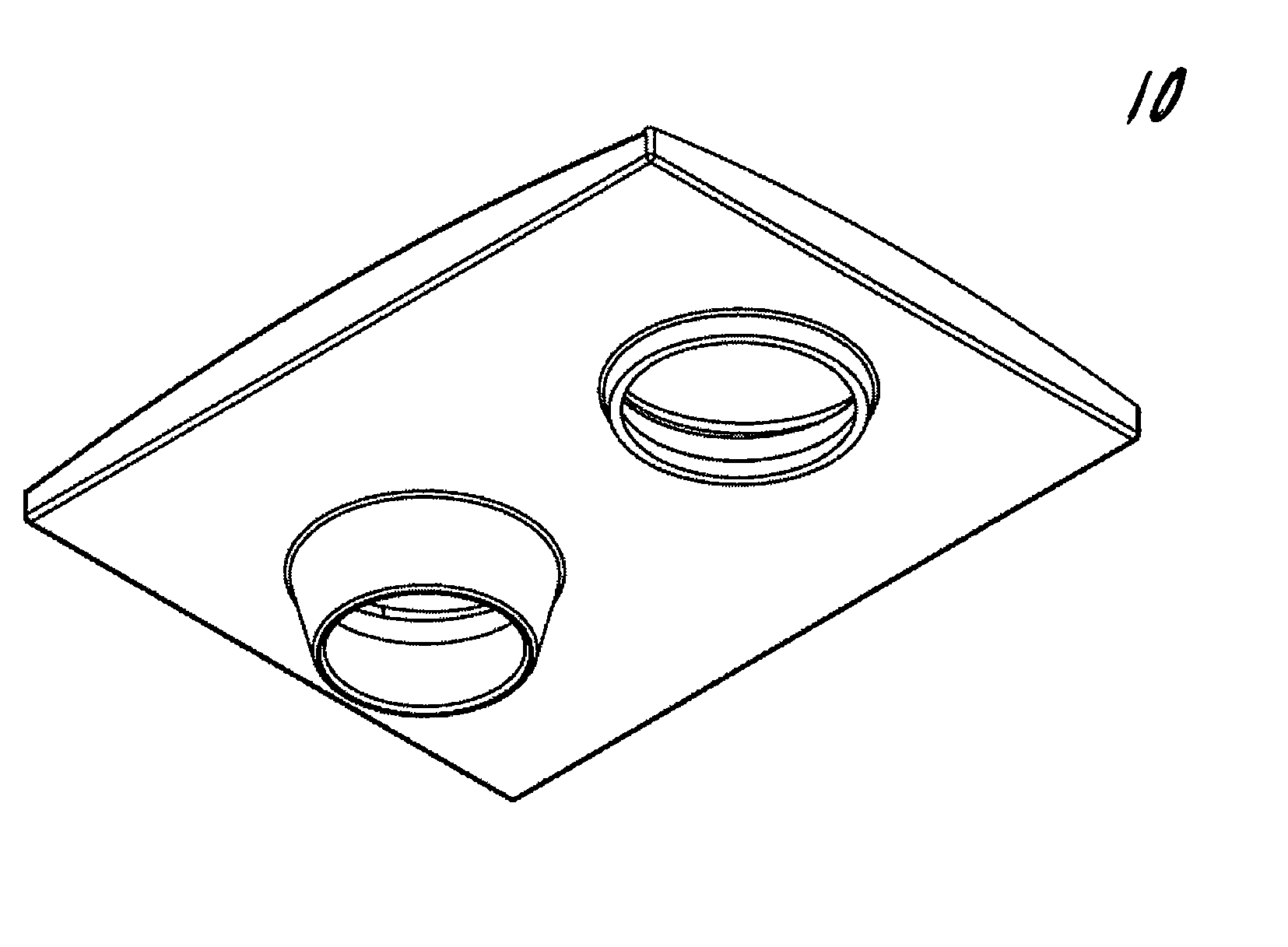
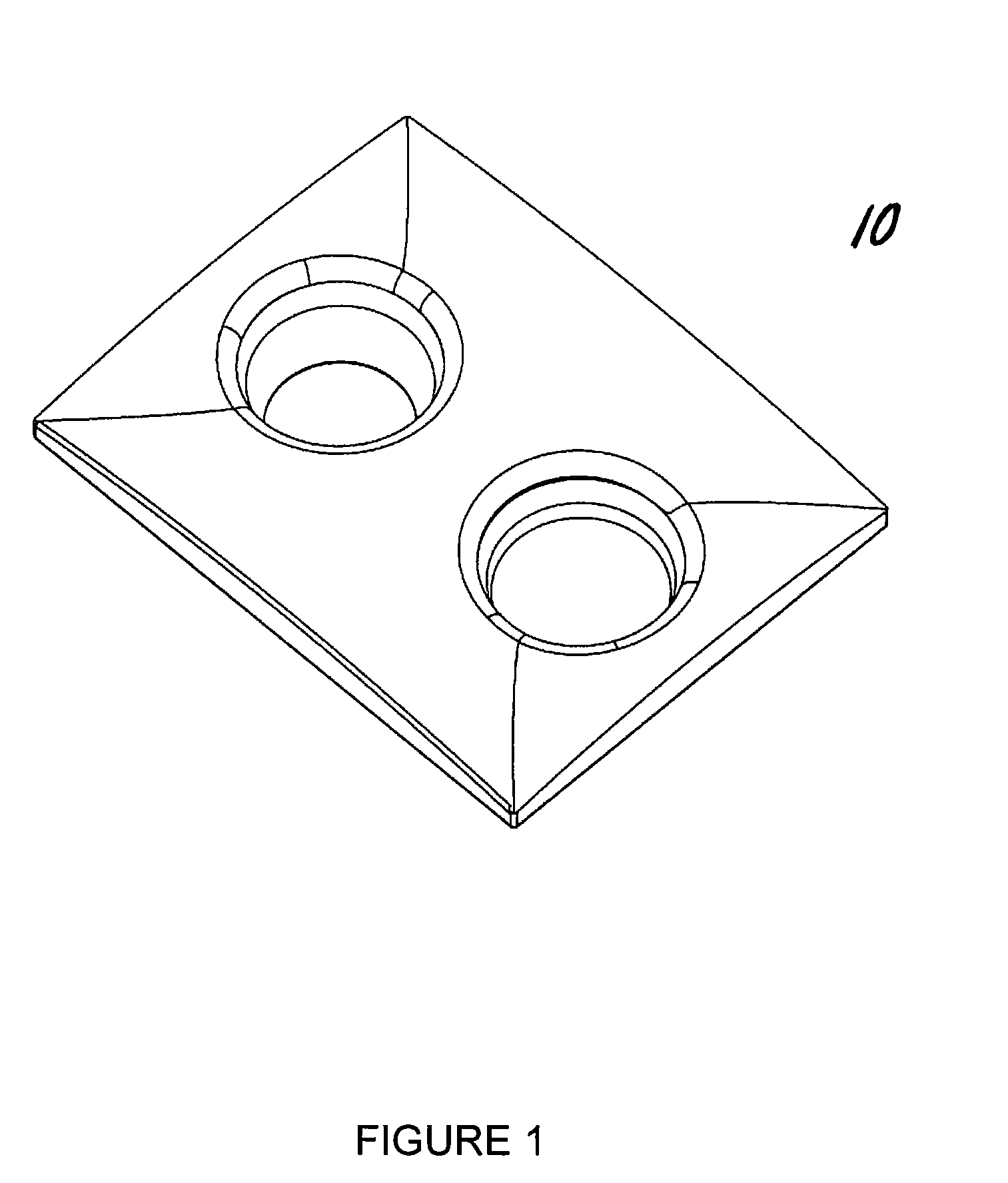
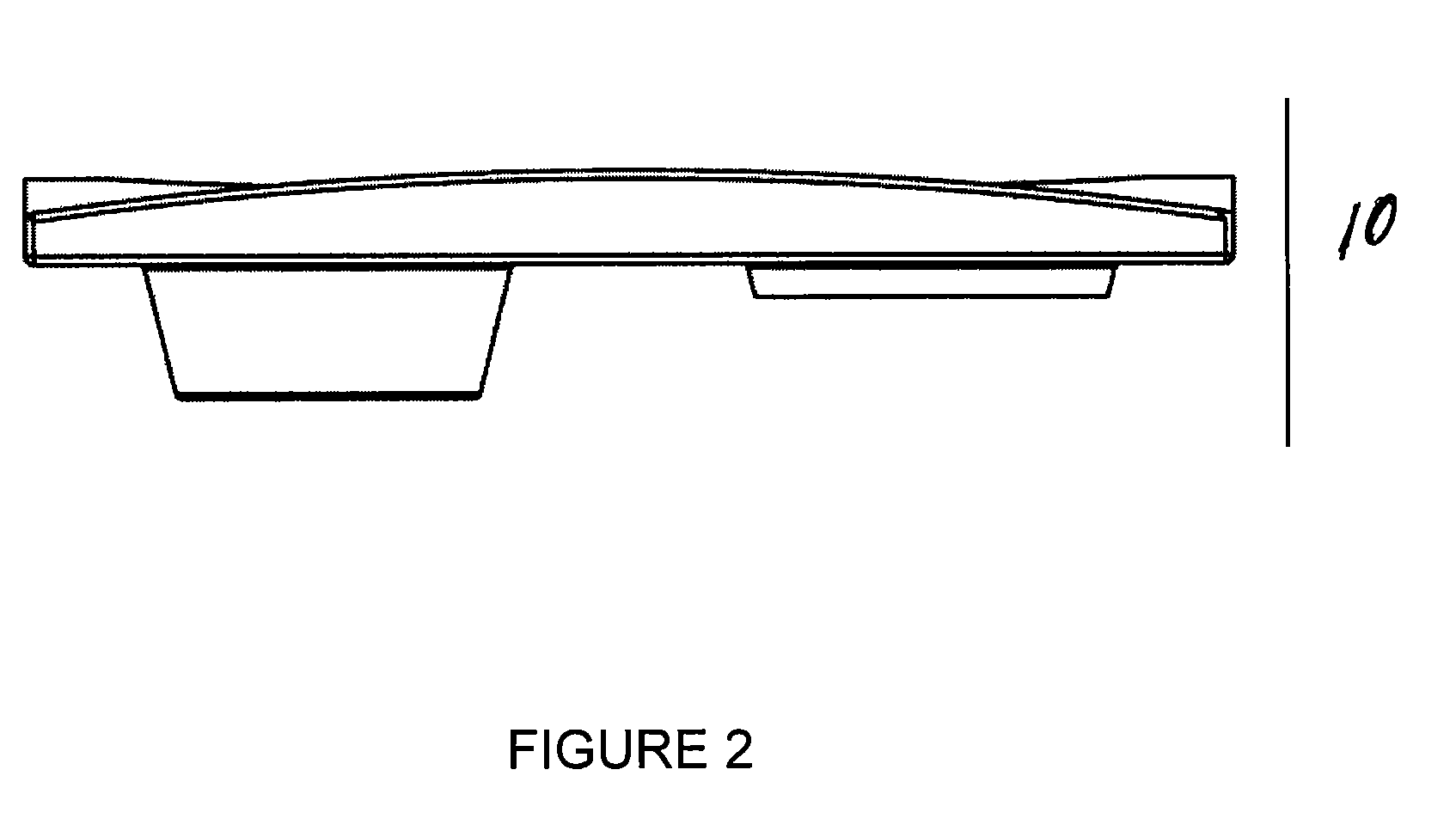
"The present invention provides a method for improving magnetic resonance imaging of breast tissue by using a device that includes a fat saturation material. This device can be used with a radio frequency coil for magnetic resonance imaging of the breasts. The device is designed to be placed on the breast coil and requires no further manipulations. The device interfaces closely with the breast and can effectively reduce the skin to air interface regardless of breast size. The technical effect of this invention is to enhance the quality and accuracy of magnetic resonance imaging of breast tissue."

Problems solved by technology

Despite the sensitivity MR imaging provides over more traditional imaging modalities such as mammography or ultrasound, there are several technologic hurdles.
While providing exceptional sensitivity, it displays variable specificity that represents a major limitation.
However, the coil pads are 2-dimensional and do not provide image enhancement by either fat saturation or by reducing the skin to air interface.
The non-uniformities in the magnetic resonance system's main magnetic field that is generated by the variations in breast geometry and magnetic susceptibility in localized areas of the breast can be problematic in obtaining good quality images.
However, the fat saturation techniques used in magnetic resonance imaging are highly sensitive to homogeneity in the magnetic field and breast anatomy is not homogenous in areas.
Fat saturation techniques while beneficial, may not uniformly suppress the fat within the breast tissue being imaged and can result in cross-suppression where water molecules may be likewise affected.
The result is unreliable fat saturation with a less than optimal image.
While contributing to image enhancement, shimming may not uniformally suppress fat and can suppress regions of water in the breast.
There are several significant drawbacks to this device.
First, it is not compatible for use with any of the breast coils being used clinically.
Secondly, the volume of fat saturation enhancing material that is re-introduced is variable without intrinsic controls to ensure there is enough material present to provide adequate image enhancement.
Thirdly, the breasts are compressed by the weight of the patient laying prone on the breast bag and subsequently further compressed when the bag is refilled with the perfluorochemical liquid after being inflated.
The resulting compression of the breast tissue negatively impacts the ability to image the vasculature and lesion angiogenesis.
Fourthly, the selectively fillable breast bags covers the breasts on all sides making biopsy impossible.
However, in the absence of a fat saturation device, artifacts are common.
The difficulty in providing surgical access is especially difficult for breasts, due to the varying sizes and shapes and desirability of minimizing manipulation which is stressful for the patient.

Method used


Image

  • Method for improving magnetic resonance imaging of the breast
  • Method for improving magnetic resonance imaging of the breast
  • Method for improving magnetic resonance imaging of the breast

Examples


example 1

[0042]In another configuration, the fat saturation enhancing pad can be constructed to provide two detachable pieces: the pad lining the top of the breast coil that the patient lays upon and the circumferential pad that covers the area surrounding the surface coil that surrounds the pendulant breast. By detaching the circumferential part of the device from the pad layering the top of the breast coil access to the breast can be achieved for the purpose of performing a biopsy.

example 2

[0043]In one embodiment, the fat saturation enhancing device can be fabricated to be used in combination with any of the various breast coils that are used clinically. In an industry where MR coils are manufacturer and system specific, the device can be configured to provide both three-dimensional circular and rectangular apertures to fit into the existing apertures in the various breast coils in use clinically.

example 3

[0044]In one embodiment, the device enhances fat saturation without compression of the breast tissue resulting in better imaging of the vasculature with enhanced identification of lesion angiogenesis. Moreover, the absence of breast tissue compression makes the device compatible with breast imaging software because its use does not alter the pendulant configuration of the breast when a patient lays prone on a breast coil.


PUM


Abstract

The present invention provides a fat saturation device to be used in conjunction with a magnetice resonance imaging (MRI) breast coil, which provides padding for the comfort of the patient being imaged while eliminating the skin-air interface, thereby reducing the level of artefacts in the image. The device comprises a coil surface pad that contacts the surface of the coil, the pad having apertures therein through which the patients breasts extend when the patient is positioned on the coil for imaging. Aperture sleeves that are removable or retractable extend from the bottom surface of the coil surface into the apertures of the breast coil. The device comprises a fat saturation material.

Description

CROSS REFERENCE TO RELATED APPLICATIONS[0001]This application is a continuation-in-part of U.S. application Ser. No. 11 / 743,467, filed May 2, 2007, now abandoned, which claims the priority of U.S. provisional application Ser. No. 60 / 797,056, filed May 2, 2006, the contents of which are hereby incorporated by reference into the present application.FIELD OF THE INVENTION[0002]The invention relates to a device and method for improving magnetic resonance imaging of the breast. More particularly, the invention relates to enhanced breast imaging by filling the void area between the breast coil aperture and breast tissue with a self-contained pad that conforms specifically to the three-dimensional surfaces of the breast coil while simultaneously accommodating differences in breast anatomy.BACKGROUND OF THE INVENTION[0003]Magnetic resonance imaging (MRI) of the breast has emerged as an important new diagnostic tool for the detection of tumors. Pre-surgical MRI can help to reduce the number ...

Claims


Application Information

Patent Timeline
Patent Type & Authority Patents(United States)
IPC IPC(8): A61B5/05
CPCA61B5/0555G01R33/28G01R33/34G01R33/3415G01R33/4828G01R33/5607A61B5/055
Inventor BRASILE, LAUREN
Owner ORGAMEND
Learn More