Method and apparatus to compensate imaging data with simultaneously acquired motion data

a technology of motion data and imaging data, applied in the field of diagnostic imaging, can solve the problems of significant time required to collect a complete set of projections, inability to provide mechanical motion detection with conventional ekg gating, and inability to correct motion errors in imaging data

Active Publication Date: 2012-04-10
GENERAL ELECTRIC CO
View PDF10 Cites 22 Cited by
  • Summary
  • Abstract
  • Description
  • Claims
  • Application Information

AI Technical Summary

Benefits of technology

[0012]According to yet another aspect of the present invention, the invention is embodied in a computer readable storage medium having a computer program stored thereon that represents a set of instructions to be executed by a computer. The instructions cause the computer to calculate a mapping function that provides a location of each sub-volume of an imaging volume as a function of time from ultrasound data. The instructions further cause the computer to determine a respective acquisition window for each sub-volume of the imaging volume for the mapping function and reconstruct an image of the imaging volume substantially free of motion artifacts from imaging data acquired from the imaging volume.
[0014]In accordance with yet another aspect, the invention is directed to an ultrasound probe attached to or integrated with a patient table. In this regard, the invention allows for “hands-free” positioning of the ultrasound probe relative to the patient during data acquisition.
[0016]According to a further aspect, the invention is embodied in a method wherein motion data and imaging data are simultaneously acquired. The motion and imaging data are acquired independently with different image modalities, e.g. ultrasound and CT, respectfully. The mechanical motion data may be used to correct for errors in the imaging data or, alternately, the motion data may be used to reduce noise in the imaging data.

Problems solved by technology

In known studies, this motion correction or compensation can add significant complexity in post processing of the images.
Because of various limitations in conventional CT scanners, the time necessary to collect a complete set of projections is significant relative to object motion.
Specifically, conventional EKG gating does not provide mechanical motion detection.
While an EKG signal can indicate that motion is occurring or is about to occur, it cannot provide accurate real-time placement data of the heart.
Conventionally, half-scan weighting is used to suppress the impact of motion; however, its effectiveness is less than optimal since half-scan weighting reduces the contribution of CT data acquired at both ends near the 180 degree projection angular range.
Further, even with a gantry speed of 0.3 s / rotation, the central region of the projection range constitutes a 150 ms temporal window, which is prohibitively slow to completely “freeze” cardiac motion.
The data acquisition window for CT systems having dual tube-detector assemblies is still between 70-80 ms which is not sufficient to eliminate heart motion.

Method used


Image

  • Method and apparatus to compensate imaging data with simultaneously acquired motion data
  • Method and apparatus to compensate imaging data with simultaneously acquired motion data
  • Method and apparatus to compensate imaging data with simultaneously acquired motion data

Examples


Embodiment Construction

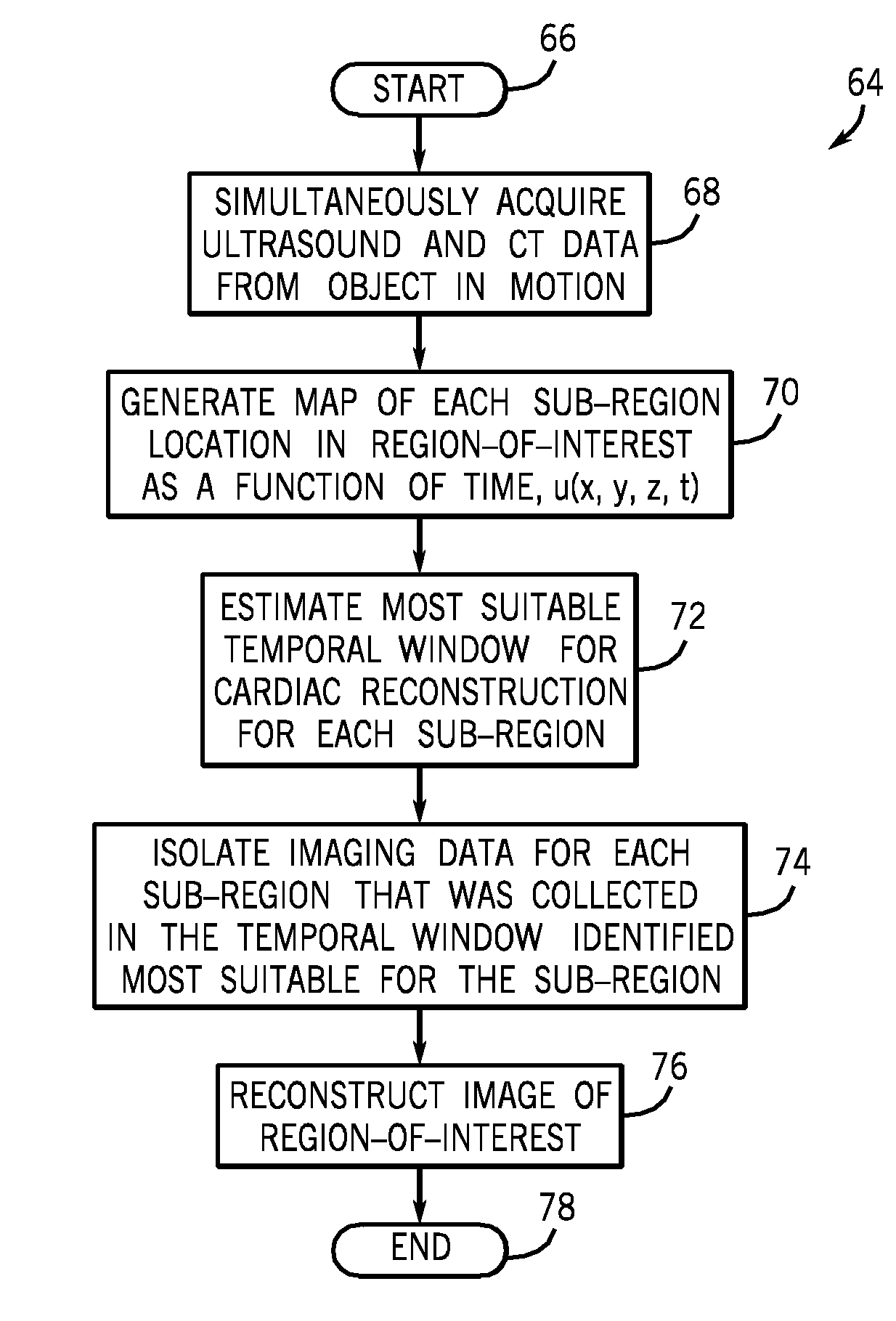
[0026]The present invention will be described with respect to a “third generation” CT scanner, but is equally applicable with other image modalities. Moreover, the present invention will be described with respect to an imaging system that includes a CT scanner that acquires image data and an ultrasound machine that acquires motion data from a patient. The CT scanner and ultrasound machine are stand-alone devices that can be used independently from one another, but, as will be described, can operate in tandem to acquire CT data and ultrasound data simultaneously. It is also contemplated that the present invention is applicable with an integrated CT / ultrasound system. It is further contemplated that the invention may be embodied in a combination ultrasound / MR system or a stand-alone ultrasound and a stand-alone MR scanner that work in tandem to acquire motion and image data.

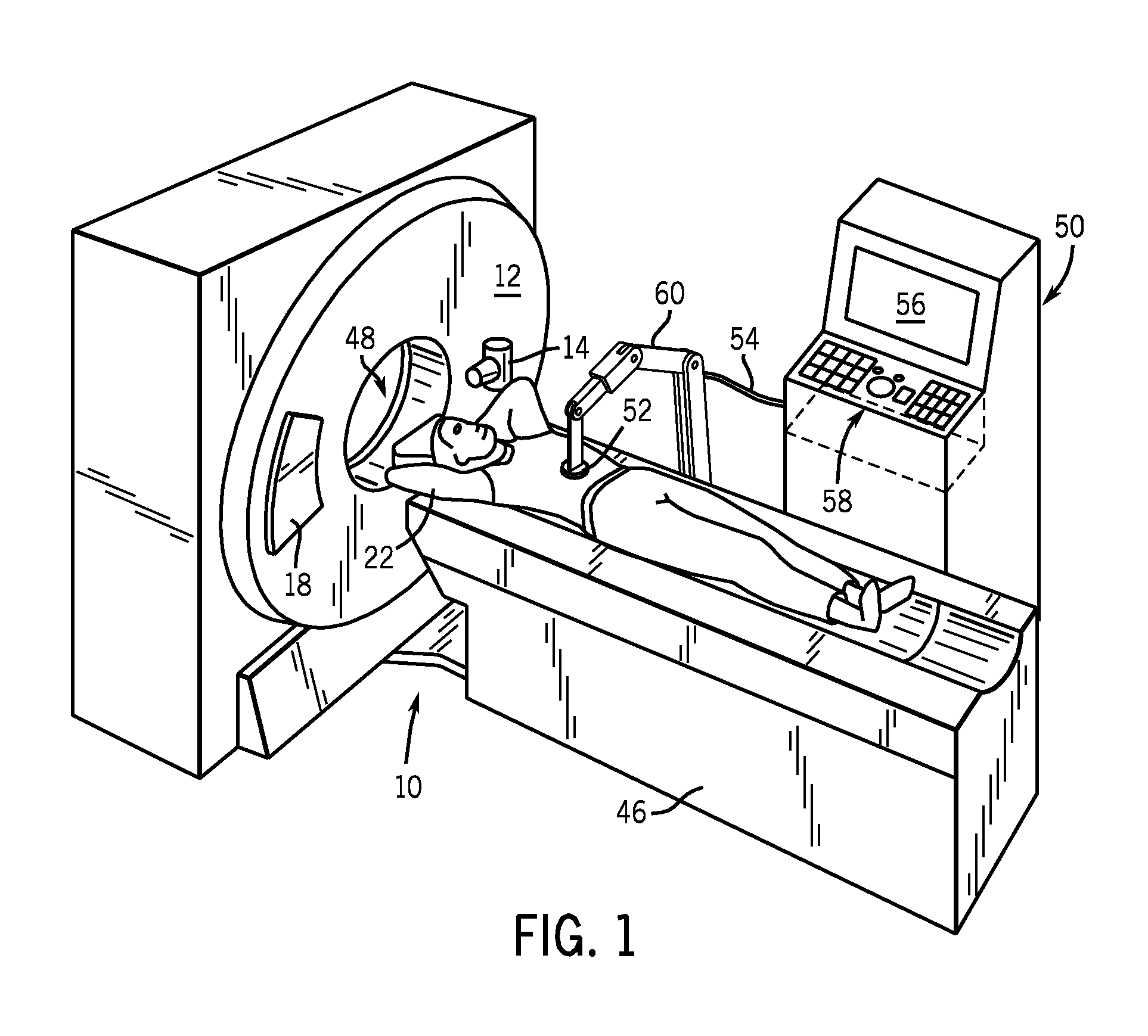
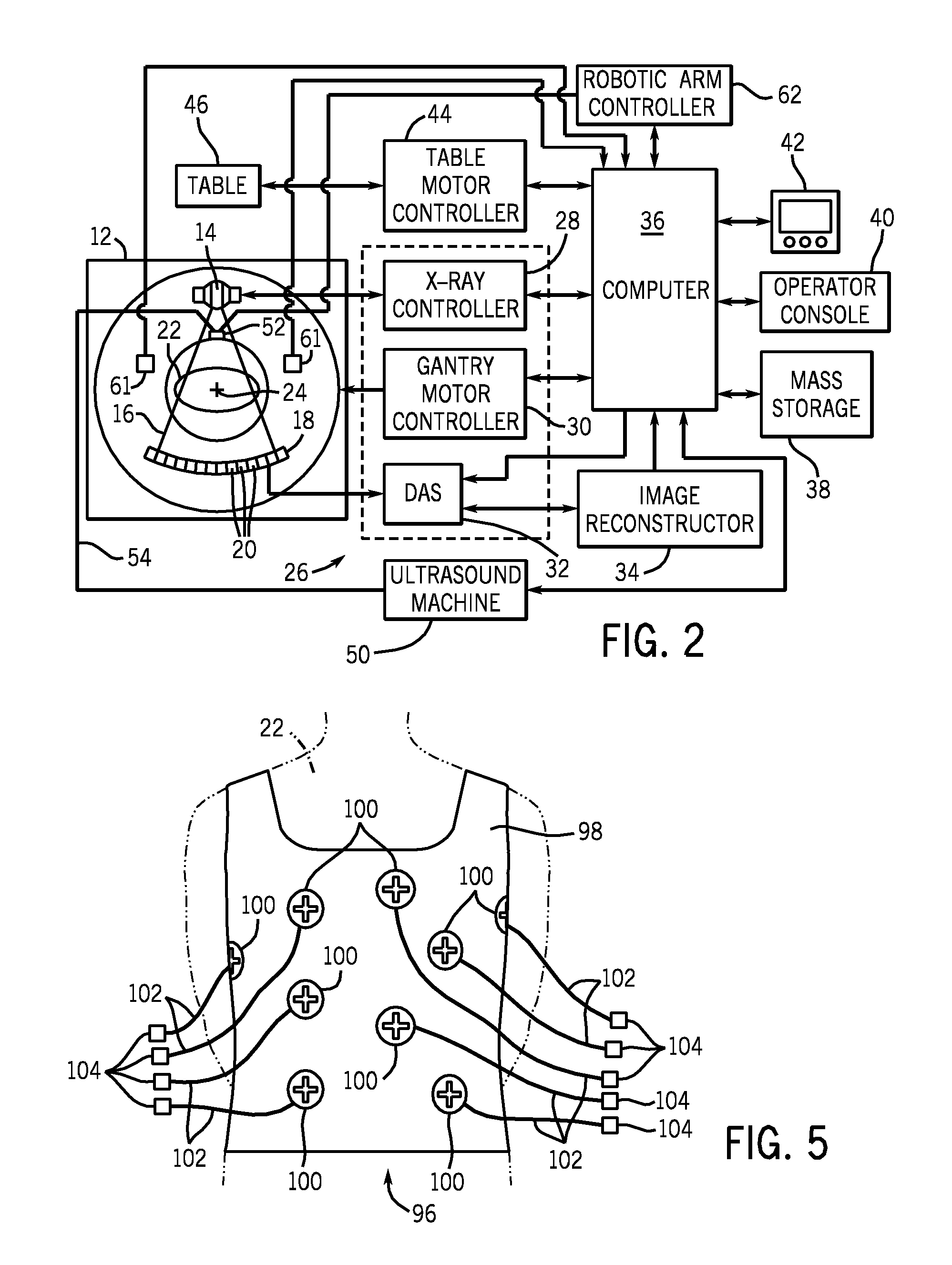
[0027]Referring to FIGS. 1 and 2, a computed tomography (CT) imaging system 10 is shown as including a gantry 12...


PUM

PropertyMeasurementUnit
imagingaaaaaaaaaa
volumeaaaaaaaaaa
imaging volumeaaaaaaaaaa

Abstract

Motion data is acquired simultaneously and in real-time with image data. The motion data provides accurate and near-instant information as to the state and position of an object prone to motion. The present invention is particularly applicable for cardiac CT or MR imaging and other physiologically gated acquisitions. In the context of cardiac imaging, the motion data includes information regarding size and location of the heart during the cardiac phases (diastole, systole, etc.) during each cardiac cycle.

Description

BACKGROUND OF THE INVENTION[0001]The present invention relates generally to diagnostic imaging and, more particularly, to a method and apparatus capable of correcting motion errors in imaging data acquired from an object prone to motion. This motion correction is advantageously achieved by using mechanical motion data acquired from the object with one modality coincident with imaging data acquisition from the object with another and different modality.[0002]Various imaging modalities are often used to image objects in or prone to motion, such as the heart in cardiac studies. For example, in cardiac computed tomography (CT), magnetic resonance imaging (MRI) and other imaging modalities directed to the acquisition of data from an object prone to motion, one or more motion correction techniques are generally used to reduce motion-induced artifacts in the reconstructed images. In known studies, this motion correction or compensation can add significant complexity in post processing of t...

Claims


Application Information

Patent Timeline
Patent Type & Authority Patents(United States)
IPC IPC(8): A61B5/05
CPCA61B5/6805A61B5/721A61B6/032A61B8/08A61B8/42A61B8/4218A61B8/4416A61B19/5244G01T1/1603G01T1/1648A61B6/4417A61B5/02007A61B5/0073Y10T29/49826A61B5/1075A61B5/7285A61B6/503A61B6/5247A61B8/0858A61B8/0883A61B8/4281A61B8/485A61B19/2203A61B2017/00694A61B2017/00991A61B2019/5236A61B2019/524A61B2019/5263A61B2019/5276A61B2034/2063A61B34/20A61B34/30A61B2090/374A61B2090/3762A61B2090/378
Inventor HSIEH, JIANGBARBER, MICHAEL JOHNKOST, BRIAN J.CLARKE, WILLIAM R.JUHL, MICHAEL A.HALL, ANNE LINDSAY
Owner GENERAL ELECTRIC CO
Why Eureka
Try Eureka