Non-invasive method to monitor microcirculation

a microcirculation and non-invasive technology, applied in the field of non-invasive methods to monitor microcirculation, can solve the problems of poor application prospects in primary care or general practitioner's office, poor application prospects, and difficult use of technology in primary care and general practitioner's offi

Inactive Publication Date: 2012-06-26
WHEELSBRIDGE
View PDF18 Cites 4 Cited by
  • Summary
  • Abstract
  • Description
  • Claims
  • Application Information

AI Technical Summary

Benefits of technology

[0009]It is another object of the present invention to provide for reliable, quick and cheap method to determine if a patient suffering from underlying disease known to produce impairments in microcirculation is susceptible to acquire such complications and thereby need a change of regiment or therapy.
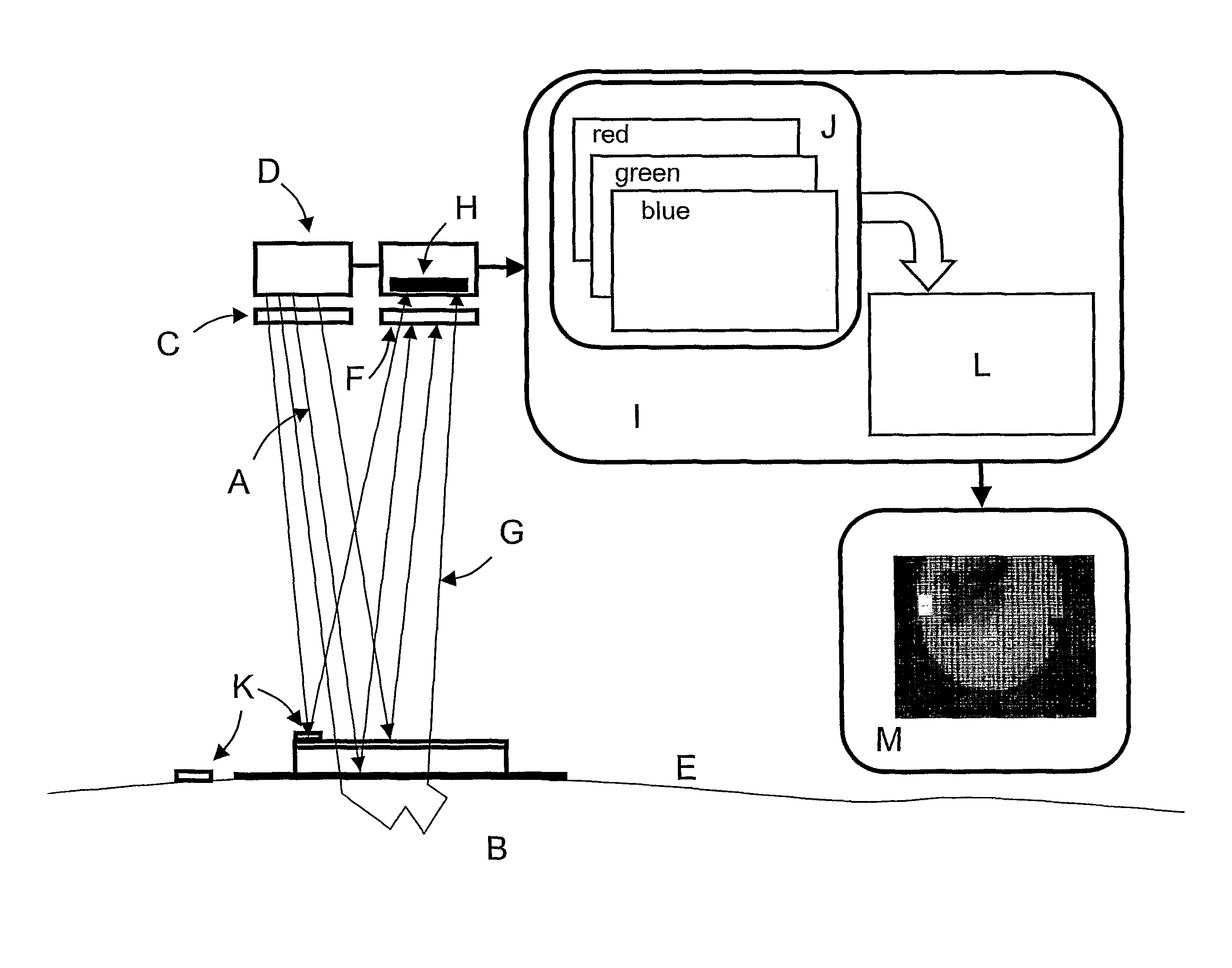
[0017]By capturing the images in a rapid sequence—as a video clip—and then compiling a movie, the fine dynamics of the microcirculation such as pulsatility can be detected. Using this aspect images representing the arterial and the venous side of the microcirculation can be generated, thereby making it possible to differentiate e.g. an arterial leg ulcer from a venous leg ulcer.
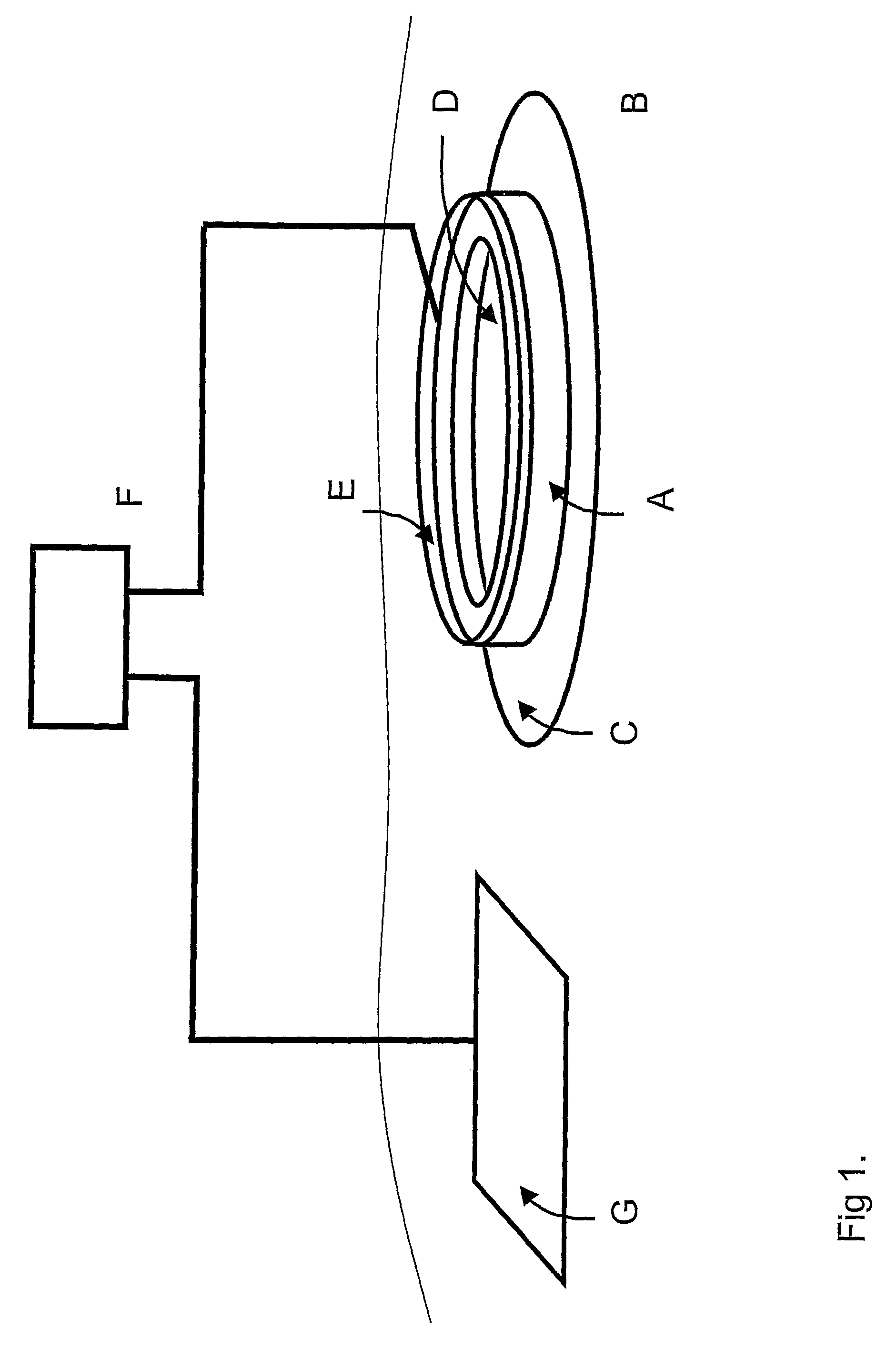
[0018]In another embodiment of the invention, the photosensitive array can be incorporated in an electrode (Camerode—Camera in an Electrode) that constitutes a wearable transducer device that facilitates continuous and mobile investigation of the most peripheral skin microcirculation in e.g. patients recovering from a heart attack or as early chock or blood pressure drop detection.
[0019]In yet another embodiment, the light from the illuminating light illuminates the tissue through flexible optical fibers. Another fiber or a bundle of oriented fibers brings the backscattered light back to the photosensitive array. By positioning the polarization filters at the distal end of the fibers—or by using polarization preserving light guides with the polarization filters positioned at the proximal end of the fibers—, the desired depth sensitivity can be attained and the microcirculation in body cavities can be investigated, thereby making it possible to detect tumours with elevated microcirculation.

Problems solved by technology

Laser Doppler perfusion imaging (and monitoring) is, however, a technology not very well suited for applications in primary care or in the office of the general practitioner, because of high cost and to some extent cumbersome handling and image interpretation.
The high degree of magnification required, however, makes this technology difficult to use in primary care and in the general practitioner's office, because of the inevitable relative movement between the microscope and the object.
Besides, the end result is in best case a structural image of the minute vessels in a small area and gives no direct information about the average vasodilatation of a more extensive skin site.

Method used


Image

  • Non-invasive method to monitor microcirculation
  • Non-invasive method to monitor microcirculation
  • Non-invasive method to monitor microcirculation

Examples


example

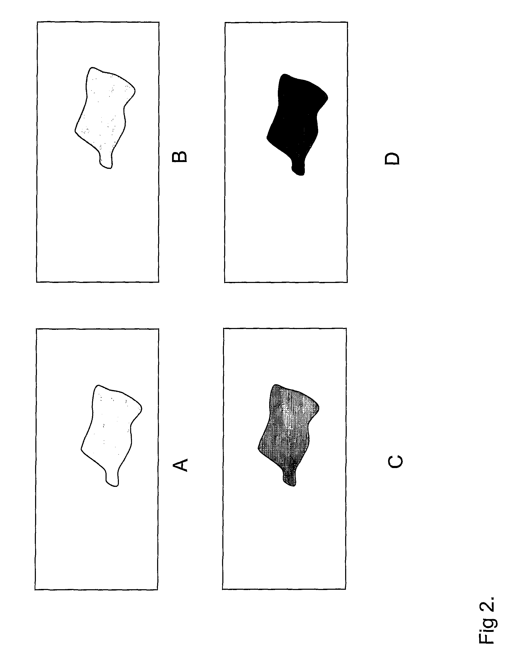
[0036]In a pilot study—following the production of vasodilatation in skin tissue—several different algorithms were applied in the generation of the output data matrix. The average value of the elements of this data output matrix is shown in FIG. 5 for the algorithms (R-G) / (G-B), (R-G) / B, RIG, RIB, (R-B) / G and (R-G) / (R-B), where R, G and B represent the average values of the data matrixes representing red, green and blue color. For each algorithm employed, the test was performed on the skin sites labelled (BDonDIL) representing a skin site Before Dilatation on DILated area, (ADonDIL) representing a skin site After Dilatation on DILated area, (BDonNOR) representing a skin reference site Before Dilatation on a NORmal not dilated area and ADonNORM representing a skin reference site After Dilatation on NORmal not dilated area respectively. As can be seen from the example in FIG. 5 the algorithm (R-G) / B gives the best discrimination among the algorithms tested. All color separations and a...


PUM

PropertyMeasurementUnit
photosensitiveaaaaaaaaaa
concentrationaaaaaaaaaa
colorsaaaaaaaaaa

Abstract

This invention relates to the non-invasive determination of the degree of vasoactivity in the microcirculation in a tissue caused by a drug, disease, injury normal or pathological regulation. More specifically, the invention relates to a method of determining the influence on microcirculation in living tissue from an irritative agent, drugs, disease, injuries normal or pathological regulation including, illuminating a tissue surface with polarized light, collecting the backscattered light through a polarizing filter, detecting the backscattered and polarized light by a photo-sensitive array, transferring the collected information in digital form to a computing device, separating the collected information into at least two data matrixes, each representing a specific wavelength range and generating an output data matrix by processing corresponding values in at least two data matrixes by an algorithm, wherein each value in said output data matrix represents the amount of influence on the microcirculation in a source point of the tissue. Thereby a representation of the tissue microcirculation is obtained.

Description

RELATED APPLICATIONS[0001]This application is a 371 of PCT / SE2005 / 000554 filed Apr. 18, 2005 and claims priority under 35 U.S.C. §119 of Swedish Application No. 0401004-7 filed Apr. 19, 2004.FIELD OF INVENTION[0002]This invention relates to the non-invasive determination of the degree of vasoactivity in the microcirculation in a tissue caused by an irritative agent, drug, disease, injury, normal or pathological regulation.BACKGROUND OF THE INVENTION[0003]In medical practice, in the development of new drugs and in skin testing procedures it is important to determine physiological responses such as the degree of vasodilatation or vasoconstriction in the microcirculation following application of a vasoactive drug directly in contact with the skin or administrated by other routes. A suitable way of transdermal drug administration is by means of iontophoresis in order to support penetration of charged drug molecules through the epidermal layer or by topical application of the drug or age...

Claims


Application Information

Patent Timeline
Patent Type & Authority Patents(United States)
IPC IPC(8): A61B5/02G01N21/21A61B5/00A61B5/026A61N1/30G01N
CPCA61B5/0261
Inventor NILSSON, GERTSJOBERG, FOLKE
Owner WHEELSBRIDGE
Why Eureka
Try Eureka