
Three-dimensional breast anatomy imaging system

a breast anatomy and three-dimensional technology, applied in the field of breast anatomy imaging system, can solve the problems of inability to accurately track the position of the optical probe as measurements are recorded, and the technology using optical probes, so as to reduce the uncertainty of measurement and increase the measurement sensitivity

Inactive Publication Date: 2012-08-14
RGT UNIV OF CALIFORNIA +1
View PDF15 Cites 7 Cited by
  • Summary
  • Abstract
  • Description
  • Claims
  • Application Information

AI Technical Summary

Benefits of technology

[0008]According to embodiments of the present invention, the same Region of Interest (ROI) in an organ such as a breast can be measured with each visit, thereby increasing measurement sensitivity by minimizing uncertainties involved with optical handheld probe position placement. Further, by tracking the rotational coordinates of the optical handheld probe on the surface of the breast, the orientation and coupling of the optical handheld probe can be consistent with each measurement and over the range of longitudinal measurements.

Problems solved by technology

However, a limitation of conventional technologies using the optical probes is the inability to accurately track the position of the optical probe as measurements are recorded.

Method used


Image

  • Three-dimensional breast anatomy imaging system
  • Three-dimensional breast anatomy imaging system
  • Three-dimensional breast anatomy imaging system

Examples


Embodiment Construction

[0025]Preferred embodiments of the present invention will now be described more fully hereinafter with reference to the accompanying drawings. This invention may, however, be embodied in different forms and should not be construed as limited to the embodiments set forth herein.

[0026]An optical imaging device uses light for imaging parts of the human body. Diffuse optical spectroscopy (DOS) is used for, for example, breast cancer detection and monitoring by measuring optical properties such as the absorption and scattering of the tissues. Diffuse optical spectroscopy typically uses red and near-red spectral region because the dominant molecular absorbers within the red or near-red spectral region in tissues include hemoglobin, water and lipids. Unlike mammography or ultrasound method, the DOS is capable of quantifying the optical properties of, for example, the hemoglobin, water and lipids.

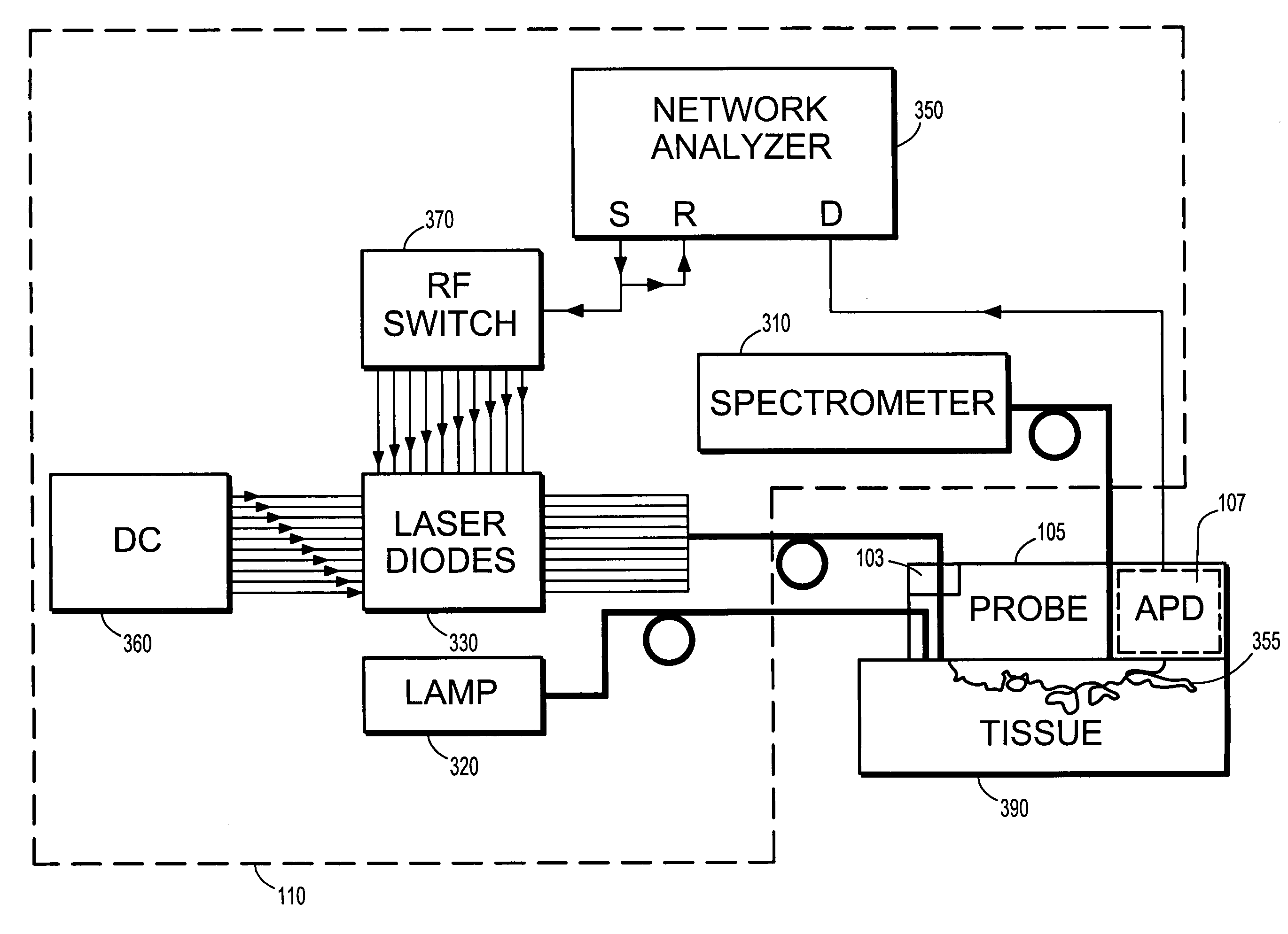
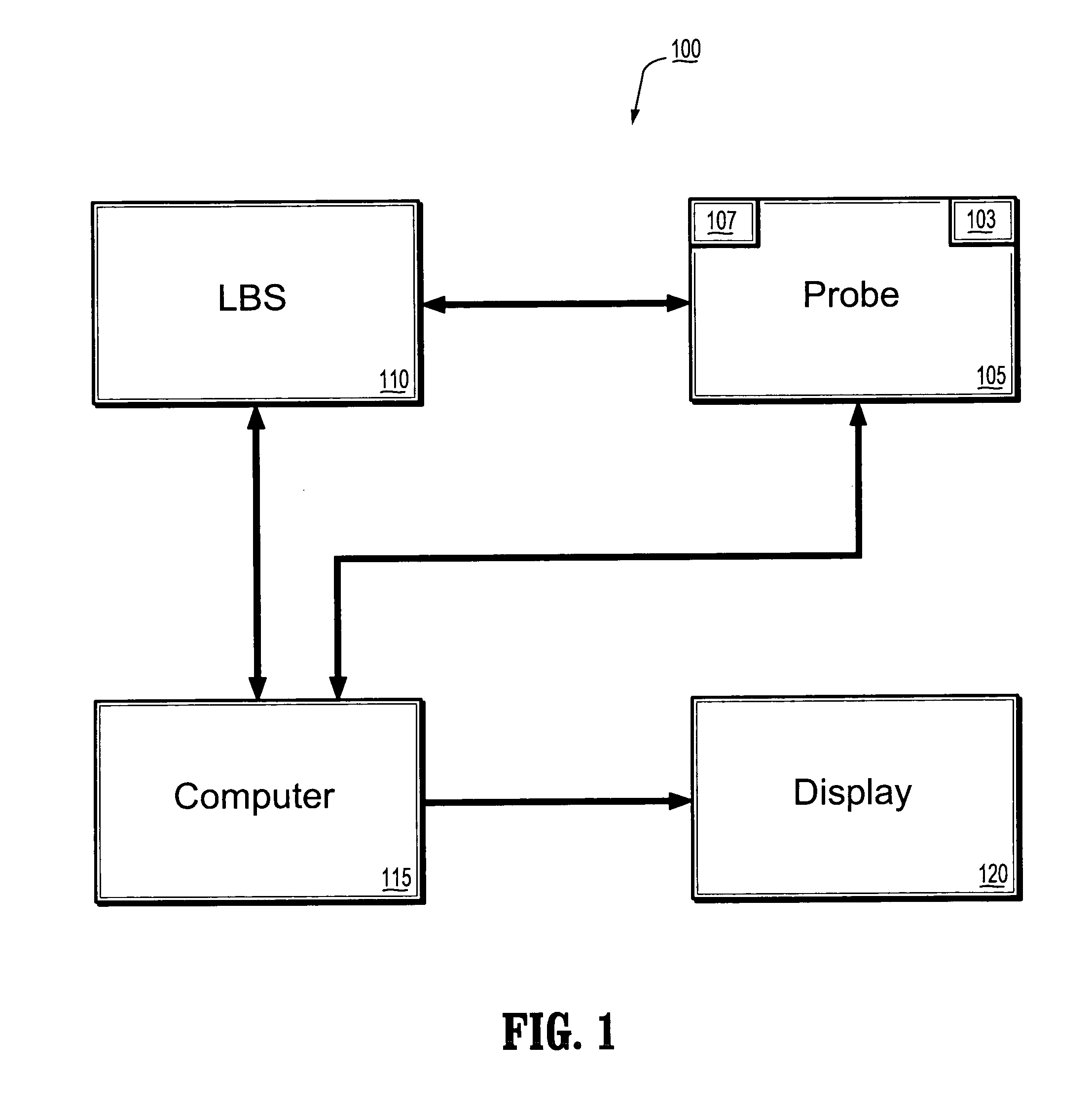
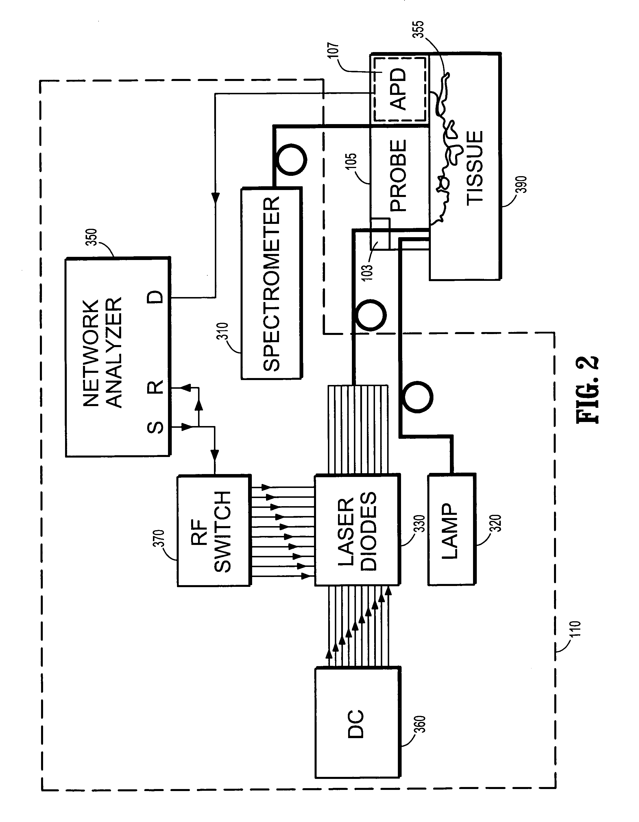
[0027]FIG. 1 shows a schematic diagram of a diffusion optical spectroscopy (DOS) system accordi...


PUM


Abstract

A diffuse optical spectroscopy system comprises a laser breast scanner, a handheld probe connected to the laser breast scanner for scanning a breast, and a tracking device coupled to the handheld probe, wherein the tracking device determines locations of the handheld probe relative to the breast. The tracking device comprises a magnetic tracking device, an optical tracking device or a laser tracking device.

Description

CROSS-REFERENCE TO RELATED APPLICATION[0001]This application claims priority to U.S. Provisional Application No. 60 / 638,231, which was filed on Dec. 22, 2004, and which is incorporated herein by reference in its entirety.BACKGROUND OF THE INVENTION[0002]1. Technical Field[0003]The present disclosure relates to a breast anatomy imaging system, and more particularly to a breast anatomy imaging system using a handheld optical imaging device combined with a position tracking system.[0004]2. Discussion of Related Art[0005]Presurgical chemotherapy is a treatment commonly used for locally advanced breast cancer. The patient's response to the presurgical chemotherapy is monitored to improve survival and reduce morbidity. A noninvasive, near-infrared technology based on diffuse optical spectroscopy (DOS) has been developed to quantitatively monitor tumor response to the presurgical chemotherapy. The quantitative, near-infrared diffuse optical spectroscopy is capable of distinguishing between...

Claims


Application Information

Patent Timeline
Patent Type & Authority Patents(United States)
IPC IPC(8): A61B6/00
CPCA61B5/0091A61B5/4312G01N21/359G01N21/474G01N21/4795A61B19/5244A61B2019/505A61B2019/5231A61B2019/5251A61B2019/5255G01N2021/1787G01N2021/4745G01N2201/0221G01N2201/0612G01N2201/0691G01N2201/08A61B34/20A61B2034/105A61B2034/2051A61B2034/2055A61B2090/373
Inventor AZAR, FRED S.KHAMENE, ALISAUER, FRANKTROMBERG, BRUCECERUSSI, ALBERT
Owner RGT UNIV OF CALIFORNIA
Learn More