System for carrying out and monitoring minimally-invasive interventions

a technology for monitoring systems and minimally invasive procedures, applied in the field of systems for carrying out and monitoring minimally invasive procedures, can solve problems such as restricted access to patients, and achieve the effect of improving patient access

Inactive Publication Date: 2012-12-18
SIEMENS HEALTHCARE GMBH
View PDF49 Cites 212 Cited by
  • Summary
  • Abstract
  • Description
  • Claims
  • Application Information

AI Technical Summary

Benefits of technology

[0006]An object of the present invention thus consists of specifying a system for carrying out and monitoring minimally-invasive interventions, especially for treatment of electrophysiological diseases, which offers improved access to the patient.
[0009]The proposed system, when the corresponding catheters and devices are used, typically enables tachycardial arrhythmias to be treated without greatly restricting access to the patient by the x-ray device. The imaging components of the x-ray device, i.e. the one or more x-ray tubes and one or more x-ray detectors, can be moved with complete flexibility around the patient in the proposed system. This is achieved by using one or more multi-axis articulated-arm robots, to the robot arms of which the x-ray tubes(s) and the x-ray detector(s) are attached. These types of multi-axis articulated-arm robots allow x-ray tubes and x-ray detectors to move on any given path, preferably circular, elliptical or spiral, around the patient, in order to create projection images from different projection directions in each case. 3D images including 3D soft tissue images can then be reconstructed from these projection images. In addition the times at which the images were created are preferably registered, so that a 4D presentation (with the time as the fourth dimension) can be reconstructed from this. The system thus allows better access to the patient, enables significantly more flexible and also new procedures to be carried out and offers an arrangement of additional tools and devices around the patient better tailored to the medical workflow.
[0012]X-ray tube and x-ray detector can be attached to the robot arm in different ways. In an advantageous embodiment x-ray tube and x-ray detector are attached to a C-arm opposite one another in the known way, with the C-arm being supported by the robot arm of the multi-axis articulated-arm robot. In this way x-ray tube and x-ray detector can move on movement paths that can also be produced with a conventional C-arm x-ray device. As well as a C-arm, similarly structure supports can naturally also be used on which x-ray tube and x-ray detector lie opposite one another. Examples of such supports are U-shaped or a V-shaped bracket, which are then in their turn supported by the robot arm.
[0020]A further significant advantage of the present system lies in the option for exchange of data between all connected devices. The operator in this case does not have to transmit any data or information from a device to the other device in each case, or enter data at this device. Instead it is ensured at all times via the central control and evaluation unit, that all connected devices have available at all times the data necessary for their use or for the use of the other devices. In particular the present system can be operated in the preferred embodiment via a central operator interface or control unit which provides all the necessary information and data. In this embodiment at least one central screen unit with one or more screens or display surfaces (e.g. a large screen with a number of sections) is provided, at which all the data created by the different devices or catheters can be displayed, overlaid if necessary.

Problems solved by technology

The application of this technology however requires 3D images to be recorded in another station before the procedure, since a computer tomograph or a magnetic resonance tomograph are generally not available in an electrophysiological laboratory.
A further problem in carrying out and monitoring minimally-invasive interventions lies in the fact that, when an x-ray device is used as an imaging system, only a restricted access to the patient is possible.

Method used


Image

  • System for carrying out and monitoring minimally-invasive interventions
  • System for carrying out and monitoring minimally-invasive interventions
  • System for carrying out and monitoring minimally-invasive interventions

Examples


Embodiment Construction

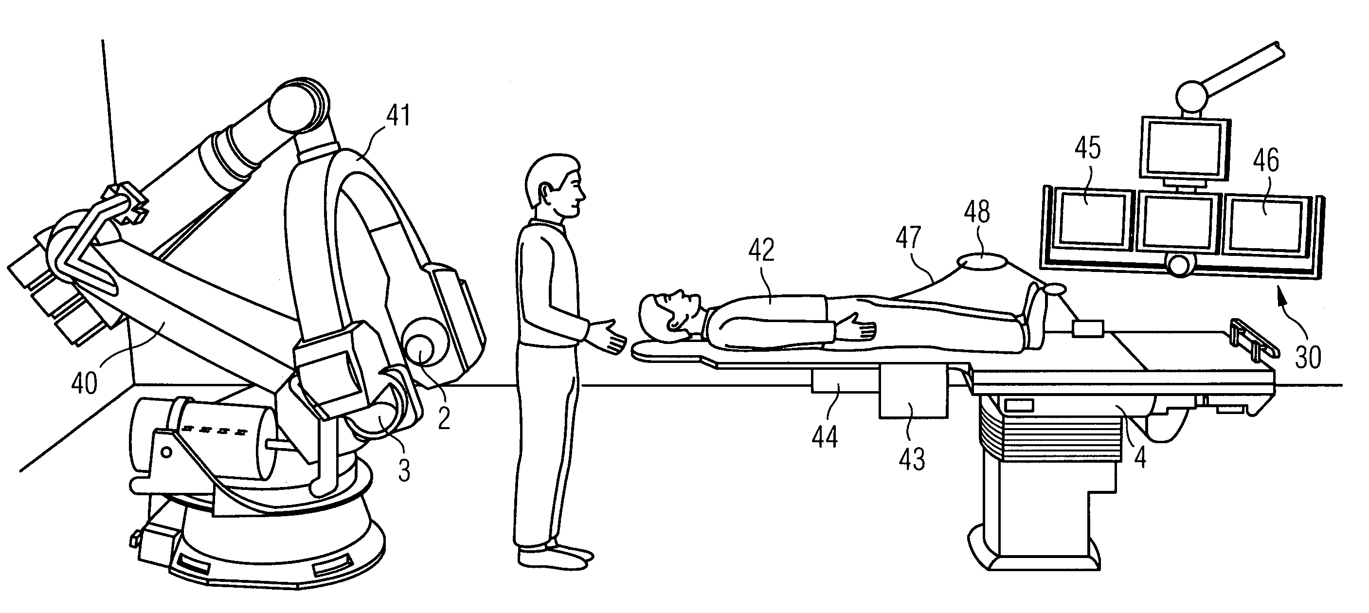
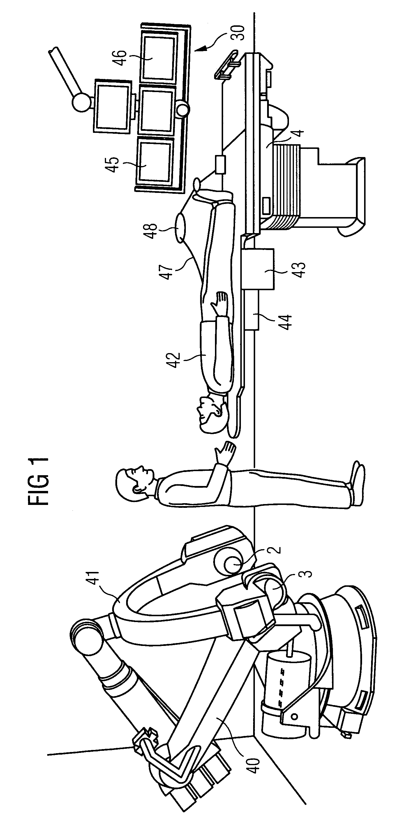
[0026]FIG. 1 shows an example for a robot-based integrated electrophysiological laboratory. In this example the articulated-arm robot 40 of the x-ray device is clearly visible, which is mounted on the floor and carries a C-arm 41 with x-ray emitter 2 and x-ray detector 3. The patient support facility 4 with the patient 42 features a control unit 43 close to the patient for the x-ray device and the catheter employed. Connected to this operating unit 43 is an interface unit 44 for connection of different catheters which has a “plug&play” functionality. The operating unit 43 provides the user with selection options, so-called organ or examination programs. If an examination program, e.g. ablation, is selected by the operator, all system components, the image processing and the associated emitter, detector and table positions are set by the system control and the devices are moved automatically to these positions. The images generated by the digital image system for the fluoroscopy reco...


PUM


Abstract

There is described a system for carrying out and monitoring minimally-invasive interventions with an x-ray device, in which at least one x-ray emitter and a x-ray detector are attached to one or more robot arms of one or more multi-axis articulated-arm robots, with which they are able to be moved for recording images from different projection directions on a predeterminable path around a patient support facility. The system includes a control and evaluation unit with interfaces for catheters and devices for carrying out the minimally-invasive intervention. The control and evaluation unit is embodied for the processing of measurement and / or image data which it receives from the catheters and devices and for control of the catheters and devices for recording the measurement and / or image data. With the proposed system the workflow is covered completely and seamlessly from the examination to the therapy, especially in the treatment of tachycardial arrythmias.

Description

CROSS REFERENCE TO RELATED APPLICATIONS[0001]This application claims priority of German application No. 10 2006 061 178.0 DE filed Dec. 22, 2006, which is incorporated by reference herein in its entirety.FIELD OF INVENTION[0002]The present invention relates to a system for carrying out and monitoring minimally-invasive interventions, especially for treatment of electrophysiological diseases. Among the most serious cardiological diseases are the tachycardial arrhythmias, such as atrial fibrillation for example. In this case conduction pathway disturbances in the heart mean that the atrium is excited at high frequency. With other, for example ventricular tachycardias, there is no complete contraction and the result is thus an insufficient pumping power of the heart. In the past attempts were either made to reduce the effect of atrial fibrillation by taking medicines continuously or to remedy the cause of the atrial fibrillation by a heart operation in which the cardiac conducting tiss...

Claims


Application Information

Patent Timeline
Patent Type & Authority Patents(United States)
IPC IPC(8): A61B5/05
CPCA61B6/12A61B6/4464A61B6/4476A61B19/52A61B6/4458A61B18/1492A61B18/24A61B19/2203A61B19/5244A61B2019/2211A61B2019/5238A61B2019/5251A61B2019/5276A61B2019/5278A61B6/4441A61B90/36A61B2090/3782A61B34/20A61B2034/301A61B2090/376A61B34/30A61B2034/2051A61B2090/378
Inventor MASCHKE, MICHAEL
Owner SIEMENS HEALTHCARE GMBH
Why Eureka
Try Eureka