Method and system to reconstruct treatment dose to a patient from integrated exit-transit images of radiation fields taken during treatment

a radiation field and integrated technology, applied in the field of radiation therapy, can solve the problems of not describing the method of reverse calculation, several problems not addressed, and the energy dependence of imaging devices

a radiation field and integrated technology, applied in the field of radiation therapy, can solve the problems of not describing the method of reverse calculation, several problems not addressed, and the energy dependence of imaging devices

US8351572B1Active Publication Date: 2013-01-08LIFELINE SOFTWARE INC

Image

  • Method and system to reconstruct treatment dose to a patient from integrated exit-transit images of radiation fields taken during treatment
  • Method and system to reconstruct treatment dose to a patient from integrated exit-transit images of radiation fields taken during treatment
  • Method and system to reconstruct treatment dose to a patient from integrated exit-transit images of radiation fields taken during treatment

Examples


Embodiment Construction

Drawing Reference Numerals

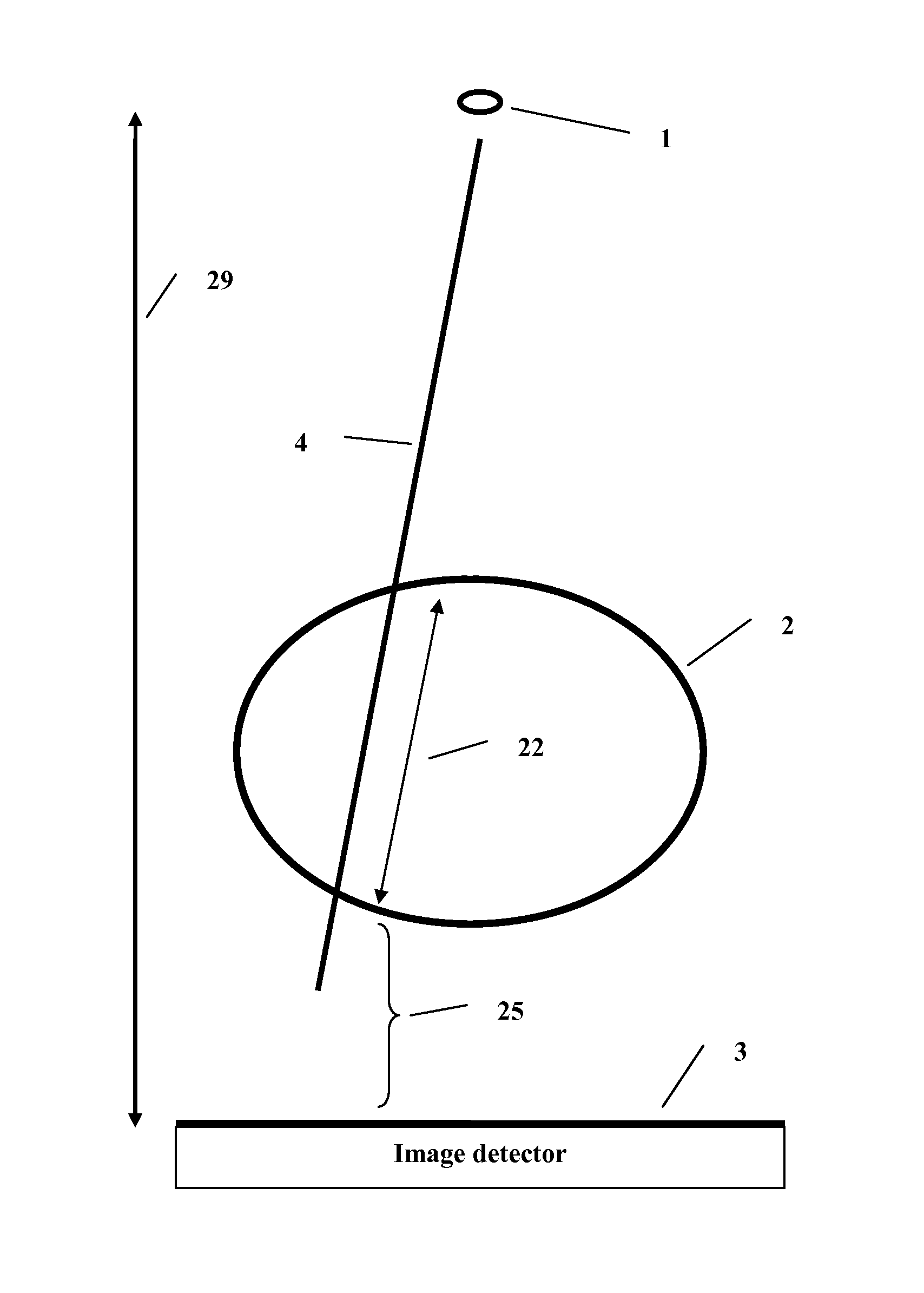
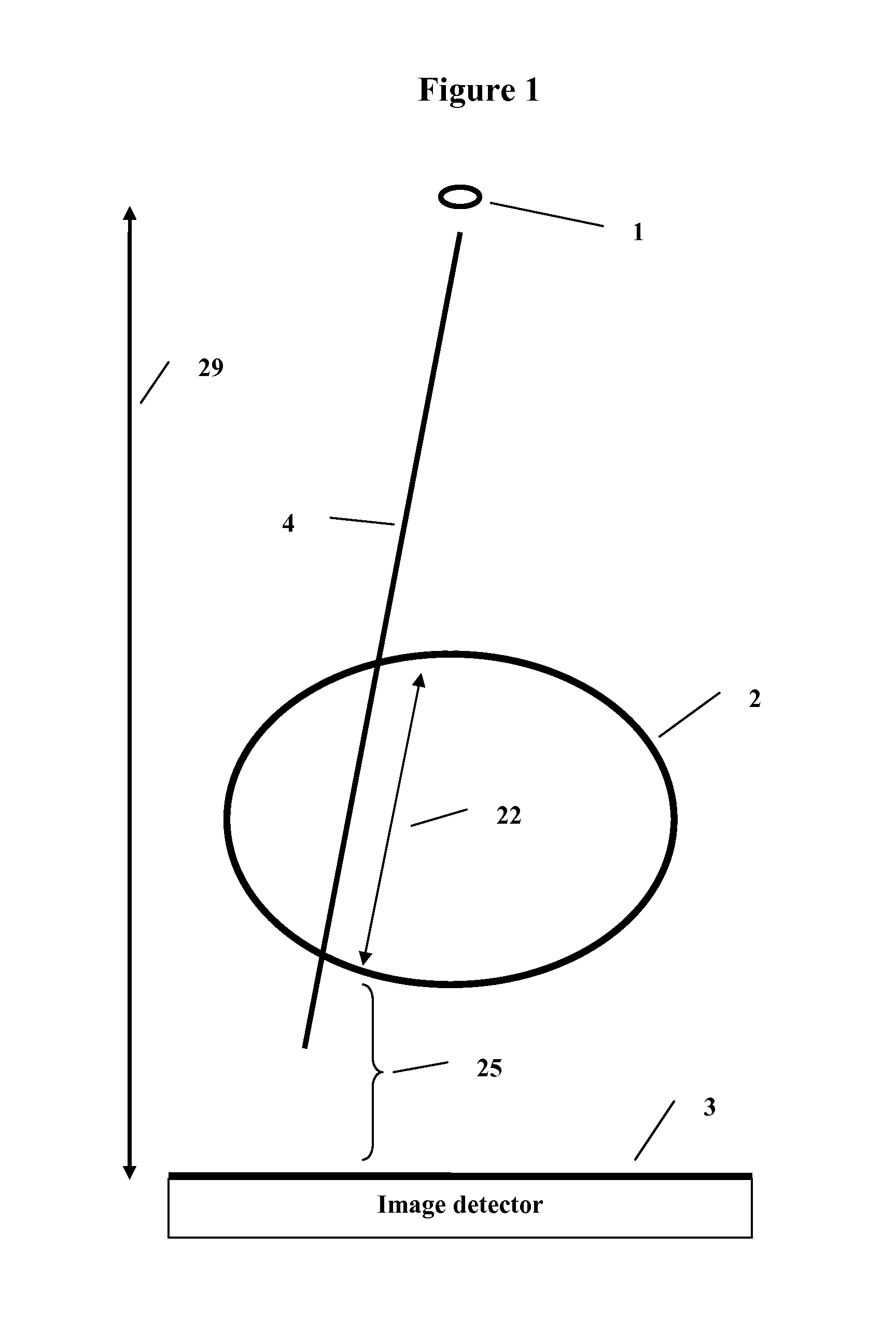
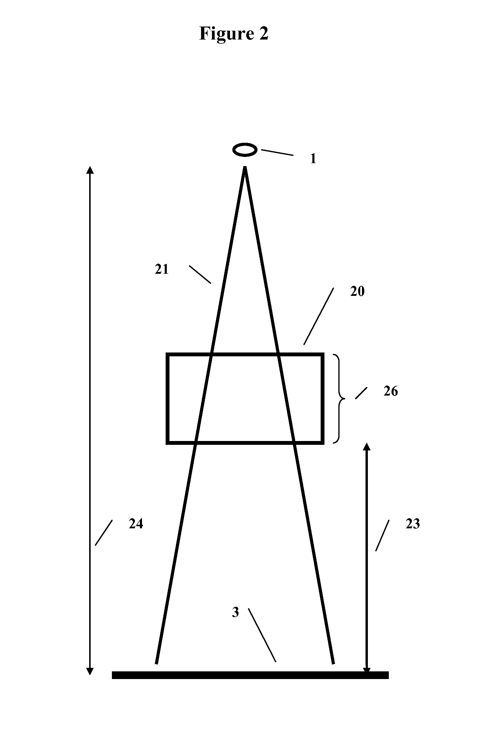
[0018]1 the source of x-rays.[0019]2 the patient.[0020]3 the image detector.[0021]4 the ray from the source of x-rays to the image detector.[0022]5 the exit image captured by the image detector.[0023]6 the deconvolution of the image (5) to an intermediate image (7) using a kernel derived for a fixed phantom thickness (26), and in a further embodiment for a fixed source to image detector distance (24) or phantom to image detector distance (23).[0024]7 the intermediate image produced from the exit image (5) by deconvolution with a kernel (6).[0025]8, 10, 12 the convolution of the image (5) to intermediate images (9, 11, 13) using the succeeding steps in thickness from the available kernels.[0026]9, 11, 13 the intermediate images produced from the deconvolution of the input exit image (5) as described above.[0027]14 the final output image which is the in air x-ray intensity fluence map to be used to compute the dose to the patient using a dose algorithm.[0028]...


PUM


Abstract

A method and system to compute the dose to a patient (2) given a captured integrated exit-transit image (5) of the radiation rays (4) traveling from the source of x-rays (1) through the patient (2) to the imaging device (3) to product the exit-transit image (5). Each radiation field image (5) is transformed (6,8,10,12) to multiple images (7,9,11,13) for each phantom thickness (26) that was measured with the imaging device (3) for a range of field sizes (21). Given the water equivalent path (22) through the patient for a ray (4) reaching a pixel (15, 16), the final pixel value (19) is interpolated from the images (9, 11) that bracket the water equivalent path through the patient (22).

Description

BACKGROUND OF THE INVENTION[0001]1. Field of the Invention[0002]This invention relates to the radiation therapy applied to patients who are typically treated for cancer or other ailments with high energy x-rays from medical linear accelerators. The invention is directed to a method and system to reconstruct the dose the patient has received using as input the radiation fields measured during treatment on the exit side of the patient with an imaging system capable of integration or a radiation detector array. This information may be used as a quality control measure of the treatment to avoid errors by comparing the reconstructed dose to the intended planned dose.[0003]2. Prior Art[0004]U.S. Pat. No. 5,754,622 describes a system for verifying the amount of radiation delivered to an object. However, this system does not describe the method to accomplish a reverse calculation. In particular, there are several problems not addressed. Radiation absorbed in the patient will cause scatter r...

Claims


Application Information

Patent Timeline
08 Jan 2013
Publication
US8351572B1
IPC
A61N5/10
CPC
A61N5/1048; A61N2005/1061
Inventors
RENNER, WENDEL DEAN