Method for creating perfusable microvessel systems

a microvessel and microvascular technology, applied in the field of physiological and pathological vascular growth and vascular growth, can solve the problems of ischemic wound healing, impaired vascular growth, and impaired vascular growth, and achieve the effect of much faster formation

Active Publication Date: 2013-05-21
NORTIS
View PDF17 Cites 4 Cited by
  • Summary
  • Abstract
  • Description
  • Claims
  • Application Information

AI Technical Summary

Benefits of technology

[0022]The presence of a luminal flow of nutrient medium may substantially increase the survival time and stability of capillary tubes in vitro. Luminal perfusion has been shown to have a positive impact on vessel growth and maturation, (French, Zuckmantel et al. 2008). This implies that the vessels would be more stable with luminal perfusion. Further, inclusion of smooth muscle cells or pericytes, endothelial progenitor cells, and even stern cells into formation of parent vessels would be believed to aid function as part of the vessel maturation process.
[0024]Additionally, contribution of the physical forces from seeding at high-density can be evaluated for the sprouting competency phenotype. Without being bound to a particular theory, for example seeding of endothelial cells at high-density results in physical compression where the endothelial cells are balled up during the process. Since endothelial cells are typically spread out laterally in vessel formation this is not initially possible during seeding where cells are tightly packed together. Thus growth into the matrix may be favored triggering the sprouting phenotype. Also, with a higher number of cells per luminal surface area, sprouts can be formed much more quickly by simple migration and coalescence of cells rather than by cell division. The contributions of genes and gene products that regulate such cellular phenotypes in vessel formation can be elucidated. The system allows the study of angiogenesis relative to wound repair, aging, cancer, psoriasis, diabetic retinopathy, inflammatory diseases, stroke, and atherosclerosis,

Problems solved by technology

In contrast, impairment of vascular growth, characteristic of aged individuals, compromises the healing of wounds and the revascularization of tissues rendered ischemic by trauma or disease.
Although published studies of angiogenesis number in the many thousands, the cellular mechanisms that mediate and regulate angiogenic growth and morphogenesis are poorly understood.
The details of angiogenic sprouting are difficult to observe in “real-time” in viva because of the opacity of most tissues.
Tissue sections are difficult to reconstruct in 3D and do not communicate the dynamic nature of vascular growth.
For all of their elegance, approaches in vivo are made difficult by (1) intra-species variation in angiogenic response from animal to animal; (2) the lack of translation of results from one species to another; (3) high costs of animal purchase and maintenance; (4) public disapproval of the use of animals for research purposes; and (5) complexities encountered in animal surgeries and in the visualization and evaluation of results.
Although the endothelial cell tubes exhibited correct polarity, the characteristics of invasion and directionality were lacking (the endothelial cells were pre-embedded and evenly dispersed in the extracellular matrix).
Each of these models has specific limitations that include difficulty in visualizing sprout formation, limited sprouting, a requirement for sectioning, or lack of effectiveness with certain types of endothelial cells.
However, this approach does not provide luminal perfusion of the microvessels.
These microvessels were of an exploratory nature and not suitable for creating a human vessel graft.
Importantly, none of the 3D models currently available reconstruct a parent blood vessel that contains a pressurized, flowing, circulatory fluid.
Consequently, none of the existing in vitro 3D models permit study of the contribution of luminal pressure and flow to vascular growth and morphogenesis,

Method used


Image

  • Method for creating perfusable microvessel systems
  • Method for creating perfusable microvessel systems
  • Method for creating perfusable microvessel systems

Examples


Embodiment Construction

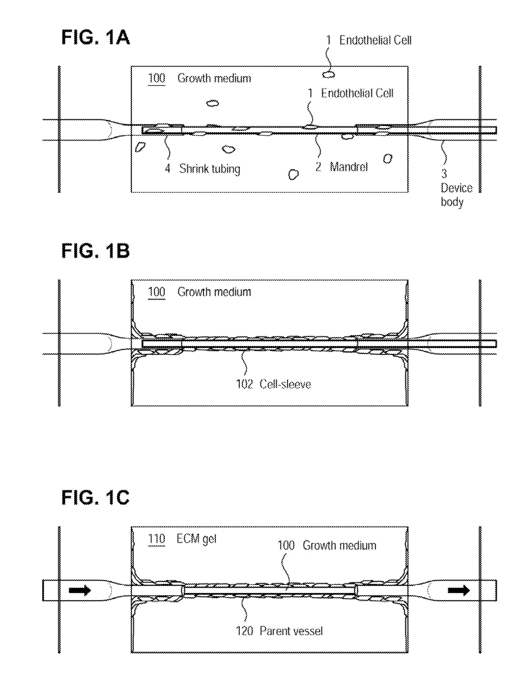
[0072]The examples presented herein are for the purpose of furthering an understanding of the invention The examples are illustrative and the invention is not limited to the example embodiments. The method of the present invention is useful for the study of physiological and pathological vascular growth, and vascular growth in response to angiogenic or angiostatic factors. Other useful applications are to methods that evaluate the angiogenic potential of cancer tissues and the response to antiangiogenic drugs. Further applications and methods are for basic research on physiology or pathology of vessel sprouting. Additionally, the method of the invention may be used to construct various wound-healing devices and for vascularization of tissue-engineered constructs.

[0073]In one example a method and device for the creation of perfusable three-dimensional microvessel networks is disclosed. As used herein “EC” refers to endothelial cells, “SMC” refers to smooth muscle cells and “CAS” refe...


PUM

PropertyMeasurementUnit
diameteraaaaaaaaaa
diameteraaaaaaaaaa
thicknessaaaaaaaaaa

Abstract

A method for creating networks of perfusable microvessels in vitro. Cells including cell types capable of sprouting are seeded 1300 into a channel in a matrix at to activate competency 1304 of the cells for sprouting as microvessels based on the seeding density. The matrix channel is perfused with medium to allow parent vessels to form and for viability 1324. The parent vessels and matrix are incubated and perfused to provide for sprouting of microvessels from parent vessels into the surrounding matrix 1328. The sprouting parent vessels are grown until network forms 1332.

Description

RELATED APPLICATION[0001]This application claims priority from and is a continuation-in-part of co-pending U.S. application Ser. No. 11 / 860,471 of Neumann, filed Sep. 24, 2007, entitled “Method for Creating Perfusable Microvessel Systems,” which is a continuation in part of U.S. application Ser. No. 11 / 388,920 of Neumann, filed Mar. 24, 2006, U.S. application Ser. No. 11 / 388,920 and U.S. application Ser. No. 11 / 860,471 of Neumann are hereby incorporated by reference.FIELD OF THE INVENTION[0002]The present invention relates to methods for the study of physiological and pathological vascular growth, and vascular growth in response to angiogenic or angiostatic factors.TECHNICAL BACKGROUND[0003]During normal processes of vascular growth (e.g., the menstrual cycle, placentation, changes in adiposity, wound repair, inflammation), the creation of new blood vessels is regulated and eventually ceases. Significantly, the deregulation of vascular growth is a critical element of pathology. For ...

Claims


Application Information

Patent Timeline
Patent Type & Authority Patents(United States)
IPC IPC(8): C12N5/07
CPCA61L27/3808A61L27/3886C12M21/08C12M25/14C12M29/10C12N5/0068C12N5/0691C12N2533/50
Inventor NEUMANN, THOMASTOUROVSKAIA, ANNAFAUVER, MARK E.YU, JULIA OI YAN
Owner NORTIS
Why Eureka
Try Eureka