

Assays and methods for fusing cell aggregates to form proto-tissues
a technology of cell aggregates and prototissues, which is applied in the field of fusion cell aggregates assays and methods, can solve the problems of little understanding of the process of tissue fusion and methods are needed to control, and the process is not well understood, so as to achieve the slowest rate of rod contraction and less cell sorting and cell mixing
- Summary
- Abstract
- Description
- Claims
- Application Information
AI Technical Summary
Benefits of technology
Problems solved by technology
Method used
Image

Examples
example 1
Design, Fabrication and Casting of Micro-Molded Agarose Gels
[0065]Micro-molds were designed, fabricated, and cast as described in Napolitano et al., Scaffold-free three-dimensional cell culture utilizing micromolded nonadhesive hydrogels, Biotechniques. 43: 494, 496-500 (2007) and in PCT Patent Publication No. WO 2007 / 087402. Briefly, molds were designed using computer-assisted design (CAD) (Solid Works Corporation—Concord, Mass.). Wax molds from the CAD files were produced with a ThermoJet® rapid prototyping machine (three-dimensional Systems Corporation—Valencia, Calif.) and replicated in polydimethyl siloxane (PDMS) (Dow Corning—Midland, Mich.). Agarose gels were cast from the PDMS molds. Powder Ultrapure© Agarose (Invitrogen—Carlsbad, Calif.) was sterilized by autoclaving, and dissolved via heating in sterile water to 2% (weight / volume). Molten agarose (2.75 ml / mold) was pipetted into each PDMS mold and air bubbles were removed via pipette suction or agitation with a sterile spa...
example 2
Cell Culture and Isolation
[0067]Normal human fibroblasts (NHFs) derived from neonatal foreskins, and H35 rat hepatoma cells (H35s) were grown in Dulbecco's Modified Eagle Medium (DMEM) (Invitrogen—Carlsbad, Calif.) supplemented with 10% fetal bovine serum (FBS) (Thermo Fisher Scientific—Waltham, Mass.) and 1% penicillin / streptomycin (Sigma—St. Louis, Mo.). NHFs were maintained in a 37° C., 10% CO2 atmosphere and H35s were maintained in a 37° C., 5% CO2 atmosphere. Cells were trypsinized, counted and re-suspended to the desired cell density. The mono-dispersed cell suspension (200 μL) was then pippetted into the rectangular recess of each gel. Medium (3 mL) was added to the gel after approximately 20 minutes and was exchanged every other day.
example 3
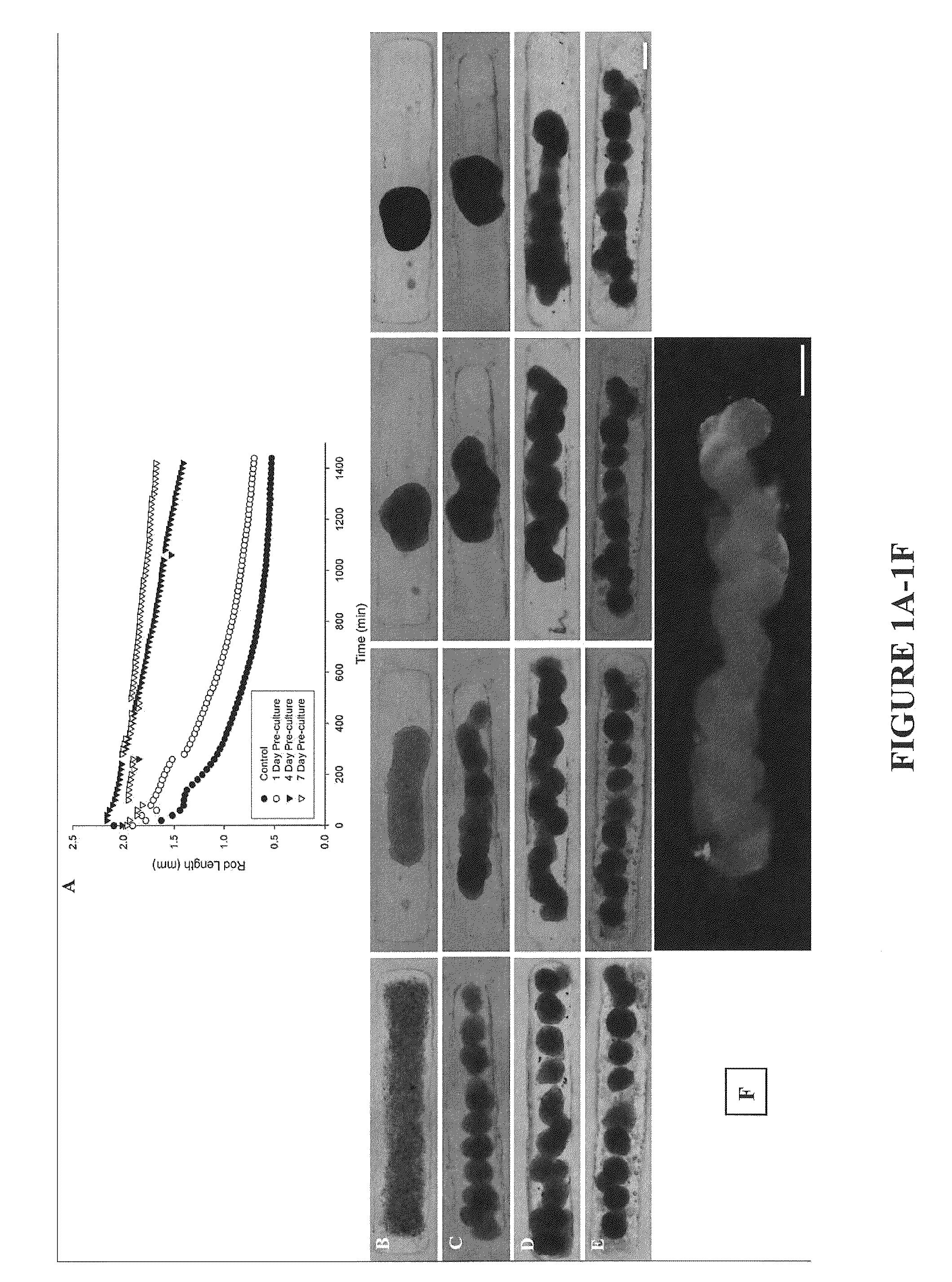
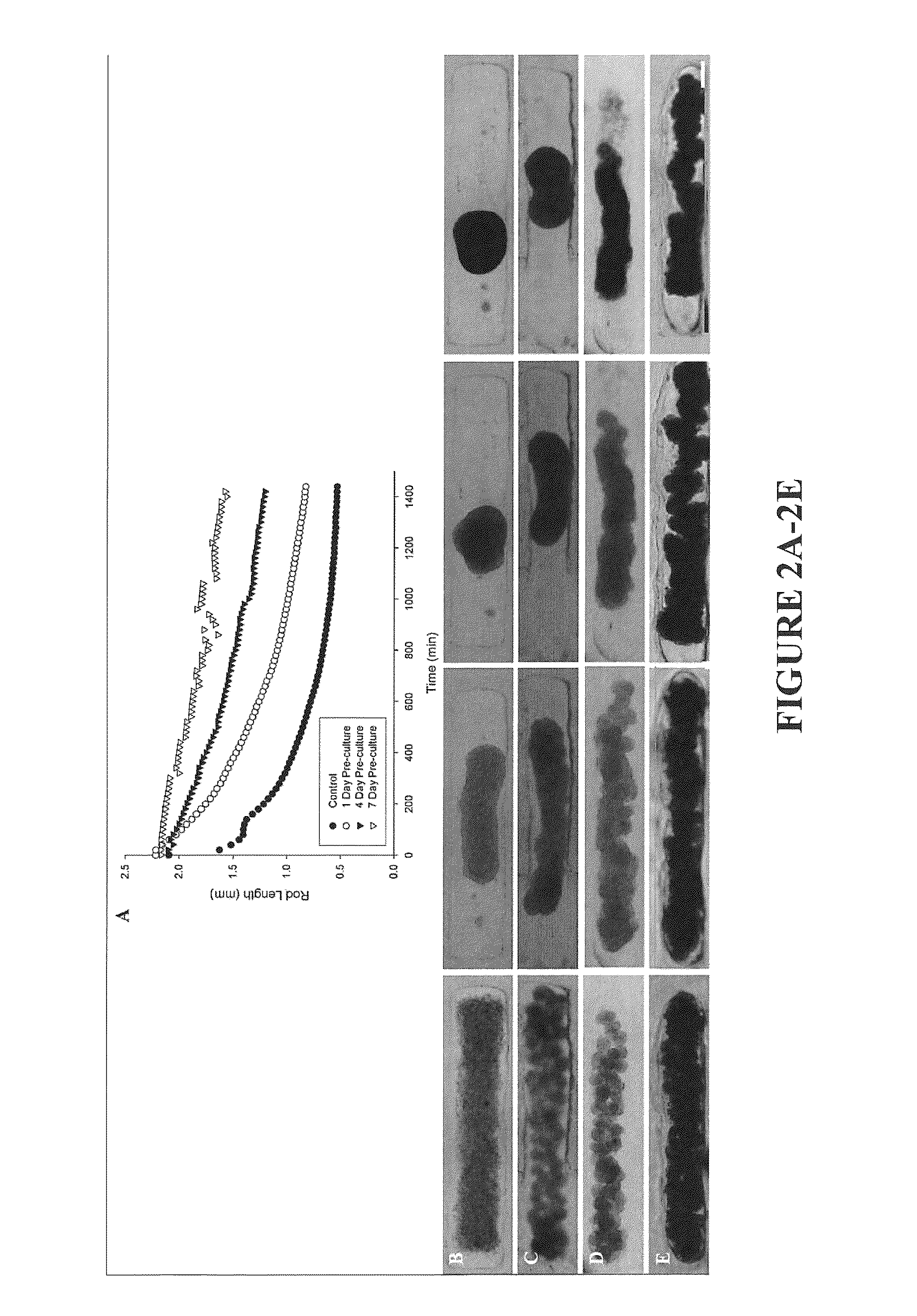
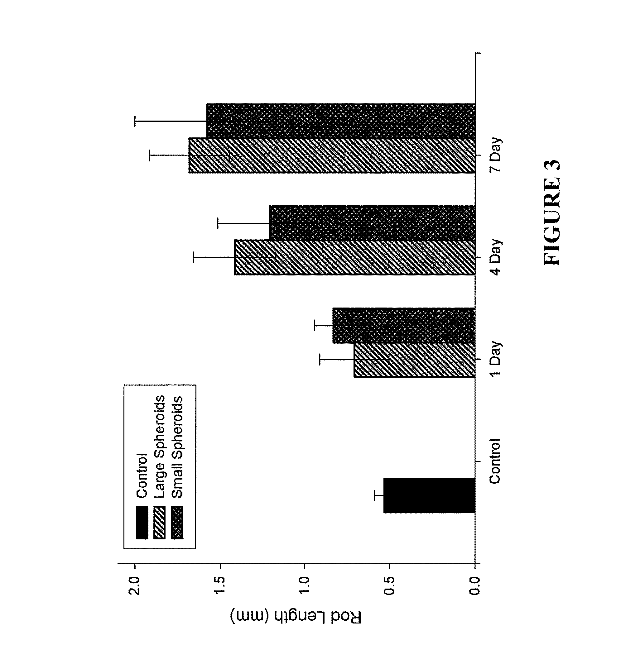
Assembly of Rod-Like Structures Using Spheroids as Building Units
[0068]Spheroids were generated using the micro-molded non-adhesive hydrogels made as described in Example 1. NHFs were seeded at a concentration of either 0.35×10⁶ cells / 822-well hydrogel or 1.0×10⁶ cells / 300-well hydrogel. Fourteen and five hydrogels, respectively, were seeded at these concentrations, resulting in microtissues of approximately 100 μm or 300 μm in diameter. The NHFs were cultured for one, four, or seven days within the hydrogels. Gels were inverted and centrifuged briefly at 800 rpm to retrieve the spheroid microtissues formed. The microtissues were resuspended in 200 μL medium and seeded onto a second micro-molded agarose gel with trough recesses and allowed to fuse for 24 hours. A corresponding control trough gel was seeded with mono-dispersed cells, 3×10⁶ cells. This seeding number was chosen to match the number of cells in each recess across experimental groups given the efficiency of spheroid tran...
PUM
| Property | Measurement | Unit |
|---|---|---|
| pre-culture time | aaaaa | aaaaa |
| pre-culture time | aaaaa | aaaaa |
| diameter | aaaaa | aaaaa |
Abstract
Description
Claims
Application Information