
Method and device for treating dysfunctional cardiac tissue

a technology of cardiac tissue and resizing ventricle, which is applied in the direction of prosthesis, surgical staples, therapy, etc., can solve the problems of reducing the effectiveness of the heart, the back-up of pressure in the vascular system behind the ventricle, and the extraordinary high cost of advanced medical care for the millions of patients suffering from hf, so as to reduce the trauma of the patient, prolong the delivery of drugs, and the effect of delivering the same amoun

Active Publication Date: 2013-08-13
BIOVENTRIX A CHF TECH
View PDF65 Cites 56 Cited by
  • Summary
  • Abstract
  • Description
  • Claims
  • Application Information

AI Technical Summary

Benefits of technology

[0020]A further advantage of the present invention is that it avoids problems associated with bolus injection of a drug, such as delivery of an amount of drug to the cardiac tissue, which is too high and which therefore may have deleterious effects on the cardiac tissue.
[0021]Another advantage is that it provides long-term delivery of a drug to the pericardium or myocardial tissue, with even delivery rate.
[0022]Still another important advantage is that extended delivery of a drug to the cardiac tissue can be achieved during the placement of clip or clamp for dysfunctional tissue exclusion, without the need for invasive surgery, thereby reducing trauma to the patient.
[0023]Further, in view of the above need of treating the dysfunctional tissue, the present invention provides a system and method incorporated into the clip or clamp employing transducers / sensors for determining the heart function and parameter and measuring heart performance of both left and right ventricles, particularly the left ventricle without requiring invasive surgery to access the left ventricle.
[0024]The sensors comprise at least a first sonomicrometer piezoelectric crystal mounted on a clip or clamp, so that when the clip or clamp is implanted on the heart chamber, so does the sensor. One or more additional sonomicrometer piezoelectric crystals can be mounted on the clip or clamp, such that the distance between the three or more sonomicrometer crystals can be determined. In each case, the sonomicrometer crystals are distributed about a heart chamber of interest such that the distance between the separated ultrasound transmitter and receiver crystal pairs changes with contraction and relaxation of the heart chamber.
[0025]The invention thus involves the incorporation of sonomicrometer piezoelectric crystals into the clip or clamp.

Problems solved by technology

Thus, it is not surprising that the cost of providing advanced medical care for the millions of patients suffering from HF is extraordinarily high—now estimated at more than $38 billion annually.
In certain pathological conditions, the ventricles of the heart become ineffective in pumping the blood, causing a back-up of pressure in the vascular system behind the ventricle.
The reduced effectiveness of the heart is usually due to an enlargement of the heart.
Acute myocardial infarction (AMI) due to obstruction of a coronary artery is a common initiating event that can lead ultimately to heart failure.
A myocardial ischemia may, for example, cause a portion of a myocardium of the heart to lose its ability to contract.
Once this tissue dies, it no longer functions as a muscle and cannot contribute to the pumping action of the heart.
As this situation worsens, the local area of compromised myocardium may in fact bulge out as the heart contracts, further decreasing the hearts ability to move blood forward.
One problem with a large dilated left ventricle is that there is a significant increase in wall tension and / or stress both during diastolic filling and during systolic contraction.
However, in a failing heart, the ongoing dilatation is greater than the hypertrophy and the result is a rising wall tension requirement for systolic contraction.
This is felt to be an ongoing insult to the muscle myocyte resulting in further muscle damage.
This vicious cycle of cardiac failure results in the symptoms of congestive heart failure such as shortness of breath on exertion, edema in the periphery, nocturnal dypsnia (a characteristic shortness of breath that occurs at night after going to bed), weight gain, and fatigue, to name a few.
The stress increase requires a larger among of oxygen supply, which can result in exhaustion of the myocardium leading to a reduced cardiac output of the heart.
Despite the major advances in both medical and surgical therapy, the management of patients with coronary artery disease and left ventricular (LV) dysfunction continues to be challenging due to the complex and multifactorial pathophysiology of this condition.
Many patients who would benefit from left ventricular reconstruction are the least likely to withstand the effects of CPB due to age, poor ejection fraction, or other cardiovascular disease.

Method used


Image

  • Method and device for treating dysfunctional cardiac tissue
  • Method and device for treating dysfunctional cardiac tissue
  • Method and device for treating dysfunctional cardiac tissue

Examples


Embodiment Construction

[0032]Although this invention is applicable to numerous and various types of cardiovascular methods, it has been found particularly useful in the field of reducing ventricular volume of the heart. Therefore, without limiting the applicability of the invention to the above, the invention will be described in such environment.

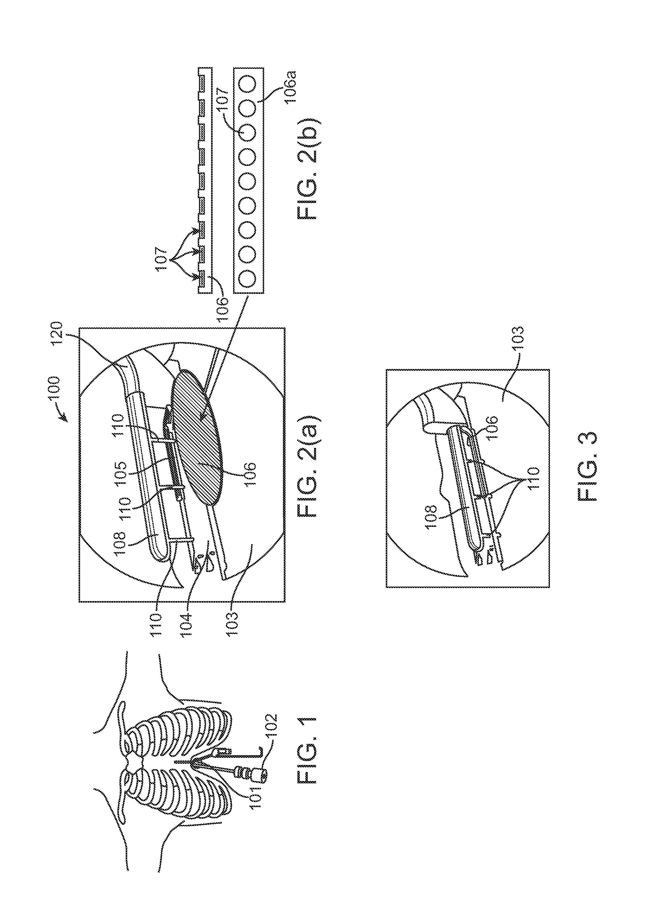
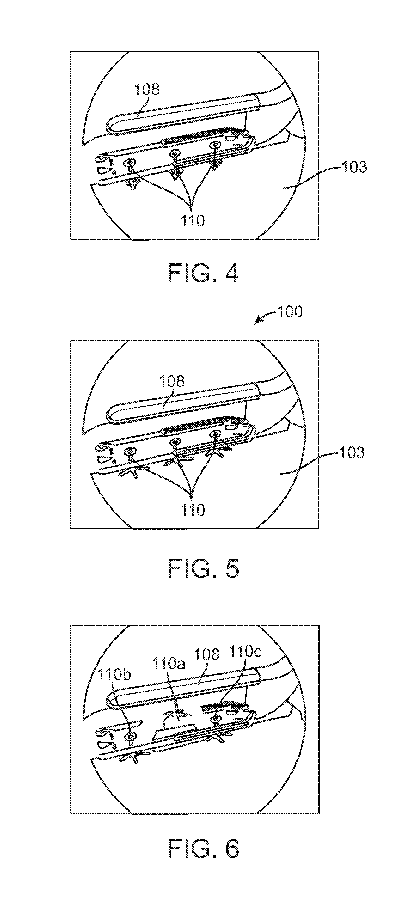
[0033]With reference to FIGS. 1-9, an anchoring system in accordance with one embodiment of the present invention will be described.

[0034]Initially, in FIG. 1, a subxiphoid incision is cut to create an opening 101 to access the ventricles of the heart. Additional access may be performed in the intercostal space for other instruments as well. Then, a double balloon catheter 102 is introduced into the heart through the opening 101 to unload the heart. The balloon catheter 102 provides inflow occlusion to decompress the ventricles, thereby reducing the systolic pressure for aid in reducing the ventricular volume or exclusion of dysfunctional cardiac tissue. The cath...


PUM


Abstract

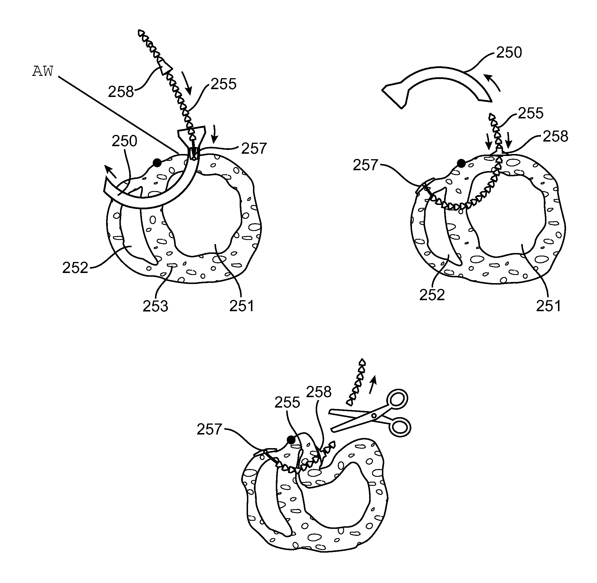
Various methods and devices are provided for reducing the volume of the ventricles of the heart. In one embodiment, a method for reducing the ventricular volume of a heart chamber is provided including the steps of inserting an anchoring mechanism onto dysfunctional cardiac tissue, deploying one or more anchors into the dysfunctional cardiac tissue, raising the dysfunctional cardiac tissue using the anchors, and securing the anchors to hold the dysfunctional cardiac tissue in place. Further, a device for reducing the volume of the ventricles of a heart chamber is provided where the device has one or more clips for placement on dysfunctional cardiac tissue of a heart, one or more anchors for deployment and securement into the dysfunctional cardiac tissue, and a lifting mechanism for raising the one or more anchors and the dysfunctional cardiac tissue.

Description

[0001]This application claims the benefit of provisional patent application Ser. Nos. 60 / 709,730 filed on Aug. 19, 2005, Ser. No. 60 / 709,690 filed on Aug. 19, 2005 and Ser. No. 60 / 711,727 filed on Aug. 26, 2005.BACKGROUND OF THE INVENTION[0002]1. Field of the Invention[0003]This invention relates generally to devices for resizing ventricle and related methods for placing the devices. More particularly, it relates to sizing devices for reducing the ventricular volume to improve the heart's pumping action. The devices and methods of the present invention are directed toward thoracoscopy or subxiphoid techniques used to facilitate sizing of the ventricles.[0004]2. Prior Art[0005]Heart failure (HF), the heart's inability to pump an adequate volume of blood to the tissues, is the only major cardiovascular condition that continues to increase in incidence in the United States. Approximately 5 million patients are currently diagnosed with HF in the United States. The American Heart Associa...

Claims


Application Information

Patent Timeline
Patent Type & Authority Patents(United States)
IPC IPC(8): A61F2/00
CPCA61B17/00234A61B17/0469A61B17/1227A61B17/1285A61F2/2487A61B17/0401A61B17/0467A61B17/06004A61B2017/00243A61B2017/0404A61B2017/06052A61B2017/06176A61B2017/306A61B2017/308A61F2/2481A61B17/0644A61B17/068A61B2017/0647
Inventor CHIN, SING-FATTANNEST, LONO'REILLY, ROBERT
Owner BIOVENTRIX A CHF TECH
Learn More