Bone graft materials and methods

a technology of bone grafting and materials, applied in the field of bone grafting, can solve the problems of increasing pain and discomfort of patients, increasing the risk of infection, and increasing the risk of disease transmission and graft rejection of patients

Active Publication Date: 2013-10-08
BIOVENTUS LLC
View PDF415 Cites 20 Cited by
  • Summary
  • Abstract
  • Description
  • Claims
  • Application Information

AI Technical Summary

Problems solved by technology

Use of autogenous bone, however, subjects a patient to increased pain and discomfort, and an increased risk of infection, because it requires the patient undergo surgery to recover the autogenous bone for use in the grafting procedure.
Both allograft bone and xenograft bone, though from natural sources, subject a patient to the risk of disease transmission and graft rejection.
Some such synthetic bone graft material is mixed with autograft, allograft, or xenograft bone, and thus still subjects a patient to the risks above.
Other disadvantages to current synthetic bone graft material are the lack of sufficient resorbability, lack of sufficient porosity, and increased manufacturing costs due to a high number of component materials.

Method used


Image

  • Bone graft materials and methods
  • Bone graft materials and methods
  • Bone graft materials and methods

Examples


example 1

Cell Preparation

[0095]Osteoblast-like MG-63 cells were prepared per American Type Culture Collection (“ATCC”) instructions. From passage three, the MG63 cells were stored frozen at −70° C. in Eagle's minimum essential medium (“EMEM”) with 20% fetal bovine serum (“FBS”). In preparation for use, the MG63 cells were thawed and then grown for at least three passages using EMEM. After three passages, the cells were transferred to Dulbecco's modified EMEM (“DMEM”). The DMEM contained no glucose and was supplemented with ascorbic acid (50 μg / mL), 10 mM β-glycerophosphate, 50 UI / mL Penicillin-Streptomycin and 10% FBS. The cells were grown for at least three more passages at these conditions. The cells were maintained at 37±1° C. in a humidified incubator with 5±1% CO2. The media was changed every 2 to 3 days. After the cells became confluent on the last passage, they were harvested and counted.

example 2

Sample Preparation

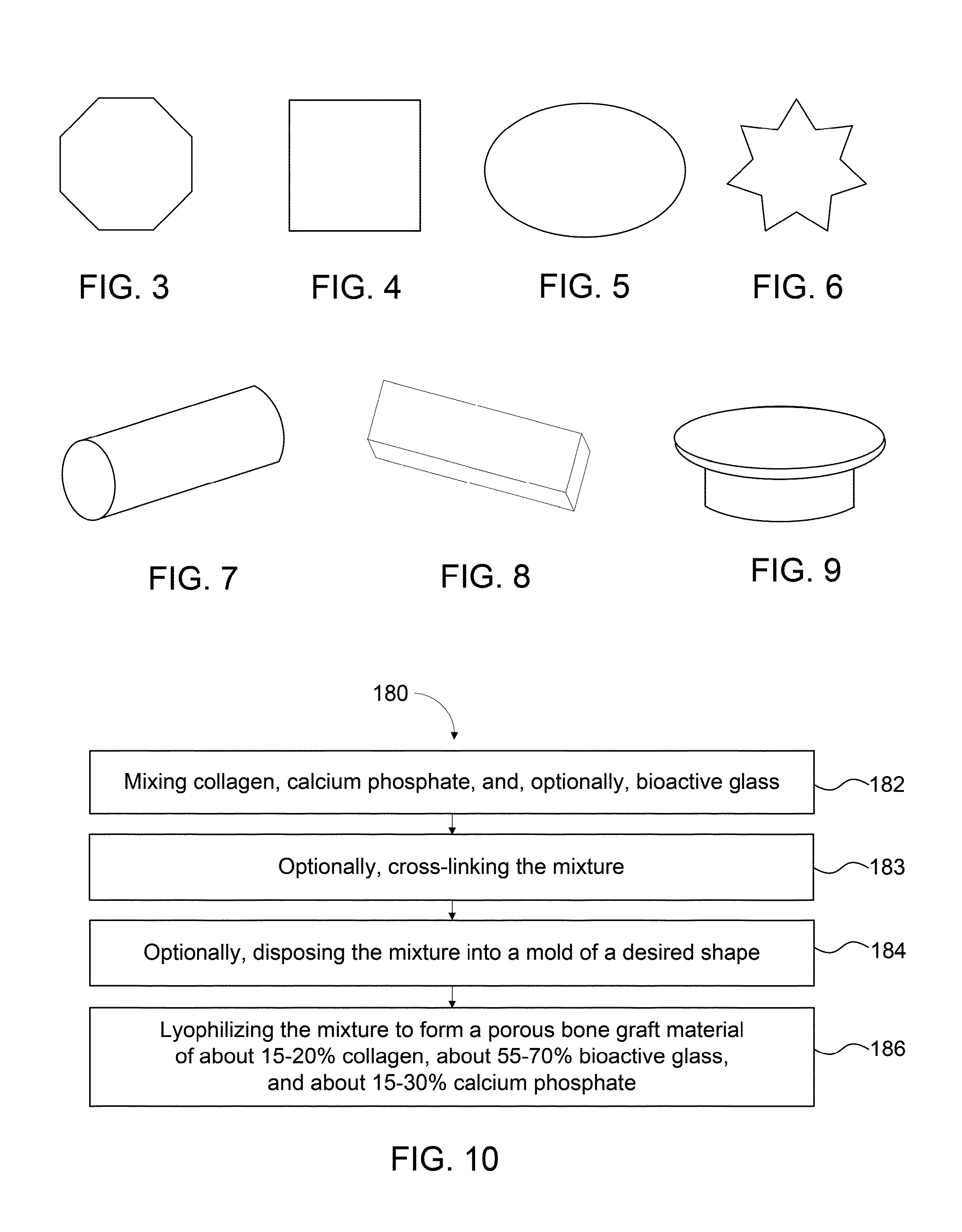
[0096]Test samples of bone graft compositions including collagen, bioactive glass, and calcium phosphate (60% HA / 40% TCP) and test samples of bone graft compositions including collagen and bioactive glass were prepared. The following preparation and testing was performed for each bone graft composition test sample. Each compound (i.e., collagen, bioactive glass, and calcium phosphate, as applicable; ratios of compounds each test sample are described below in Examples 3 through 8) was weighed aseptically using an analytical scale under a biological safety cabinet and transferred to a sterile non-treated tissue culture Petri dish (60×15 mm). A 30 mg mixture of the compounds in the specified ratio was aseptically prepared. The 30 mg mixture was transferred to and divided amongst four Petri dishes, each non-treated. A 1 mL PBS was added to each dish. Each dish was then vigorously swirled by hand on a desk surface. Each dish was also spun in a centrifuge at 3000 rpm for...

example 3

Collagen:Bioactive Glass:Calcium Phosphate (15:65:20)

[0102]A test sample of a bone graft composition including 15% collagen, 65% bioactive glass, and 20% calcium phosphate (60% HA / 40% TCP) was prepared according to Example 2. Cell confluence was determined based on visual observation of the sample without staining, as shown in the images of the sample taken on day 2, day 5, and day 11 in FIGS. 11A-11C, respectively. Cell confluence reached nearly an average of 93% by day 5. Cell confluence reached about 100% by day 11. No cytotoxicity was observed when compared with cell controls. A chart of cell confluence of this Example 3 compared to Examples 4 and 5 below is shown in FIG. 13.

[0103]Referring to FIG. 12A, visual observation under light microscopy of the Van Kossa stained cells showed pink, large, prolonged cells with dark-red big nuclei. The cells were in a fairly thick multilayer with many aggregations. Some crystals and phosphates appeared intact, but with grayish aureole. Some ...


PUM

PropertyMeasurementUnit
sizeaaaaaaaaaa
sizeaaaaaaaaaa
porosityaaaaaaaaaa

Abstract

Compositions, materials, methods and kits for bone grafting are described. In some embodiments, a bone graft composition includes about 15% to about 20% by weight collagen, about 55% to about 70% by weight bioactive glass, and about 15% to about 30% by weight a calcium phosphate. The bioactive glass and the calcium phosphate together are about 80% to about 85% by weight of the bone graft composition. In some embodiments, a bone graft composition includes a collagen matrix and a plurality of bioactive glass particulates dispersed throughout the collagen matrix. The collagen matrix is about 20% to about 60% by weight of the bone graft composition, and the bioactive glass is about 40% to about 80% of the bone graft composition. In some embodiments, a majority of the bioactive glass particulates are about 53 μm to about 425 μm in size.

Description

BACKGROUND[0001]The present invention generally relates to bone grafting, and more particularly to repairing and / or filling a void or gap in a bone or bony structure of a patient.[0002]A need exists for improved bone graft materials. Current bone grafting includes the use of autogenous bone as a graft material (i.e., “autografting”). Use of autogenous bone, however, subjects a patient to increased pain and discomfort, and an increased risk of infection, because it requires the patient undergo surgery to recover the autogenous bone for use in the grafting procedure. Current bone grafting also includes the use of bone from a donor as a graft material (e.g., “allografting” from the same species or “xenografting” from a different species). Both allograft bone and xenograft bone, though from natural sources, subject a patient to the risk of disease transmission and graft rejection.[0003]Current bone grafting further includes the use of synthetic bone graft material. Some such synthetic b...

Claims


Application Information

Patent Timeline
Patent Type & Authority Patents(United States)
IPC IPC(8): A61K35/32A61K38/00
CPCA61L27/46C08L89/06A61F2/28A61L2400/06A61L2430/02A61F2310/00293A61F2310/00329A61F2310/00371A61F2002/2835A61L27/24A61L27/446
Inventor COOK, RUSSELL L.ANTONE, DURAID S.
Owner BIOVENTUS LLC
Why Eureka
Try Eureka