Method for evaluation of renal vascular perfusion using power doppler ultrasonography

a technology of power doppler and ultrasonography, which is applied in the field of methods, can solve the problems of complex organ dysfunction or even death, difficult to elucidate the dynamic blood flow state with a static image, and limited clinical application, so as to enhance the accuracy of quantification of renal perfusion and enhance the evaluation of renal function.

Active Publication Date: 2014-07-15
R&B LEASING +1
View PDF4 Cites 6 Cited by
  • Summary
  • Abstract
  • Description
  • Claims
  • Application Information

AI Technical Summary

Benefits of technology

[0008]To solve the foregoing disadvantages present in the prior art, the object of the present invention is to provide a non-invasive evaluation method for examination of renal perfusion using Power Doppler ultrasonography to detect the real-time status of renal perfusion, and to provide a more dynamic, haemodynamic-matching information for quantification of renal perfusion and evaluation of renal function.
[0010]The present invention provides a non-invasive, inclusive of complete heartbeat cycles, dynamic, haemodynamic-matching analysis method using the power Doppler ultrasound. The curves obtained by the method according to the present invention show a regular pattern. In addition, the accuracy of quantification of renal perfusion and evaluation of renal function is greatly enhanced with the use of WPDVI in combination with PDVI.

Problems solved by technology

Declines in renal function may accumulate metabolite wastes in the body and consequently cause multiple-organ dysfunction or even death unless renal dialysis treatment or kidney transplantation is performed.
However the clinical application is limited.
It's difficult to elucidate the dynamic blood flow state with a static image.
However, color Doppler is not sensitive in depicting small vessels or vessels with low velocity.
Power Doppler sonography is based on the total integrated energy of Doppler shifted echoes and has higher sensitivity for much slower blood flow, but it can not address the flow direction or flow velocity.
In addition, these methods showed poor specificity and sensitivity.
However, all these applications collected “static” information on renal perfusion without details on haemodynamics, which were not sufficient to reflect the actual dynamic condition of renal perfusion.

Method used


Image

  • Method for evaluation of renal vascular perfusion using power doppler ultrasonography
  • Method for evaluation of renal vascular perfusion using power doppler ultrasonography
  • Method for evaluation of renal vascular perfusion using power doppler ultrasonography

Examples


Embodiment Construction

[0028]To enable persons skilled in the art to better understand the aforementioned objective, features and advantages of the present invention, embodiments are quoted and explained in combination with attached Figs. as follows.

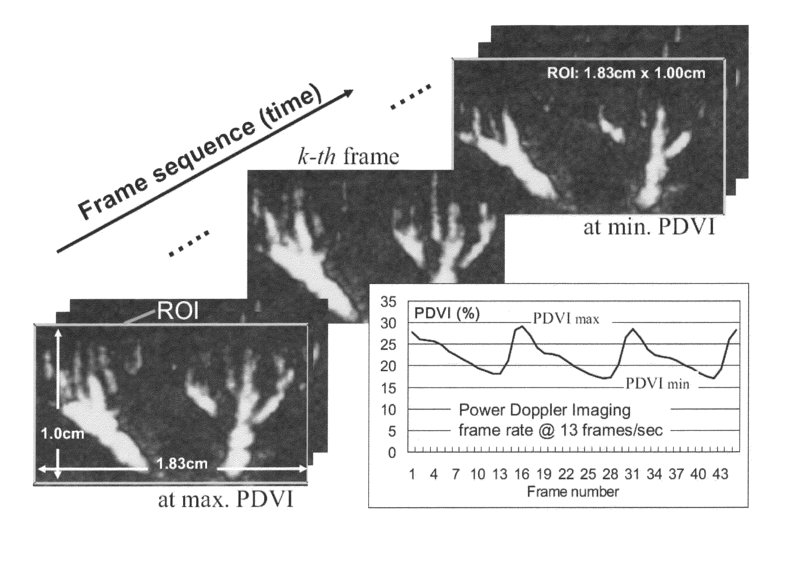
[0029]The present invention relates to a method for quantification of renal perfusion using power Doppler ultrasonography. The techniques of the invention principally comprises the steps of: using a Power Doppler ultrasound and consecutively capturing serial images (n=45) on kidney for at least two complete heartbeat cycles; selecting a region of interest (ROI) for vascular analyses; demarcating and summing pixels showing reflected signals of blood flow to obtain the area of blood perfusion per image; and calculating the ratio of the quantity of the pixels having reflected signals of blood flow to the total of pixels in the ROI to obtain a power Doppler vascularity index (PDVI). Based on the defined PVDI of each image, a curve that lasted for at least two hear...


PUM


Abstract

A method for evaluation of renal perfusion with power Doppler ultrasonography is disclosed in the present invention. Serial renal vascular images at different vascular areas including the whole vascular tree, interlobar, arcuate, and interlobular vessels were captured. Imaging processing software was designed to analyze the changes of power Doppler intensity of colored pixels within regions of interest (ROI). Power Doppler Vascularity index (PDVI) has been defined as the percentage of vascular perfusion within a region of interest (ROI). The renal vascular perfusion index (RVPI) is defined as the maximal power Doppler vascular index divided by minimal power Doppler vascular index (PDVImax / PDVImin) among the serial images. The mean of weighted power Doppler vascular index (WPDVImean) is defined as the average of the intensity of color pixels among the ROI within the serial images. By using the RVPI and WPDVImean, a more dynamic sense of vascular perfusion and a novel approach for the evaluation of renal vascular function in clinical practice can be provided.

Description

BACKGROUND OF THE INVENTION[0001]1. Field of the Invention[0002]The present invention relates to a method for evaluation of renal perfusion, in particular to a method for evaluation of renal perfusion by capturing and analyzing consecutive serial images in renal vessels using power Doppler ultrasonography.[0003]2. The Prior Arts[0004]The main function of kidney is to remove metabolic wastes (mainly nitrogenous wastes) in the body. Declines in renal function may accumulate metabolite wastes in the body and consequently cause multiple-organ dysfunction or even death unless renal dialysis treatment or kidney transplantation is performed. A large amount of blood flow supply is necessary for the renal function, so that 25˜40% cardiac output comes into kidney for the renal perfusion. Therefore the measurement of renal blood flow has become an important tool for the assessment of renal functions. Power Doppler ultrasonography provides a non-invasive method among the various measurement met...

Claims


Application Information

Patent Timeline
Patent Type & Authority Patents(United States)
IPC IPC(8): A61B8/00A61B5/20A61B8/06A61B8/13A61B18/00G06T7/00
CPCG06T7/0016A61B5/201A61B8/06A61B8/13A61B8/469A61B2018/00511G06T2207/10016G06T2207/10132G06T2207/20104G06T2207/30084G06T2207/30104A61B5/413
Inventor SHYU, JEOU-JONGPAO, SUN-HUASHAU, YIO-WHAWANG, SHUO-MENGCHOU, NAI-KUAN
Owner R&B LEASING
Why Eureka
Try Eureka