Normalization of retinal nerve fiber layer thickness measurements made by time domain-optical coherence tomography

a time domain, optical coherence tomography and normalization technology, applied in the field of normalization of retinal nerve fiber layer thickness measurements made by time domain optical coherence tomography, can solve the problem of measurement variability in long-term follow-up of td-oct circle scan

Active Publication Date: 2014-12-16
UNIVERSITY OF PITTSBURGH
View PDF4 Cites 3 Cited by
  • Summary
  • Abstract
  • Description
  • Claims
  • Application Information

AI Technical Summary

Benefits of technology

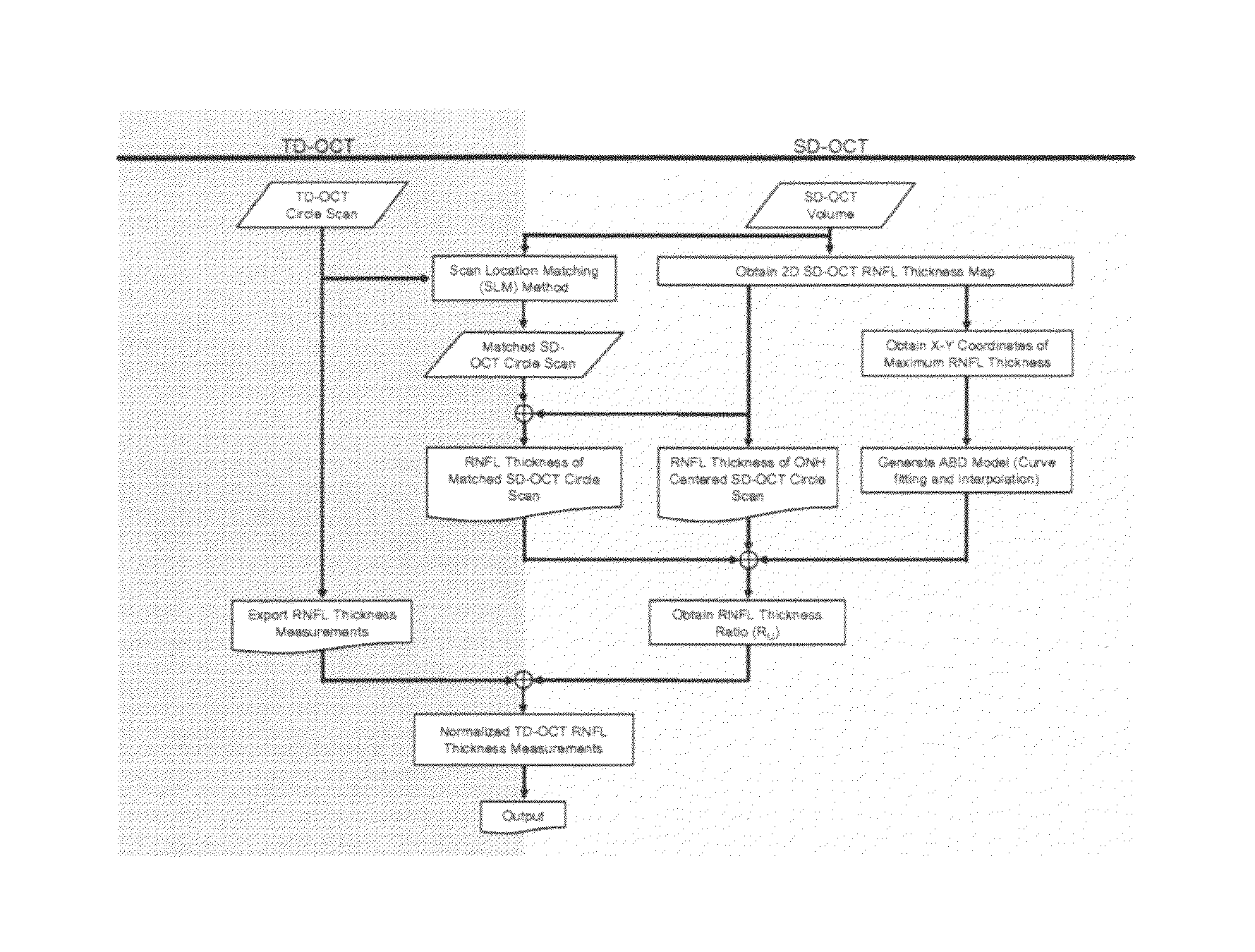
[0006]The present invention makes possible the normalization of RNFL thickness measurements of an off-centered TD-OCT circle scan to a virtual location, centered on the ONH, by using both a mathematical model of the retinal tissue pattern and scan location matching algorithm.14 Once one normalizes the RNFL thickness measurements of TD-OCT circle scan, pursuant to the invention, clinicians can track, compare, and finally detect any abnormal retinal change over a long period of time, without RNFL thickness-measurement variability caused by the off-centered TD-OCT scans.
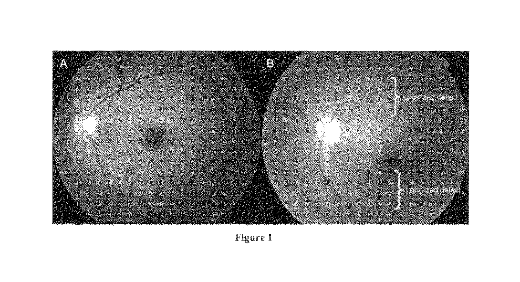
[0039]The present inventors thus have developed and evaluated an automated algorithm to normalize the RNFL thickness measurement of off-centered TD-OCT circle scan to a virtual properly centered location. The results show that the mathematical RNFBD model, in accordance with the invention, properly identified the anatomical RNFBD pattern, by using the corresponding 3D SD-OCT volume data, and improved the reproducibility of TD-OCT RNFL thickness measurements in all sectors except global mean (Table 3). FIG. 11 shows that RNFBD model agrees well with localized defect due to glaucoma. This indicates that the multiple TD-OCT RNFL measurements from longitudinal repeated TD-OCT scans can be compared in ideally the same location, regardless the accuracy of the actual scan location (FIG. 5). In other words, the approach of the invention makes it possible for clinicians to track, compare, and detect any retinal changes over a longer period of time with relatively low measurement variation, which may enable more reliable and sensitive glaucomatous progression analysis.
[0040]With the present invention, RNFL thickness-measurement normalization effected by combining individual mathematical RNFBD models and the SLM algorithm improves the reproducibility of TD-OCT RNFL thickness measurements. In addition, the inventive methodology constitutes a useful tool for study of longitudinal glaucoma progression.

Problems solved by technology

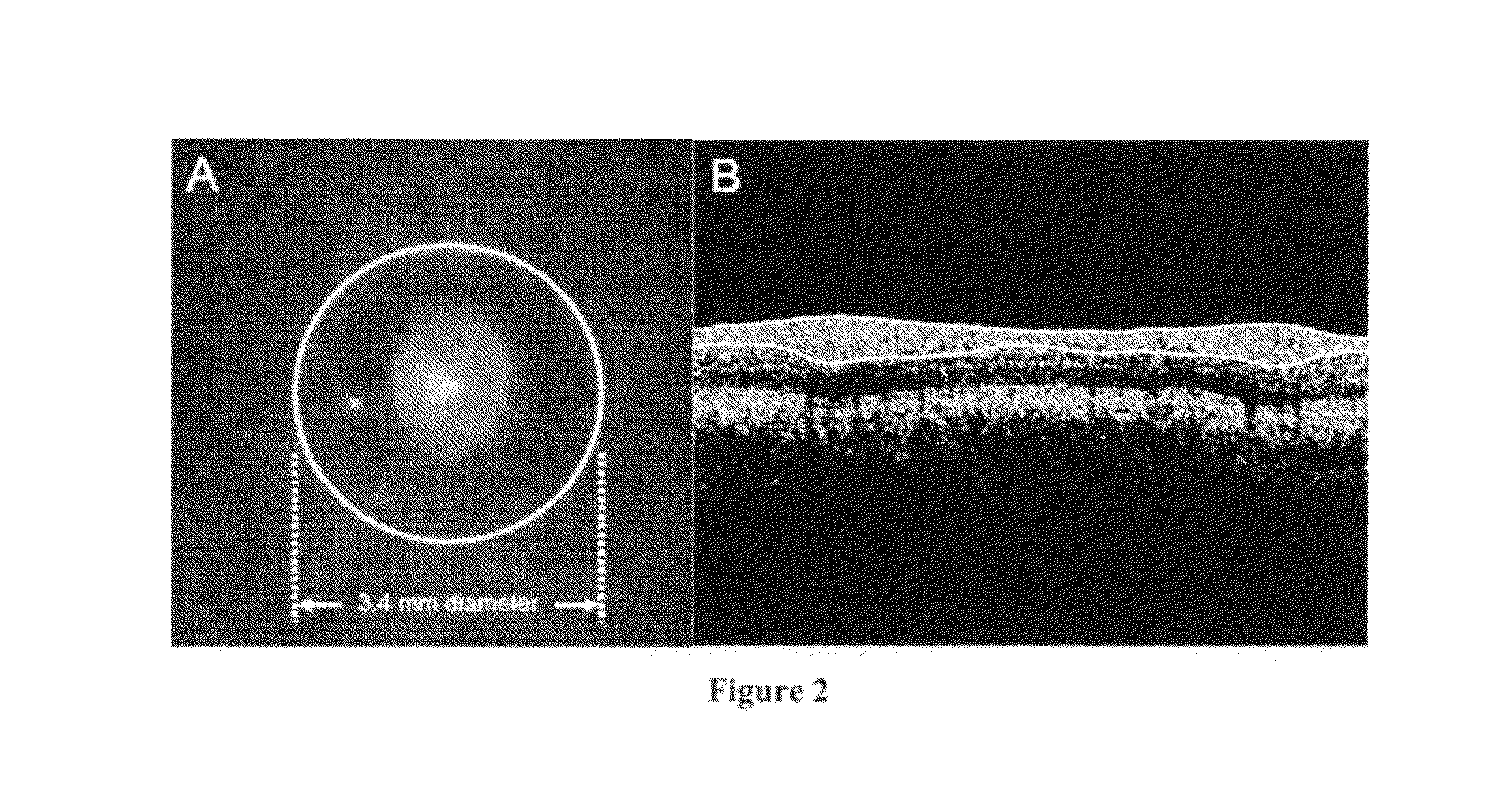
One limitation of TD-OCT circle scan is measurement variability in long term follow-up because operator manually places the TD-OCT circle scan around the ONH (FIG.

Method used


Image

  • Normalization of retinal nerve fiber layer thickness measurements made by time domain-optical coherence tomography
  • Normalization of retinal nerve fiber layer thickness measurements made by time domain-optical coherence tomography
  • Normalization of retinal nerve fiber layer thickness measurements made by time domain-optical coherence tomography

Examples


Embodiment Construction

Introduction

[0052]Optical coherence tomography, which was developed in 1991 by D. Huang, J. Schuman, et al at the Massachusetts Institute of Technology (MIT), Cambridge, is a low-coherence, interferometer-based, noninvasive medical imaging modality that can provide non-contact, real-time, high-resolution, cross-sectional images of biological tissue.1 According to a new market research report (Optical Coherence Tomography—Technology, Markets and Applications: 2008-2012 published by US media company PennWell (Tulsa, Okla.)), the optical coherence tomography (OCT) market will grow with revenues expected to top $800 million by 2012. The report estimates that the global market for OCT systems currently stands at around $200 million and is growing at an annual rate of 34% (Medical Physics Web; Jan. 16, 2008; http: / / medicalphysicsweb.org / cws / article / research / 32456). The first conventional time domain OCT (TD-OCT; Stratus OCT) was commercialized by Carl Zeiss Meditec, Inc. (CZMI), Dublin, C...


PUM


Abstract

A scan location matching (SLM) method identifies conventional time domain optical coherence tomography (TD-OCT) circle scan locations within three-dimensional spectral domain OCT scan volumes. A technique uses both the SLM algorithm and a mathematical retinal nerve fiber bundle distribution (RNFBD) model, which is a simplified version of the anatomical retinal axon bundle distribution pattern, to normalize TD-OCT thickness measurements for the retinal nerve fiber layer (RNFL) of an off-centered TD-OCT circle scan to a virtual location, centered on the optic nerve head. The RNFBD model eliminates scan-to-scan RNFL thickness measurement variation caused by manual placement of TD-OCT circle scan.

Description

CROSS-REFERENCE TO RELATED PATENT APPLICATIONS[0001]This application claims priority to U.S. provisional application No. 61 / 263,036, filed Nov. 20, 2009, the entire contents of which are incorporated herein by reference.STATEMENT REGARDING FEDERALLY SPONSORED RESEARCH[0002]This invention was made with United States government support in part under grant numbers R01-EY13178-09, R01-EY11289-23, and P30-EY08098-20, awarded by the National Institutes of Health (Bethesda, Md.), The Eye and Ear Foundation (Pittsburgh, Pa.), and unrestricted grants from Research to Prevent Blindness, Inc. (New York, N.Y.). The United States government has certain rights in the invention.BACKGROUND OF THE INVENTION[0003]Glaucoma is a progressive optic neuropathy, which induces irreversible structural damage of the retina that is manifested, for example, in retinal ganglion cell loss and thinning, due to axon loss, of the retinal nerve fiber layer (RNFL).1,2 Glaucomatous damage occurs along axon bundle and c...

Claims


Application Information

Patent Timeline
Patent Type & Authority Patents(United States)
IPC IPC(8): A61B3/14A61B3/10A61B3/00G01B9/02G01B11/06
CPCG01B9/02027G01B9/02087G01B11/06G01B2290/65G01B9/02091A61B3/102
Inventor KIM, JONG S.ISHIKAWA, HIROSHISCHUMAN, JOEL S.WOLLSTEIN, GADI
Owner UNIVERSITY OF PITTSBURGH
Why Eureka
Try Eureka