Method for assessing cancerous state
a cancerous state and mammalian technology, applied in the field of mammalian cancerous state assessment, can solve the problems of unsatisfactory diagnosis methods and treatment methods, and the mortality rate of cancer patients is still high, and achieve the effect of significantly reducing the expression level of neurofilament3 gene in the cancer cell line and high frequency
- Summary
- Abstract
- Description
- Claims
- Application Information
AI Technical Summary
Benefits of technology
Problems solved by technology
Method used
Image
Examples
example 1
Test of Confirming Methylation Status of Neurofilament3 Gene in Pancreatic Cancer Cell Line
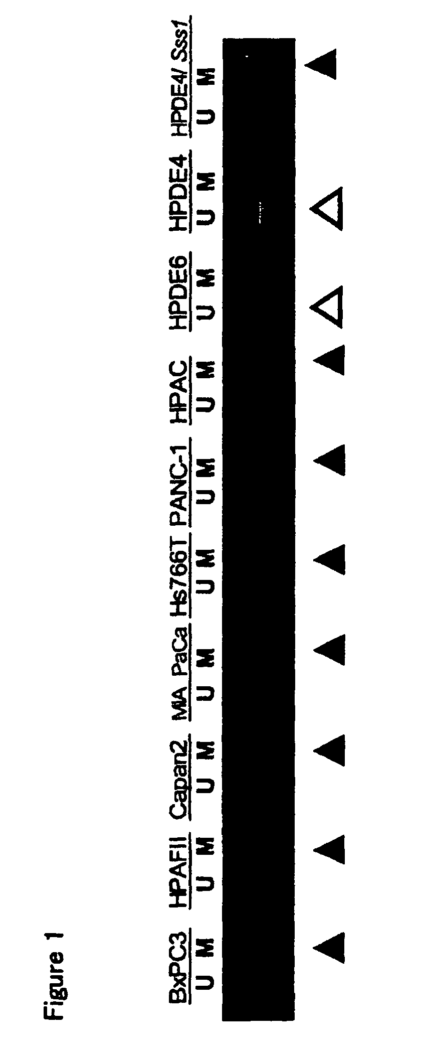
[0144]Seven kinds of human-derived pancreatic cancer cell lines [BXPc3, HPAF-II, Capan-2, MiaPaCa-2, Hs766T, PANC-1 and HPAC (all obtained from ATCC)] were cultured to sub-confluent in a medium exclusively used for each cell line described in catalogs of ATCC (American Type Culture Collection), and thereafter about 1×10⁷ cells were collected, respectively. Two kinds of immortal (normal) pancreatic ductal epithelial cells (HPDE-4 / E6E7 and HPDE6-E6E7c7) [these are maintained and managed by Dr. Tsao (Ontario Cancer Institute and Department of Pathology, University of Toronto) and available from the researcher] were cultured to sub-confluent in Keratinocyte-SFM, liquid medium (Invitrogen) containing 50 U / ml of penicillin and 50 μg / ml of streptomycin, and thereafter about 1×10⁷ cells were collected, respectively. 10-Fold volume of a SEDTA buffer [10 mM Tris / HCl (pH 8.0), 10 mM EDTA (pH 8.0), 100 mM...
example 2
Test of Confirming Methylation Status of Neurofilament3 Gene in Pancreatic Cancer Tissues
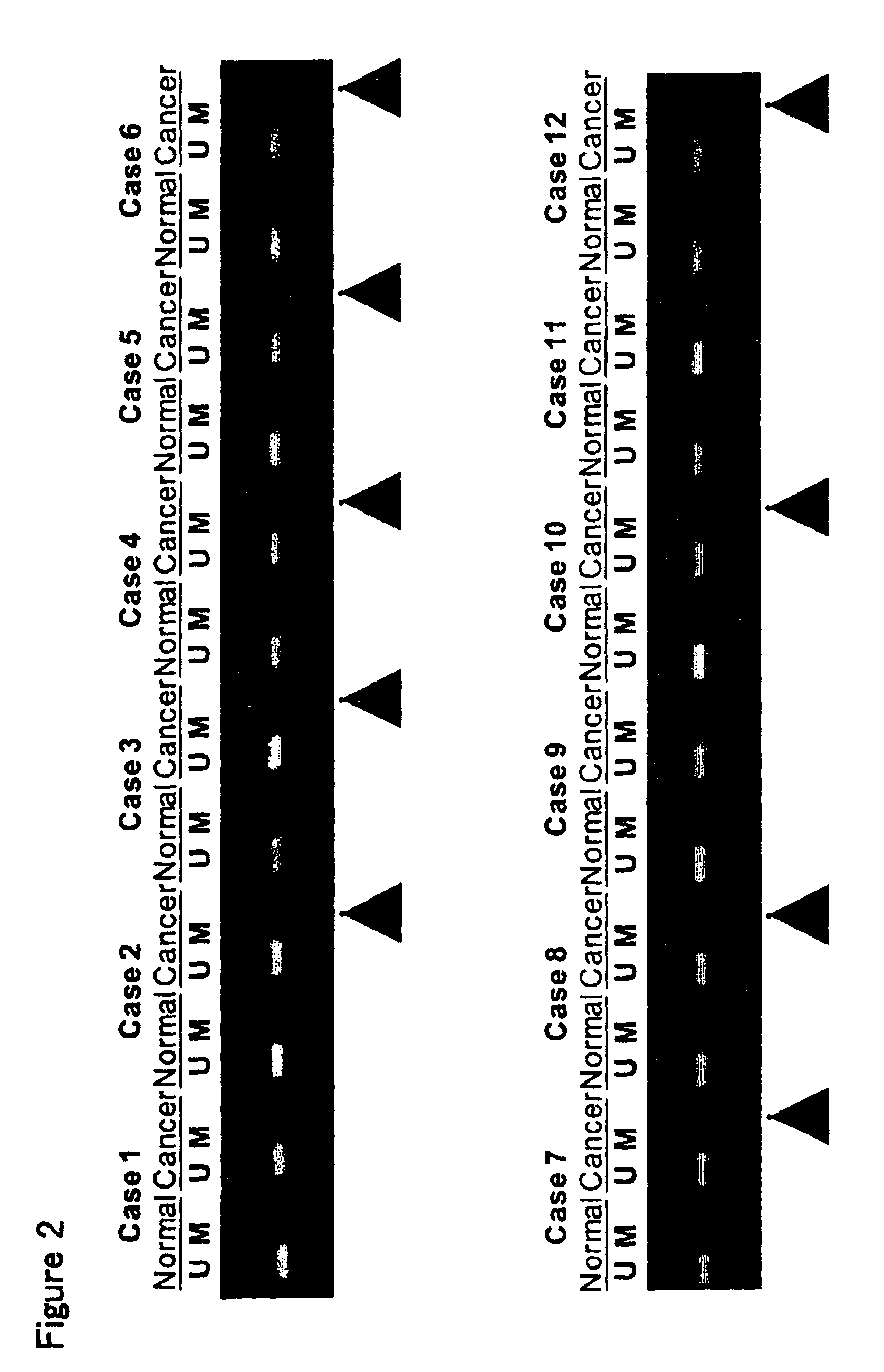
[0153]To each of 12 specimens (Case 1 to Case 12) of pancreatic cancer tissues and the surrounding pancreatic normal tissues (obtained from patients with their informed consent), 10-Fold volume of a SEDTA buffer [10 mM Tris / HCl (pH 8.0), 10 mM EDTA (pH 8.0), 100 mM NaCl] was added, and this was homogenized. To the resulting mixture were added proteinase K (Sigma) of 200 μg / ml and sodium dodecylsulfate of the amount to give a concentration of 1% (w / v), and this was shaken at 55° C. for about 16 hours. After completion of shaking, the mixture was treated by phenol [saturated with 1M Tris / HCl (pH 8.0)]-chloroform extraction. The aqueous layer was recovered, and NaCl was added thereto to give a concentration of 0.5N, and this was ethanol-precipitated to recover the precipitates. The recovered precipitates were dissolved in a TE buffer (10 mM Tris, 1 mM EDTA, pH 8.0), and RNase A (Sigma) was added th...
example 3
Test of Confirming Expression Status of Neurofilament3 Gene in Pancreatic Cancer Cell Line and Effect of Methylation Inhibitor on Expression of the Gene
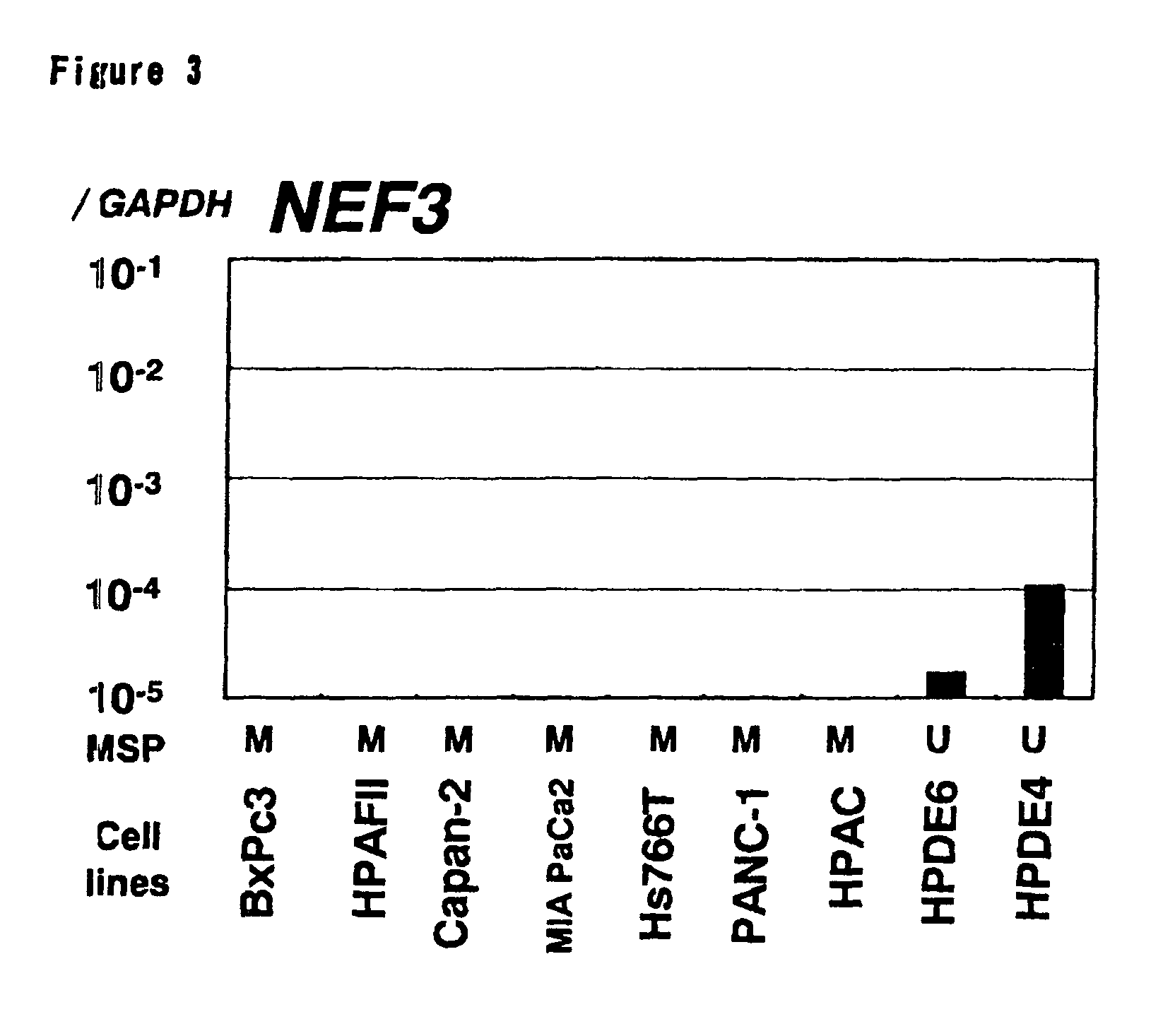
[0161]seven kinds of human-derived pancreatic cancer cell lines (BXPc3, HPAF-II, Capan-2, MiaPaCa-2, Hs766T, PANC-1 and HPAC) and immortal (normal) pancreatic ductal epithelial cells lines (HPDE-4 / E6E7 and HPDE6-E6E7c7) were cultured to 70 confluent in an exclusively used medium, and thereafter, each cell was corrected. 1 ml of an ISOGEN solution (Nippon Gene) was mixed with collected each cell (wet weight about 100 mg), this was homogenized, and 0.2 ml of chloroform was added thereto to suspend them. After suspending, the mixture was centrifuged (4° C., 15000×g, 15 minutes) to recover the supernatant. 0.5 ml isopropanol was added to the recovered supernatant to suspend them, and the suspension was centrifuged (4° C., 15000×g, 15 minutes) to recover the precipitates (RNA). The recovered precipitates were rinsed with 75% ethanol, and ...
PUM
| Property | Measurement | Unit |
|---|---|---|
| pH | aaaaa | aaaaa |
| temperature | aaaaa | aaaaa |
| temperature | aaaaa | aaaaa |
Abstract
Description
Claims
Application Information
 - R&D
- Intellectual Property
- Life Sciences
- Materials
- Tech Scout
- Unparalleled Data Quality
- Higher Quality Content
- 60% Fewer Hallucinations
Browse by: Latest US Patents, China's latest patents, Technical Efficacy Thesaurus, Application Domain, Technology Topic, Popular Technical Reports.
© 2025 PatSnap. All rights reserved.Legal|Privacy policy|Modern Slavery Act Transparency Statement|Sitemap|About US| Contact US: help@patsnap.com


