Image-based risk score—A prognostic predictor of survival and outcome from digital histopathology

a risk score and digital histopathology technology, applied in image enhancement, image analysis, instruments, etc., can solve the problems of inability to accurately, efficiently and reproducibly automate image analysis tools, and the tedious and time-consuming process of manual detection of bc nuclei in histopathology, etc., to achieve the effect of long time and high cos

Active Publication Date: 2015-04-07
RUTGERS THE STATE UNIV
View PDF6 Cites 4 Cited by
  • Summary
  • Abstract
  • Description
  • Claims
  • Application Information

AI Technical Summary

Benefits of technology

[0014]In an embodiment of the present invention, an image-based computer-aided prognosis (CAP) system and method that seeks to replicate the prognostic power of molecular assays in cancer histopathology is developed. The system and method of the present invention involves first semi-automatically detecting BC nuclei via an Expectation Maximization driven algorithm. Using the nuclear centroids, two graphs (Delaunay Triangulation and Minimum Spanning Tree) are constructed and a total of 12 features are extracted from each image. A non-linear dimensionality reduction system and method, Graph Embedding, projects the image-derived features into a low-dimensional space, and a Support Vector Machine classifies the BC images in the reduced dimensional space. Using only the tissue slide samples, a mechanism for digital slide scanning, and a computer, the image-based CAP system and method of the present invention aims to overcome many of the drawbacks associated with Oncotype DX, including the high cost associated with the assay; limited laboratory facilities with specialized equipment, and length of time between biopsy and prognostic prediction.
[0015]The present invention involves key methodological contributions and the use of several state of the art machine learning system and methods, including, but not limited to: a robust, efficient method to automatically detect BC nuclei; image features to describe the architectural arrangement of BC nuclei and hence, quantitatively describe cancer grade, and the use of non-linear dimensionality reduction to classify and visualize the underlying biological data structure in a low-dimensional representation.

Problems solved by technology

The manual detection of BC nuclei in histopathology is a tedious and time-consuming process that is unfeasible in the clinical setting.
Previous approaches to cell segmentation—thresholding [2], clustering [3], and active contour models [4]—are not very robust to the highly variable shapes and sizes of BC nuclei, as well as artifacts in the histological fixing, staining, and digitization processes.
A possible reason for this is that pathologists currently lack the automated image analysis tools to accurately, efficiently, and reproducibly quantify the presence and degree of LI in BC histopathology.
While several researchers have been developing algorithms for detection of nuclei [18], [23]-[29] in digitized histopathology, there have been no attempts to automatically detect or quantify extent of LI on BC histopathology.
However, they are not appropriate for the task of LI detection due to the similarity in appearance between BC and lymphocyte nuclei (FIG. 4(a)).
These methods, however, might be limited in their ability to handle variations in the appearance of BC nuclei (FIGS. 4(b), (c)) and image acquisition artifacts (FIGS. 4(e), (f)).
Probabilistic models, however, are usually limited by the availability of expert-annotated training data.
Detection of lymphocytes alone, however, cannot completely characterize the abnormal LI phenotype because a baseline level of lymphocytes is present in all tissues.

Method used


Image

  • Image-based risk score—A prognostic predictor of survival and outcome from digital histopathology
  • Image-based risk score—A prognostic predictor of survival and outcome from digital histopathology
  • Image-based risk score—A prognostic predictor of survival and outcome from digital histopathology

Examples


exemplary embodiment 1

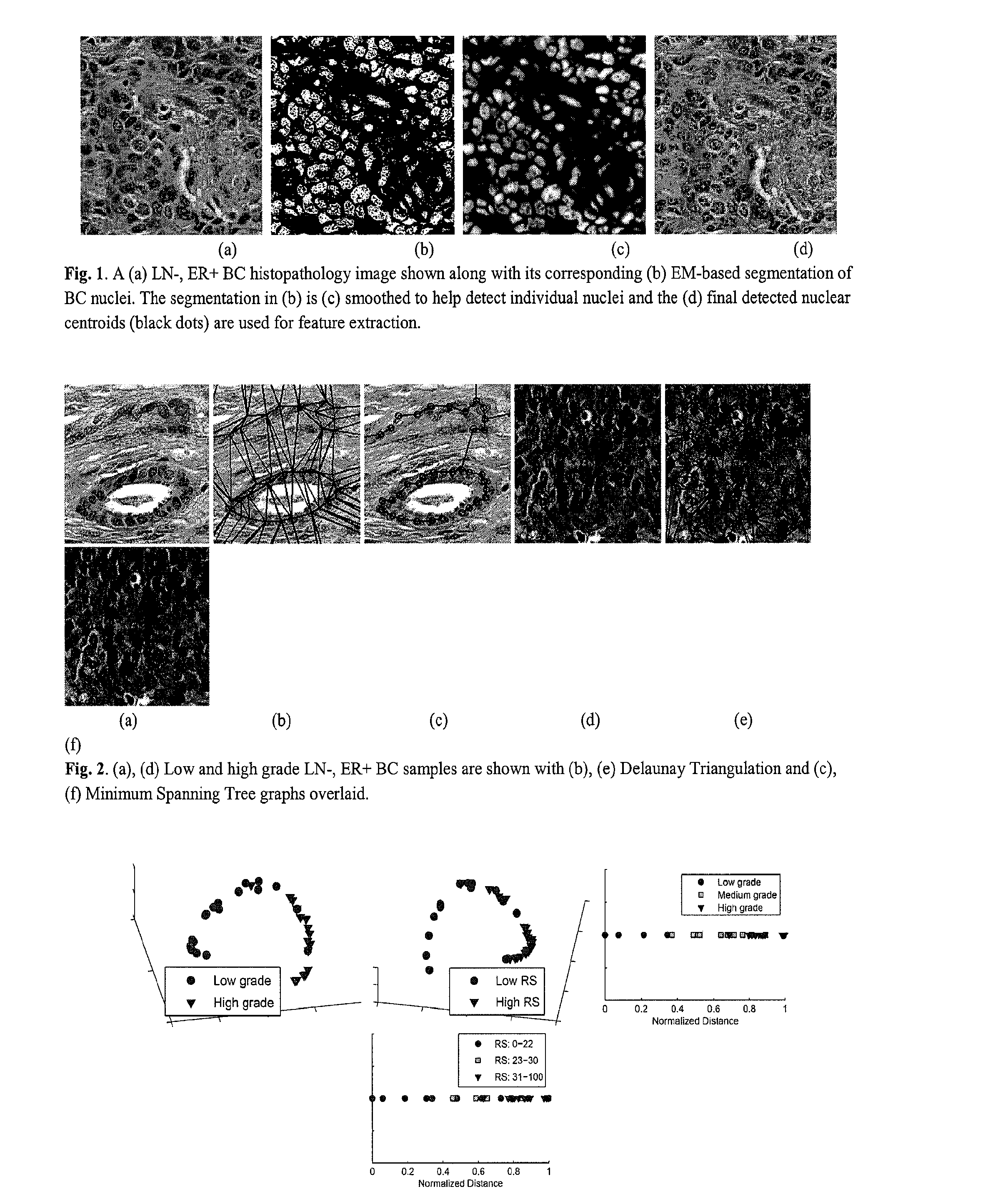
Automated Detection of Nuclei Using EM-Based Gaussian Mixture

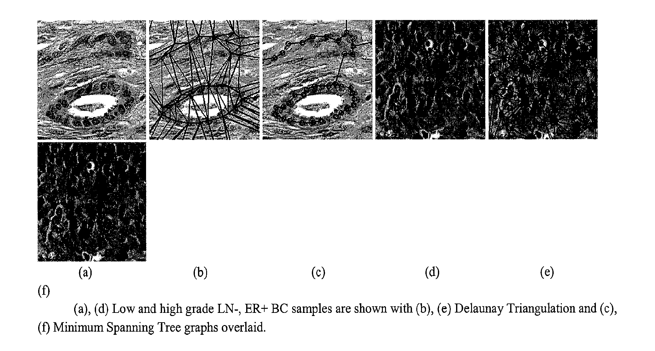
[0046]The present invention relates to an image-based computer-aided prognosis (CAP) system and method that seeks to replicate the prognostic power of molecular assays in BC histopathology. Using only the tissue slide samples, a mechanism for digital slide scanning, and a computer, our image-based CAP system and method aims to overcome many of the drawbacks associated with Oncotype DX, including the high cost associated with the assay; limited laboratory facilities with specialized equipment, and length of time between biopsy and prognostic prediction.

Dataset

[0047]A total of 37 hematoxylin and eosin (H & E) stained breast histopathology images were collected from a cohort of 17 patients and scanned into a computer using a high resolution whole slide scanner at 20× optical magnification. For all methods, we define the data set Z={C1, C2, . . . , CM} of M images, where an image C=(C, g) is a 2D set of pixels c∈C and g is the...

exemplary embodiment 2

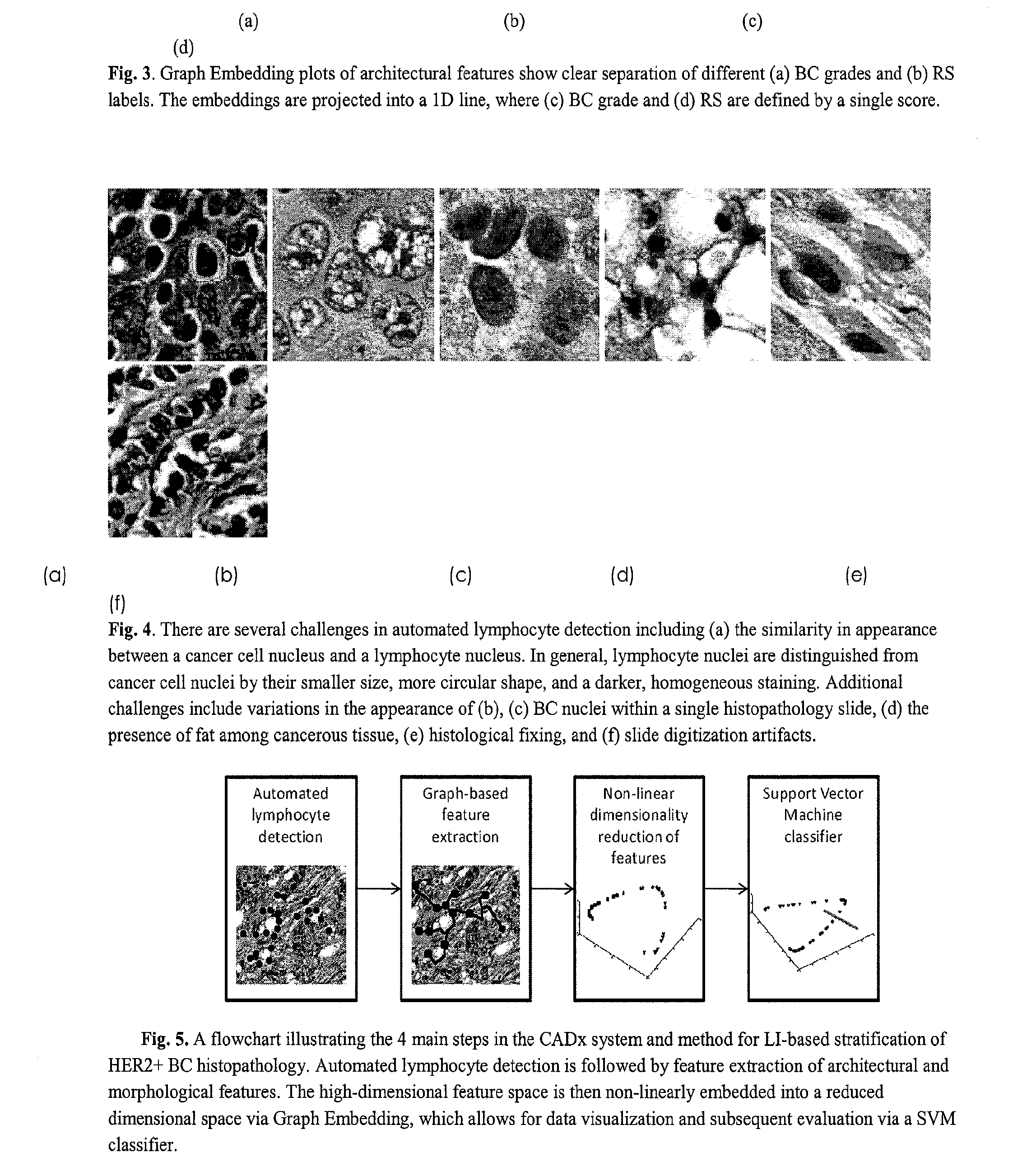
[0066]The ability to automatically detect LI would be invaluable to BC pathologists and oncologists, since manual detection of individual lymphocyte nuclei in BC histopathology is a tedious and time-consuming process that is not feasible in the clinical setting. The availability of a computerized image analysis system and method for automated quantification of LI extent in HER2+ BC will enable development of an inexpensive image-based system for predicting disease survival and outcome.

[0067]An important prerequisite for extracting histopathological image attributes to model BC appearance is the ability to automatically detect and segment histological structures, including nuclei and glands. Consequently the ability of an image analysis system and method to grade the extent of LI in a BC histopathology image is dependent on the algorithm's ability to automatically detect lymphocytes. Automated lymphocyte detection, however, is a non-trivial task complicated by the intrinsic similarit...


PUM


Abstract

The present invention relates to an image-based computer-aided prognosis (CAP) system and method that seeks to replicate the prognostic power of molecular assays in histopathology and pathological processes, including but not limited to cancer. Using only a tissue slide samples, a mechanism for digital slide scanning, and a computer, the present invention relates to an image-based CAP system and method which aims to overcome many of the drawbacks associated with prognostic molecular assays (e.g. Oncotype DX) including the high cost associated with the assay, limited laboratory facilities with specialized equipment, and length of time between biopsy and prognostic prediction.

Description

CROSS REFERENCE TO RELATED APPLICATIONS[0001]This application claims priority to U.S. Provisional Patent Application No. 61 / 149,158 filed on Feb. 2, 2009, the disclosure of which is hereby incorporated by reference in its entirety.FIELD OF THE INVENTION[0002]This invention concerns a computer-aided prognosis (CAP) system and method that employs quantitatively derived image information to predict pathological processes, disease outcome and patient survival. While digital pathology has made tissue specimens amenable to computer-aided diagnosis (CAD) for disease detection, the CAP system and method of the present invention predicts disease outcome and patient survival.BACKGROUND[0003]The current gold standard in identifying many disease states is the subjective visual interpretation of microscopic histology of fixed tissue sections of the involved organ. Examples of this include the diagnosis of cancer as well as many inflammatory and degenerative diseases. Over the past decade, the in...

Claims


Application Information

Patent Timeline
Patent Type & Authority Patents(United States)
IPC IPC(8): G06K9/00G06T7/00G06V10/764
CPCG06T7/0012G06K9/00147G06T2207/30024G06T2207/30096G06V20/698G06V10/764G06F18/2415
Inventor MADABHUSHI, ANANTBASAVANHALLY, AJAYGANESAN, SHRIDAR
Owner RUTGERS THE STATE UNIV
Why Eureka
Try Eureka