Device for processing eye tissue by means of femtosecond laser pulses

a technology of eye tissue and laser pulses, applied in optics, medical science, instruments, etc., can solve the problems of laser switching off, significant limits on cut guidance implementation, and the pulse rate has to be actively reduced

Active Publication Date: 2015-04-28
SIE SURGICAL INSTR ENG
View PDF16 Cites 0 Cited by
  • Summary
  • Abstract
  • Description
  • Claims
  • Application Information

AI Technical Summary

Benefits of technology

[0008]It is an object of the present invention to propose an ophthalmological device for processing eye tissue by means of femtosecond laser pulses which does not have at least some disadvantages of the known systems. In particular, it is an object of the present invention to propose an ophthalmological device for processing eye tissue by means of femtosecond laser pulses which manages without mechanical movement of lenses for the scanning of a processing region and allows the use of laser sources having high pulse frequencies, in particular pulse frequencies in the MHz range with more than one million pulses per second in order to reduce the processing time, and a more flexible cut guidance.
[0013]The cascading of the two beam-deflecting scanner systems with movable deflection mirrors enables eye tissue to be processed in a flexibly configurable and controllable manner, wherein the first scanner system covers an extended processing region, for example the entire eye, and the second scanner system disposed upstream superimposes a fast fine scanning movement, the form, size and orientation of which are flexibly adjustable, without overly large projection and transmission optical units having to be used, and without a mechanical movement of lenses or projection objectives being required for scanning, with the result that high scanning frequencies or scanning rates can be realized. The arrangement of the comparatively faster second scanner system, the “ultrafast scanner system”, between the laser source and the first scanner system, the “fast main scanner system”, allows the use of smaller beam apertures, e.g. mirrors, and thus enables fast, high-frequency scanning of the eye tissue. In this case, the faster second scanner system can be optimized for high-frequency scanning and processing, i.e. cutting, of the eye tissue with a relatively small excursion, and the first scanner system can be optimized for rapidly moving to any addressable points in the extended processing region (image region).
[0015]In one embodiment variant, the control module is designed to determine the curve orientation of the scanning curve by coupled control of the excursion amplitude of the deflection about the first mirror axis, of the excursion amplitude of the deflection about the second mirror axis, and of the relative phase of the deflection about the first mirror axis and of the deflection about the second mirror axis. The coupled control of the excursion amplitudes and relative phase makes it possible, for example, to control the orientation of an ellipse.
[0024]In a further embodiment variant, the control module is designed to control the deflection about the first mirror axis and the deflection about the second mirror axis such that the deflection about the first mirror axis is effected with a deflection frequency having a doubled magnitude relative to the deflection about the second mirror axis, and the deflection about the first mirror axis brings about at least partial compensation of the deflection brought about by the first scanner system in the direction of the processing line.
[0028]In one embodiment variant, the control module is designed to control the deflection of the femtosecond laser pulses about the first mirror axis and the deflection of the femtosecond laser pulses about the second mirror axis such that the scanning curve has a round curve shape, and the control module is designed to control the scanning speed of the first scanner system such that a resulting scanning trajectory having a defined trajectory profile arises from the superimposition of the scanning curve onto the processing line. When a circular scanning curve is superimposed onto the processing line, control of the scanning curve with regard to its orientation becomes dispensable on account of the symmetry.

Problems solved by technology

In particular the mechanical movement of light projectors or lenses for scanning working regions, and also the mass inertia of galvanometer scanner systems, which does not permit arbitrarily high accelerations, limit the possible scanning patterns and scanning trajectories to the effect that greater changes in direction have to be avoided, and that the pulse rate has to be actively reduced (in a complicated manner) or the laser has to be switched off when the minimum scanning speed is undershot, for example at reversal points.
Consequently, the known scanner systems impose significant limits on cut guidances that can be implemented.
In contrast to surface processing, when cutting soft tissue, for example eye tissue, it is not always possible to employ simple pulse scanning patterns, for example line or spiral patterns, since tissue deformations can be caused by internal evolution of gas or release of stresses, and it is necessary to avoid said tissue deformations by means of suitably more complex scanning patterns taking account of the expected biomechanical behaviour of the tissue.
Although the known scanner systems make it possible to process simple scanning patterns, for example to cut a tissue flap, this generally being performed as a large area segment with a simple edge geometry, in the case of isolated processing regions with a complicated edge geometry, such as are required for refractive correction, for example, or in the case of other, biomechanically governed more complex scanning patterns, that is no longer possible in such a simple manner.
In other words: for fundamental physical reasons, large image fields cannot be scanned with small (i.e. rapidly deflectable) mirrors and at the same time have spot diameters that are desirably small for the processing quality.
As a result of the simple control of the curve orientation on the basis of the excursion amplitudes, a costly and slow mechanical and / or optical image rotator becomes invalid, since the control is effected entirely by electronic and / or programming means.

Method used


Image

  • Device for processing eye tissue by means of femtosecond laser pulses
  • Device for processing eye tissue by means of femtosecond laser pulses
  • Device for processing eye tissue by means of femtosecond laser pulses

Examples


Embodiment Construction

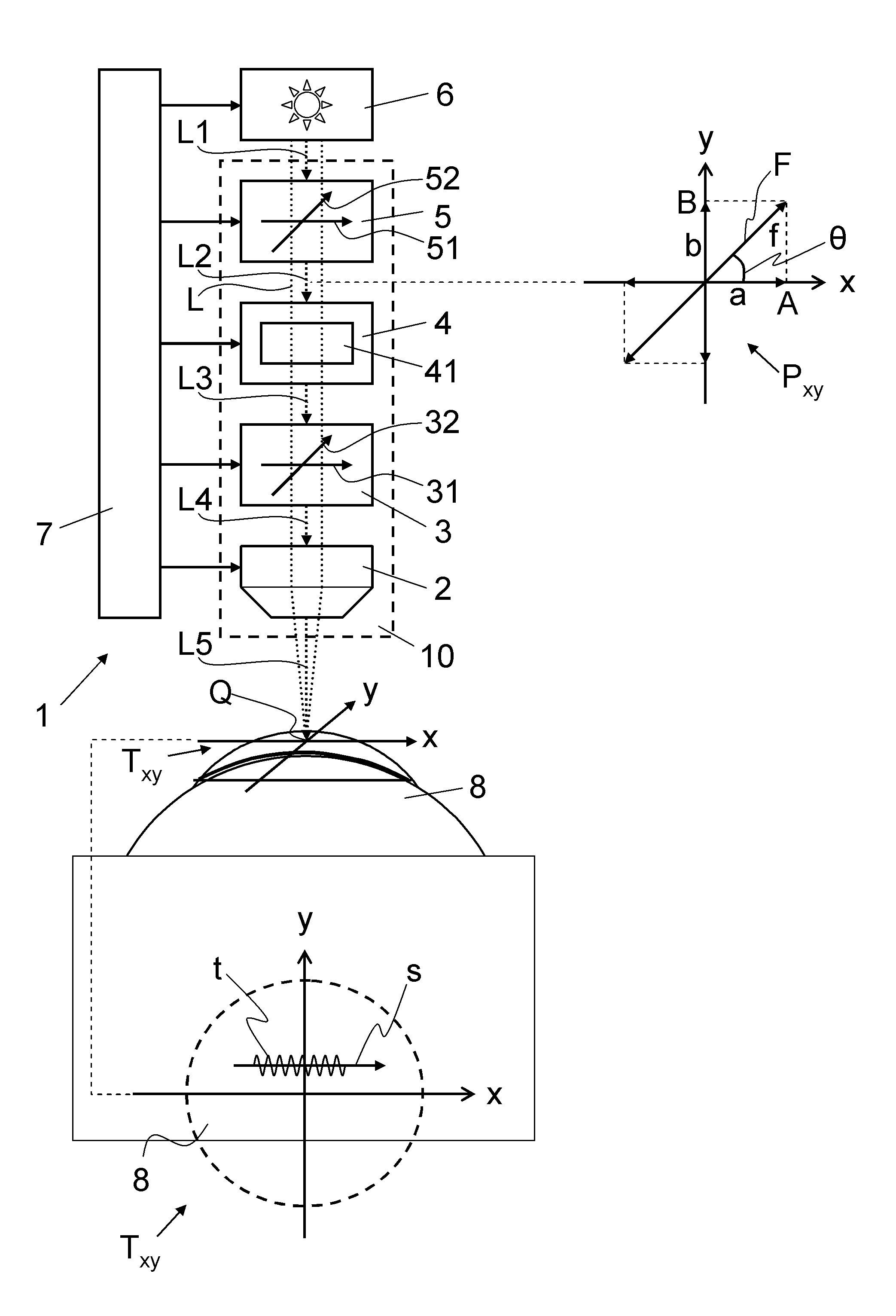
[0041]In FIG. 1, the reference symbol 1 refers to an ophthalmological device for processing eye tissue 8 by means of a pulsed laser beam L1 with femtosecond laser pulses. The pulsed laser beam L1, preferably having pulse frequencies in the MHz range with more than one million pulses per second, is supplied by a beam source 6 and, in a manner focused by means of an optical transmission system 10, projected along a scanning trajectory t as a pulsed processing beam L5 onto or into the eye tissue 8. Depending on the embodiment, the beam source 6 is part of the optical transmission system 10 or configured as a separate unit connected to the optical transmission system 10 via a light transmission system, for example a fibre-optic line and / or a mirror / lens system.

[0042]As is illustrated schematically in FIG. 1, the ophthalmological device 1 or the optical transmission system 10 includes two beam-deflecting scanner systems 3, 5 and an optional filter module 4, which are arranged in the beam...


PUM


Abstract

For processing eye tissue (8) by means of femtosecond laser pulses, an ophthalmological device (1) includes a projection optical unit (2) for the focused projection of the femtosecond laser pulses into the eye tissue (8). Disposed upstream of the projection optical unit (2) is a first beam-deflecting scanner system (3) for scanning the eye tissue (8) with the femtosecond laser pulses along a processing line (s). A second beam-deflecting scanner system (5) is disposed upstream of the first scanner system (3) and is designed for scanning the eye tissue (8) with the femtosecond laser pulses on a scanning curve (f) superimposed on the processing line (s). The second scanner system (5) has a scanner speed that is a multiple of the scanning speed of the first scanner system (3), and comprises at least one deflection mirror, a first scanning axis (51) and a second scanning axis (52). A control module (8) controls the deflection of the femtosecond laser pulses about the first scanning axis (51) and the deflection of the femtosecond laser pulses about the second scanning axis (52) in accordance with a defined curve shape, a defined curve amplitude and a defined curve orientation of the scanning curve (f).

Description

CROSS REFERENCE TO RELATED APPLICATIONS[0001]The present application claims benefit of and priority to U.S. Provisional Patent Application Ser. No. 61 / 368,731 filed Jul. 29, 2010 entitled VORRICHTUNG ZUM BEARBEITEN VON AUGENGEWEBE MITTELS FEMTOSEKUNDENLASERPULSEN, the entire contents of which are hereby incorporated by reference herein.BACKGROUND[0002]1. Field of the Disclosure[0003]The present invention relates to an ophthalmological device for processing eye tissue by means of femtosecond laser pulses. The present invention relates, in particular, to an ophthalmological device including a projection optical unit for the focused projection of the femtosecond laser pulses into the eye tissue.[0004]2. Related Art[0005]For processing eye tissue by means of laser beams, an image region or a working region is scanned with laser pulses by means of the pulsed laser beam being deflected in one or two scanning directions by means of suitable scanner systems (deflection devices). The deflect...

Claims


Application Information

Patent Timeline
Patent Type & Authority Patents(United States)
IPC IPC(8): A61F9/011A61F9/008
CPCA61F9/00827A61F9/00836A61F2009/00872A61F2009/00897
Inventor RATHJEN, CHRISTIAN
Owner SIE SURGICAL INSTR ENG
Why Eureka
Try Eureka