Method for diagnosing a hemoglobin-related disorder

a hemoglobin-related disorder and disorder technology, applied in the field of hemoglobin-related disorder diagnosis and/or staging, can solve the problems of triggering cell apoptosis, overwhelming by defective production, damage to cells,

Inactive Publication Date: 2015-08-04
INST NAT DE LA SANTE & DE LA RECHERCHE MEDICALE (INSERM) +3
View PDF3 Cites 2 Cited by
  • Summary
  • Abstract
  • Description
  • Claims
  • Application Information

AI Technical Summary

Benefits of technology

[0110]In another example of competitive immunoassay, the quantification of free α-Hb is achieved by the antibody coupled with or conjugated with a ligand, said ligand binding to an additional antibody added to the sample. One example of said ligand is fluorescein. The additional antibody may be bound to a solid support. In this example of competitive immunoassay, the additional antibody binds to the ligand coupled with the antibody that binds in turn to (i) free α-Hb present in the sample and (ii) labelled α-Hb added to the sample. Said mass complex formed allows isolation and measurement of the signal generated by the label coupled with the labelled α-Hb.
[0121]In one embodiment, one antibody that binds to α-Hb is the “capture” antibody, and is bound to a solid support, such as protein coupling or protein binding surface, colloid metal particles, iron oxide particles, or polymeric beads. One example of polymeric beads is a latex particle. In such an embodiment, the capture antibody is bound to or coated on a solid phase support using standard non-covalent or covalent binding methods, depending on the required analytical and / or solid phase separation requirements. The solid support may be in the form of test-tubes, beads, microparticles, filter paper, membrane, glass filters, magnetic particles, glass or silicon chips or other materials known in the art. The use of microparticles, particularly magnetisable particles, that have been directly coated with the antibody or particles that have been labelled with a universal binder (such as avidin or anti-species antibody) is useful for significantly shortening the assay incubation time.
[0122]Alternatively, the capture anti-α-Hb antibody may be coupled with a ligand that is recognized by an additional antibody that is bound to or coated on the solid support. Binding of the capture antibody to the additional antibody via the ligand then indirectly immobilizes the capture antibody on the support. An example of such a ligand is fluorescein. Alternatively, the binding partner may also be detected indirectly by a secondary detection system. Said secondary detection system is based on several different principles known in the art such as antibody recognition and other forms of immunological or non-immunological bridging and signal amplification detection systems (for example, the biotin-streptavidin system). When a signal amplification system is used, the label includes a first protein such as biotin coupled with the capture antibody, and a second protein such as streptavidin that is coupled with an enzyme. The second protein binds to the first protein. The enzyme produces a detectable signal when provided with substrate(s), so that the amount of signal measured corresponds to the amount of binding partner that is bound α-Hb. Examples of enzymes include, without limitation, alkaline phosphatase, amylase, luciferase, catalase, beta-galactosidase, glucose oxidase, glucose-6-phosphate dehydrogenase, hexokinase, horseradish peroxidase, lactamase, urease and malate dehydrogenase. Suitable substrates include, without limitation, TMB (3,3′,5,5′-tetramethyl benzidine), OPD (o-phenylene diamine), and ABTS (2,2′-azino-bis(3-ethylbenzthiozoline-6-sulfonic acid). The signal amplification approach may be used to significantly increase the assay sensitivity and low level reproducibility and performance.

Problems solved by technology

In the red blood cell of β-thalassemic patients, AHSP acts as a scavenger against the pool of free α-chain but may be overwhelmed by a defective production or level of availability of β-like chains.
Thus, the free α-Hb monomers in the red cells overload the AHSP capacity and precipitate, damaging the cell and triggering cell apoptose.
Such a method could certainly not be considered as routine laboratory practice.
It results that there is currently no simple and rapid test to evaluate this parameter.
It is cumbersome, expensive and time consuming procedure.
Furthermore, until very recently, it was considered that was impossible to detect or quantify free α-Hb since the excess α-Hb either precipitates in erythroid precursors in the bone marrow (resulting in ineffective erythropoiesis), causing their premature destruction, and although partly proteolyzed binds to the cell membrane of adult erythroid cells, leading to their hemolysis and promoting apoptosis (Bank, 2007 and Yu et al., 2007).

Method used


Image

  • Method for diagnosing a hemoglobin-related disorder
  • Method for diagnosing a hemoglobin-related disorder
  • Method for diagnosing a hemoglobin-related disorder

Examples


example

Material & Methods

[0148]Patients:

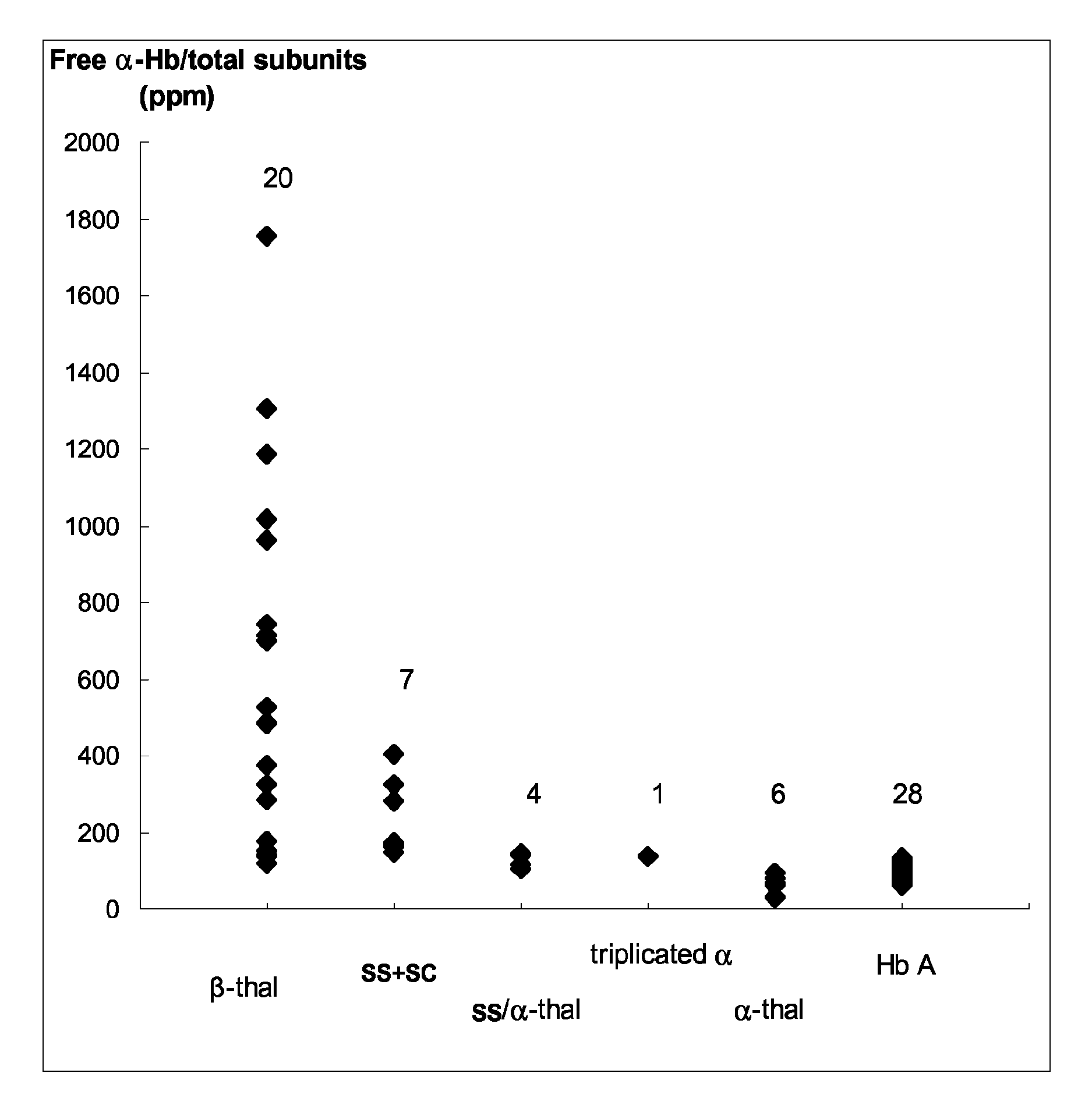
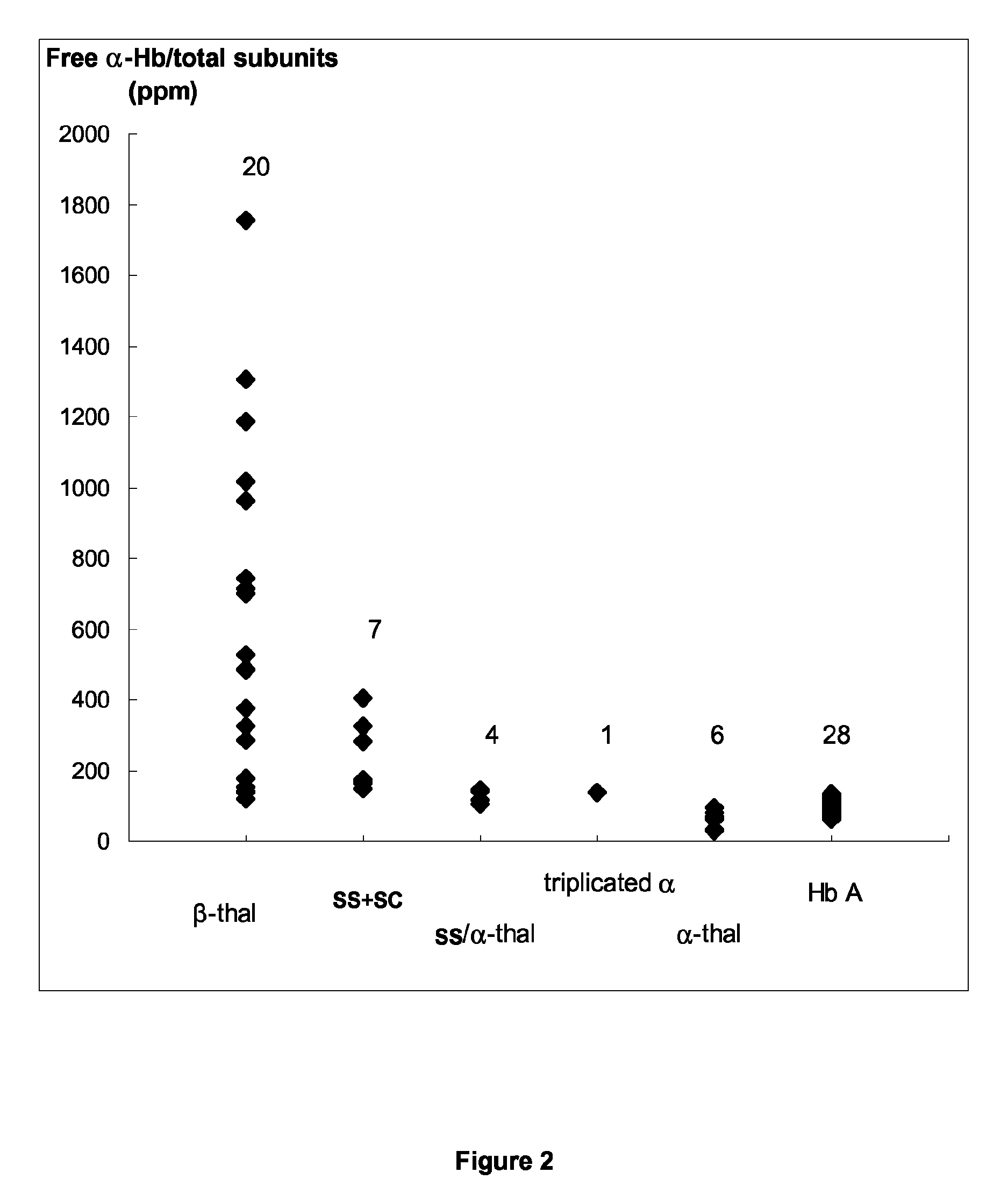
[0149]This study concerns 66 patients (43 males and 23 females) including 28 patients without apparent Hb disorder (reference group) (21 males and 7 females; mean age, 63±11 years), 20 β-thalassemic patients (12 males and 8 females; mean age, 39±15 years), 6 α-thalassemic patients (2 males and 4 females; mean age, 50±15 years), 7 SS or SC patients (4 males and 3 females; mean age, 36±9 years), 4 patients SS / α-thal (3 males and 1 female, mean age, 39±8 years) and 1 male triplicated α patient (69 years) For patients, except the controls without apparent Hb disorder, the β- and α-thal genotypes were determined. An informed consent was obtained from all participants to this study according to the international Helsinki declaration and French ethical regulations.

[0150]Hematological and Genotyping Investigations:

[0151]EDTA-anticoagulated Venous Blood samples was collected during routine sampling for the current follow-up of their clinical conditions. Hb ph...


PUM

PropertyMeasurementUnit
pHaaaaaaaaaa
pHaaaaaaaaaa
timeaaaaaaaaaa

Abstract

A method for diagnosing and / or staging a hemoglobin disorder such as β-thalassemia involves contacting a blood sample of a subject with an Alpha-Hemoglobin Stabilizing Protein which may be present on a solid support, and detecting and / or quantifying free α-Hb. Detection or quantification can be achieved by photometry such as HTRF or immunological procedures such as EIA and ELISA. The free α-Hb is compared to a reference value, and can be used to correlate with the diagnosis and / or staging of the hemoglobin disorder for the subject.

Description

FIELD OF THE INVENTION[0001]The invention relates to a method for diagnosing and / or staging a hemoglobin-related disorder, such as β-thalassemias, in a subject in need thereof.[0002]The invention also relates a method for monitoring a treatment against said hemoglobin-related disorder in a subject in need thereof.BACKGROUND OF THE INVENTION[0003]The normal development of red blood cells requires a coordinated synthesis of the hemoglobin (Hb) subunits, the α- and β-globins in the case of adult hemoglobin (Hb A). The α- and β-globin chains are encoded by genes on different chromosomes, 16 and 11 respectively, and their expression is controlled independently. In the normal red blood cell, slightly more α-chains than β-chains are produced. Unlike the β-hemoglobin chains (β-Hb) which are soluble and form homologous tetramers, the free α-hemoglobin chains (α-Hb) are highly instable, and when in excess, form precipitates and act as active oxidants causing apoptosis and inefficient erythrop...

Claims


Application Information

Patent Timeline
Patent Type & Authority Patents(United States)
IPC IPC(8): G01N33/53G01N33/72
CPCG01N33/721G01N33/53G01N2800/52G01N2800/22G01N2800/56
Inventor BAUDIN-CREUZA, VERONIQUEVASSEUR, CORINNEGALACTEROS, FREDERIC
Owner INST NAT DE LA SANTE & DE LA RECHERCHE MEDICALE (INSERM)
Why Eureka
Try Eureka