Method of diagnosing cancer and reagents therefor

a cancer and reagent technology, applied in the field of cancer diagnosis and reagents therefor, can solve the problems of difficult diagnosis of cancer, increased cost, and clear as to the extent and nature of epigenetic changes, and achieve the effect of facilitating the determination of the expression level of nucleic acids

Active Publication Date: 2015-10-13
GARVAN INST OF MEDICAL RES
View PDF0 Cites 0 Cited by
  • Summary
  • Abstract
  • Description
  • Claims
  • Application Information

AI Technical Summary

Benefits of technology

[0063]The present inventors have also shown that compounds useful for the treatment of a cancer also return the modified chromatin to a relatively normal or healthy state. For example, the present inventors have shown that treatment of a cancer cell with a histone de-acetylase inhibitor and / or a methylation inhibitor reduces the degree of modified chromatin in the region identified by the inventors. Accordingly, these findings also provide the basis of screening method for determining the efficacy of treatment of a subject for cancer. For example, in one embodiment, the present invention provides a method for monitoring the efficacy of treatment of a subject receiving treatment for a cancer, said method comprising identifying and / or detecting in a sample from the subject:
[0092]The present inventors have also demonstrated that modified expression of a gene or nucleic acid within Chromosome 2 from about map position 2q14.1 to about map position 2q14.3 of the human genome in a sample from a subject relative to a non-cancerous cell is indicative of an enhanced degree of chromatin modification. For example, modified expression is determined by performing a method comprising:(i) determining the level of expression of a nucleic acid positioned within Chromosome 2 from about map position 2q14.1 to about map position 2q14.3 of the human genome in the sample derived from a subject;(ii) determining the level of expression of a nucleic acid positioned within Chromosome 2 from about map position 2q14.1 to about map position 2q14.3 of the human genome in a suitable control sample,wherein a reduced level of expression at (i) compared to (ii) is indicative of an enhanced degree of chromatin modification.
[0095]Suitable probes and / or primers will be apparent to the skilled artisan based on the description herein. For example, the probe or primer comprises a nucleotide sequence selected from the group consisting of SEQ ID NOs: 200-219. Preferably, such a probe or primer is labeled with a detectable marker (e.g., a fluorescent marker) to thereby facilitate determining the level of expression of a nucleic acid.

Problems solved by technology

This diversity in clinical and molecular characteristics make cancer difficult to diagnose.
The detection of microsatellite instability as a diagnostic for cancer, requires the patient to have a detectable tumor beforehand and, as a consequence, is not an early test that can lead to early effective treatment.
This leads to increased cost, as a number of samples must be assayed for each diagnosis performed.
However, it is not clear as to the extent and nature of these epigenetic changes in cancer cells.
However, the 5-methylcytosine content of a cell is not necessarily a measure of the level or extent of chromatin modification in these cells.
Accordingly, the assay of Gama-Sosa et al., does not provide an accurate measurement of chromatin changes that are associated with cancer.
However, the methylation of these genes is limited to discrete regions of these genes and shown to be useful only in relation to the detection of specific cancers (Plass, Hum. Mol. Genet., 11: 2479-2488, 2002).
In view of the heterogeneity of cancers, it is difficult to produce a single diagnostic that is useful for different cancer types.

Method used


Image

  • Method of diagnosing cancer and reagents therefor
  • Method of diagnosing cancer and reagents therefor
  • Method of diagnosing cancer and reagents therefor

Examples


example 1

Identification of a Hypermethylated Region of Chromosome 2 in Colon Cancer

[0617]Samples used in the identification of hypermethylated DNA in colon cancer were derived from 112 colorectal carcinomas with paired non-adjacent areas of normal colonic mucosa. Samples were collected as and frozen within 2 hours of removal and stored at −80° C. until analysis. All samples were obtained from the Hospital de la Santa Creu u Sant Pau (Barcelona, Spain).

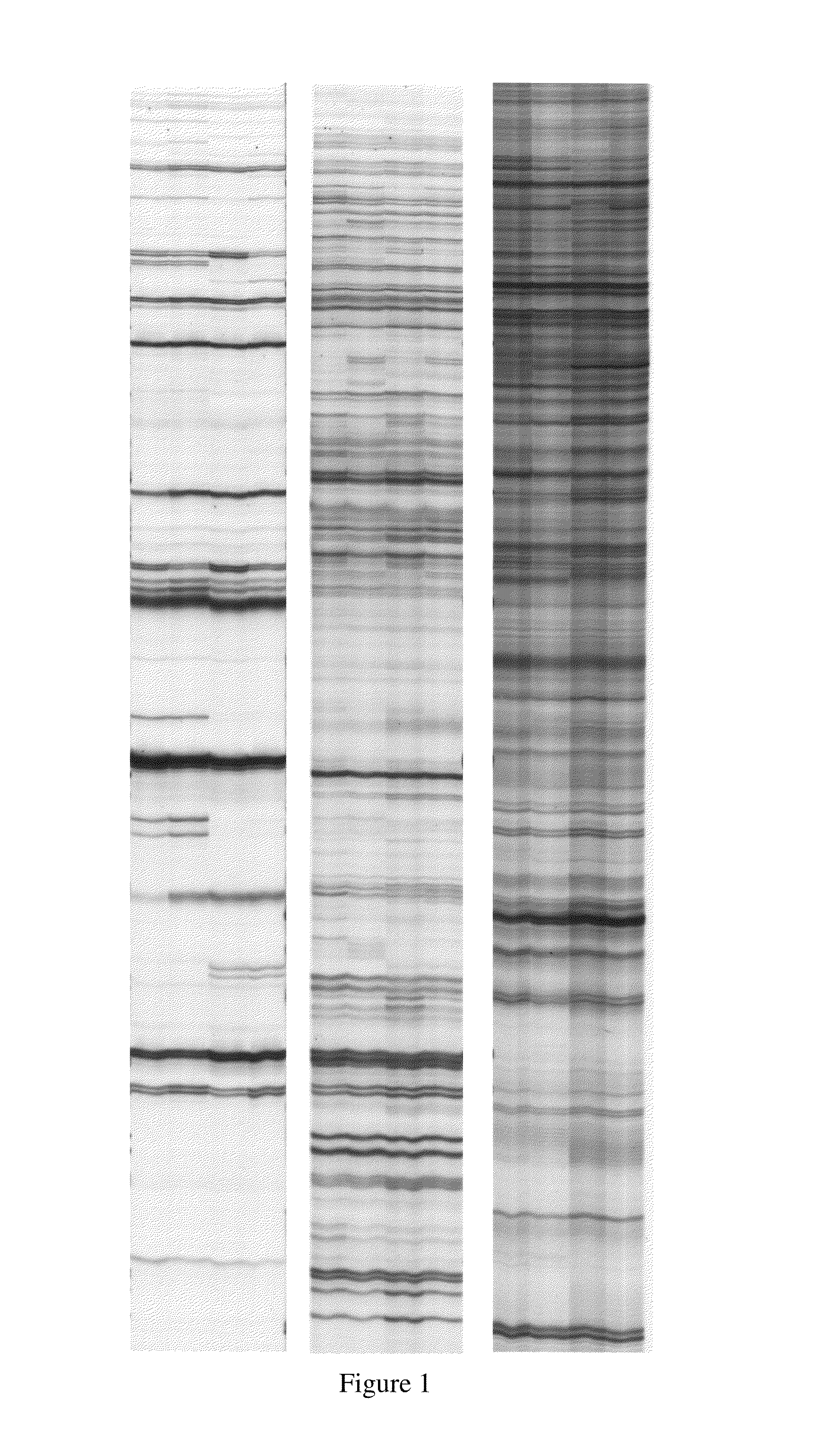
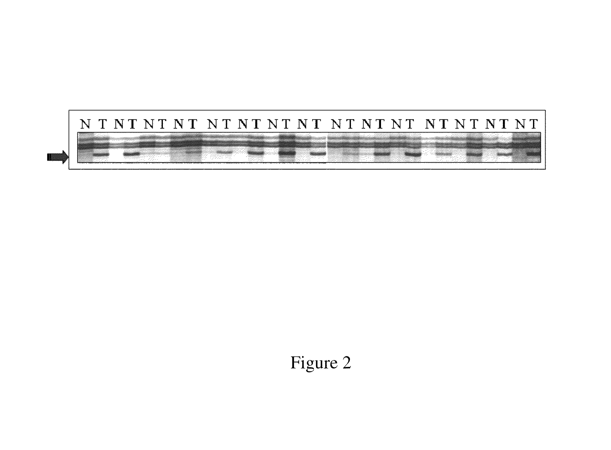
[0618]Using the global methylation approach, amplification of inter-methylated sites (AIMS) assay (essentially as described in Frigole et al., Nucleic Acids Res. 30: e28, 2002) the inventors identified a SmaI fragment, designated the Z fragment, that was differentially methylated in 71 out of 112 (63%) of colorectal / normal tumor matched pairs. An example of a gel showing an AIMS assay identifying the Z band is shown in FIGS. 1 and 2.

[0619]The Z fragment was isolated from an acrylamide gel, sequenced and mapped to human Chromosome 2 map position...

example 2

DNA Hypermethylation of CpG Islands Neighbouring Engrailed-1

2.1 Methods

Bisulfite Treatment

[0621]DNA was extracted from the HCT116 / SW480 cells using the Puregene extraction kit (Gentra Systems) and Trizol reagent (Invitrogen) from colorectal cancer or normal matched samples according to the manufacturer's protocol. The bisulfite reaction was carried out using 2 μg of restricted DNA for 16 h at 55° C. under conditions essentially as previously described (Clark et al., Nuc. Acids Res., 22: 2990-2997, 1994). After neutralization, the bisulphite treated DNA was ethanol precipitated, dried, resuspended in 50 μl of H2O and stored at −20° C. Approximately 2 μl of DNA was used for each of the nested PCR amplifications. The primers used for the amplifications are set forth in SEQ ID NOs: 71 to SEQ ID NO: 198. The combinations used are described herein. At least three independent PCR reactions were performed to ensure a representative methylation profile.

Direct PCR Sequence Analysis

[0622]Poole...

example 3

DNA Methylation Across Chromosome 2q14.2 in Cancer

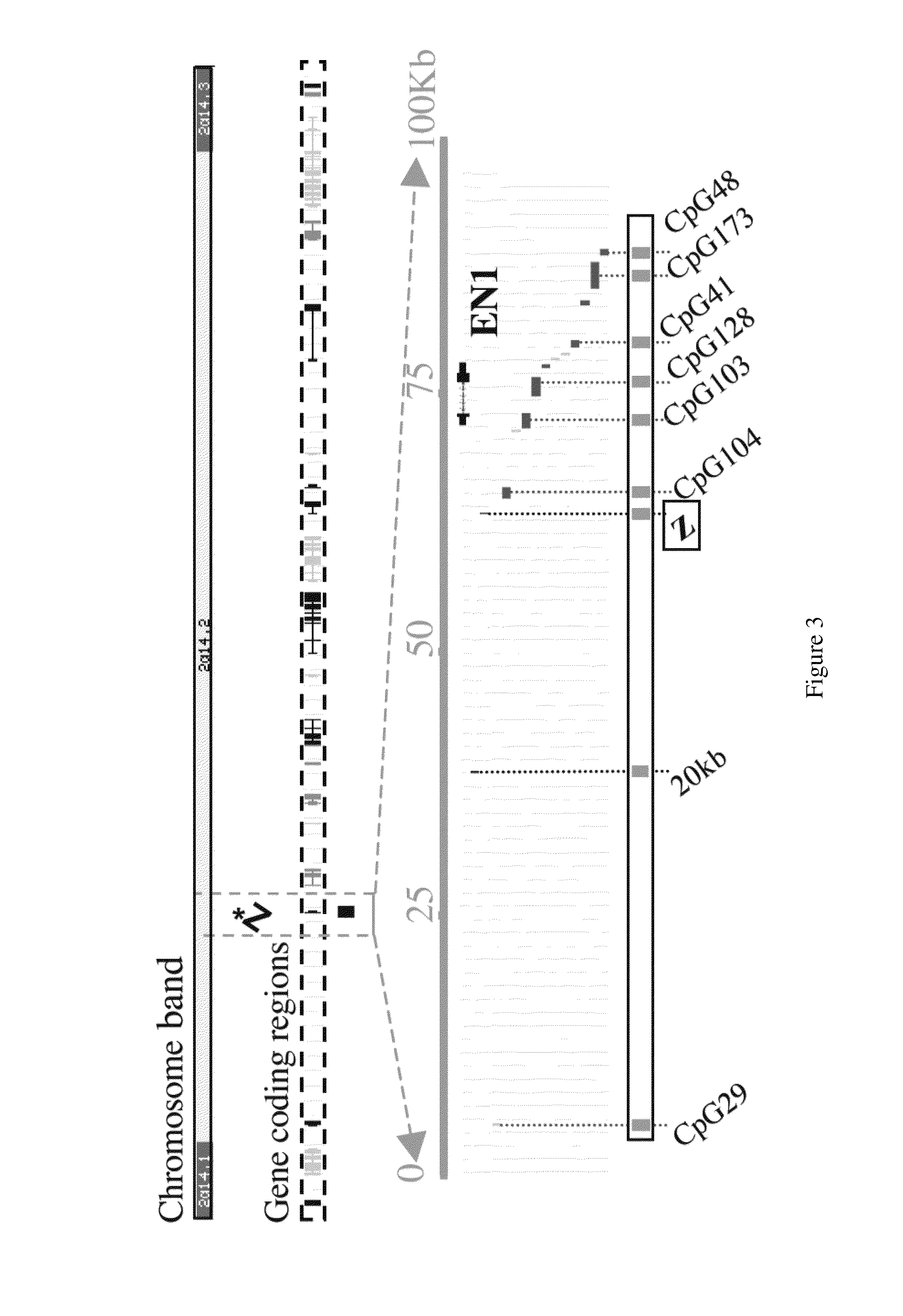
[0630]The analysis of the methylation status of DNA on either side of the 83 Kb methylated region described in Example 2 was then extended to determine the length of the differentially methylated region and to define the boundaries of the CpG island hypermethylation across the Chromosome 2q14.2 cytogenetic band. As shown in FIGS. 8a and 8b, Chromosome 2q14.2 is a 4 Mb region that is both gene rich and rich in CpG islands.

[0631]FIG. 8c shows the locations of defined and predicted genes and associated CpG islands localized to Chromosome 2q14.2. Ten defined genes reside in 2q14.2; of these eight have CpG island associated promoters, one (GLI2) has a 3′ CpG island and MARCO has no associated CpG island.

[0632]To determine the methylation status of Chromosome 2q14.2 direct bisulfite PCR sequencing and clonal analysis were used to analyze CpG islands associated with known genes (eight in total), in addition to a number of intervening CpG is...


PUM

PropertyMeasurementUnit
temperatureaaaaaaaaaa

Abstract

The present invention provides methods for diagnosis and monitoring the efficacy of treatment of a cancer. More particularly, the methods of the invention comprise detecting an enhanced degree of chromatin modification within Chromosome 2 of the human genome from about map position 2q14.1 to about map position 2q14.3 in a sample derived from a subject. The methods include detecting an enhanced level of methylation, or detecting an enhanced level of modification of a histone positioned within the chromatin within the region of about 2q14.1 to 2q14.3 of Chromosome 2. The methods also include detecting a modulated level of expression of a gene within the region of about 2q14.1 to 2q14.3 of Chromosome 2. The gene may be selected from the group consisting of DEAD box polypeptide 18 (DDX18), translin (TSN), v-ral simian leukaemia viral oncogene homolog B (RALB), secretin recepto (SCTR), engrailed homolog 1 (EN1), macrophage receptor with collagenous structure (MARCO), protein tyrosine phosphatase non-receptor type 4 (PTPN4), insulin induced gene 2 (INSIG2), inhibin beta B (INHBB), GLI-Kruppel family member 2 (GLI2), FLJ10996, STEAP3, diazepam binding inhibitor (DBI), MGC10993, erythrocyte membrane protein band 4.1 like 5 (EPB41L5), FLJ14816, transcription factor CP2-like 1 (TFCP2L1).

Description

FIELD OF THE INVENTION[0001]The present invention relates to the epigenetic state of a region of chromatin within the region of Chromosome 2 from about map position 2q14.1 to about 2q14.3 and its use to diagnose and / or monitor and / or prognose cancerBACKGROUND OF THE INVENTION[0002]1. General[0003]This specification contains nucleotide and amino acid sequence information prepared using PatentIn Version 3.3, presented herein after the claims. Each nucleotide sequence is identified in the sequence listing by the numeric indicator <210> followed by the sequence identifier (e.g. <210>1, <210>2, <210>3, etc). The length and type of sequence (DNA, protein (PRT), etc), and source organism for each nucleotide sequence, are indicated by information provided in the numeric indicator fields <211>, <212> and <213>, respectively. Nucleotide sequences referred to in the specification are defined by the term “SEQ ID NO:”, followed by the sequence identifier...

Claims


Application Information

Patent Timeline
Patent Type & Authority Patents(United States)
IPC IPC(8): C12Q1/68
CPCC12Q1/6886C12Q2523/125C12Q2600/118C12Q2600/16C12Q2600/154C12Q2600/156C12Q2600/136
Inventor MAS, JORDI FRIGOLAPEINADO, MIGUEL A.CLARK, SUSAN JOY
Owner GARVAN INST OF MEDICAL RES
Why Eureka
Try Eureka