
CD20 antibodies and uses thereof

a technology of cd20 and antibodies, which is applied in the field of cd20 antibodies, can solve the problems of not being able to find novel molecules having unique physical and functional features addressing the problems known in the art, and the type i antibodies typically do not have effective pro-apoptotic activity on their own, so as to improve the adcc function, improve stability, and increase stability

Inactive Publication Date: 2015-11-03
IMMUNOGEN INC
View PDF12 Cites 7 Cited by
  • Summary
  • Abstract
  • Description
  • Claims
  • Application Information

AI Technical Summary

Benefits of technology

These antibodies demonstrate high potency in inducing apoptosis and cytotoxicity in CD20-expressing cells, including lymphoma cells, with improved stability and efficacy, overcoming resistance and toxicity issues of existing therapies.

Problems solved by technology

Type I antibodies typically do not have effective pro-apoptotic activity on their own, unless the antibodies are cross-linked.
However, many patients do not respond to rituximab treatment and responding patients taking rituximab eventually relapse and often develop resistance to rituximab treatment.
However, until now, no novel molecule having unique physical and functional features addressing the problems known in the art was available.
Although CD20 is known in the art, CD20 is not a favorable target for antibody-drug conjugation, since known anti-CD20 antibodies are very poorly internalized.
Conjugates of CD20 antibodies have been studied previously but have not demonstrated significantly strong potency, especially when non-disulfide or acid stable linkers are used.
Similarly, it has been reported that calicheamicin conjugates of anti-CD20 antibody made with an acid-stable amide linker did not show improved in vivo efficacy over rituximab in a Ramos xenograft model in nude mice.
Acid stable adriamycin conjugates of the same antibody were shown to be completely ineffective.
While these clinical data suggest that anti-CD20 radio-immunoconjugates can be more effective than rituximab, they are not widely used because of additional toxicities and difficulty in administration associated with using radioactive compounds.

Method used


Image

  • CD20 antibodies and uses thereof
  • CD20 antibodies and uses thereof
  • CD20 antibodies and uses thereof

Examples


example 1

Production of Murine CD20 Antibodies

[0502]An expression plasmid pSRa-CD20 was constructed that contained the entire CD20 coding sequence (CDS) flanked by XbaI and BamHI restriction sites that allowed expression of human CD20 (GI 23110989). 300-19 cells, a pre-B cell line derived from a Balb / c mouse (Reth et al., Nature, 317:353-355 (1985)), was transfected with this expression plasmid to stably express high levels of human CD20 on the cell surface and used for immunization of Balb / c VAF mice. Mice were subcutaneously immunized with approximately 5×10⁶ CD20-expressing 300-19 cells per mouse every 2-3 weeks by standard immunization protocols known to those of skill, for example, such as those used at ImmunoGen, Inc. Immunized mice were boosted with antigen three days before being sacrificed for hybridoma generation. Spleens from mice was collected according to standard animal protocols, such as, for example grinding tissue between two sterile, frosted microscopic slides to obtain a si...

example 2

Binding Characterization by Flow Cytometry

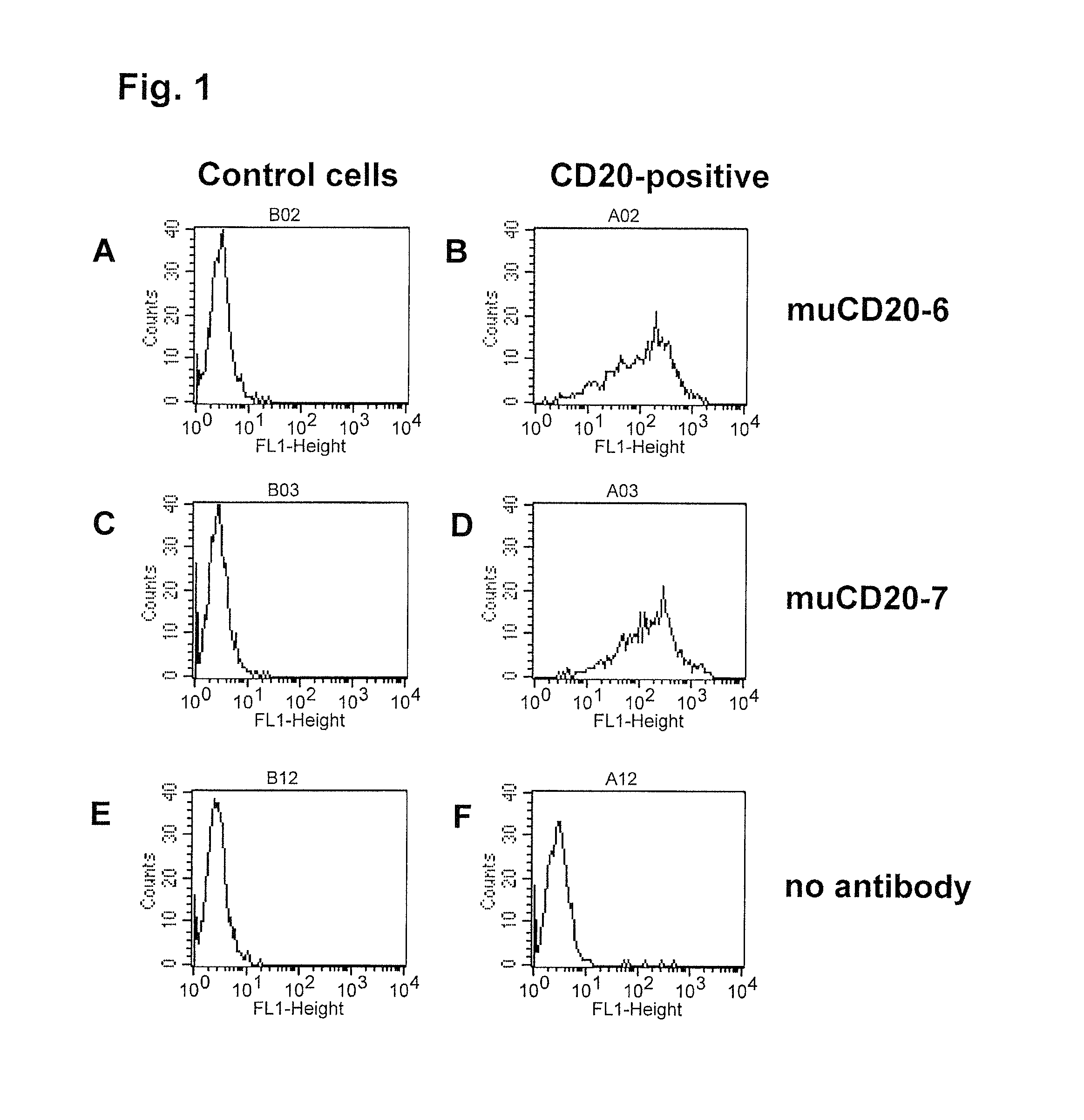
[0509]Binding specificity was tested by flow cytometry using purified antibodies. FACS histograms demonstrating the binding of muCD20-7 and muCD20-6 to CD20-expressing 300-19 cells and the absence of binding to the parental 300-19 cells are shown in FIG. 1. Either muCD20-7 or muCD20-6 antibody was incubated for 3 h with either CD20-expressing 300-19 cells or the non-transfected 300-19 cells (1×10⁵ cells per sample) in 100 μL FACS buffer (RPMI-1640 medium supplemented with 2% normal goat serum). Then, the cells were pelleted, washed, and incubated for 1 h with 100 μL of FITC-conjugated goat anti-mouse IgG-antibody (such as is obtainable from, for example Jackson Laboratory, 6 μg / mL in FACS buffer). The cells were pelleted again, washed with FACS buffer and resuspended in 200 μL of PBS containing 1% formaldehyde. Samples were acquired using a FACSCalibur flow cytometer with the HTS multiwell sampler or a FACS array flow cytometer and analyzed ...

example 3

Pro-Apoptotic Activity of Murine Anti-CD20 Antibodies

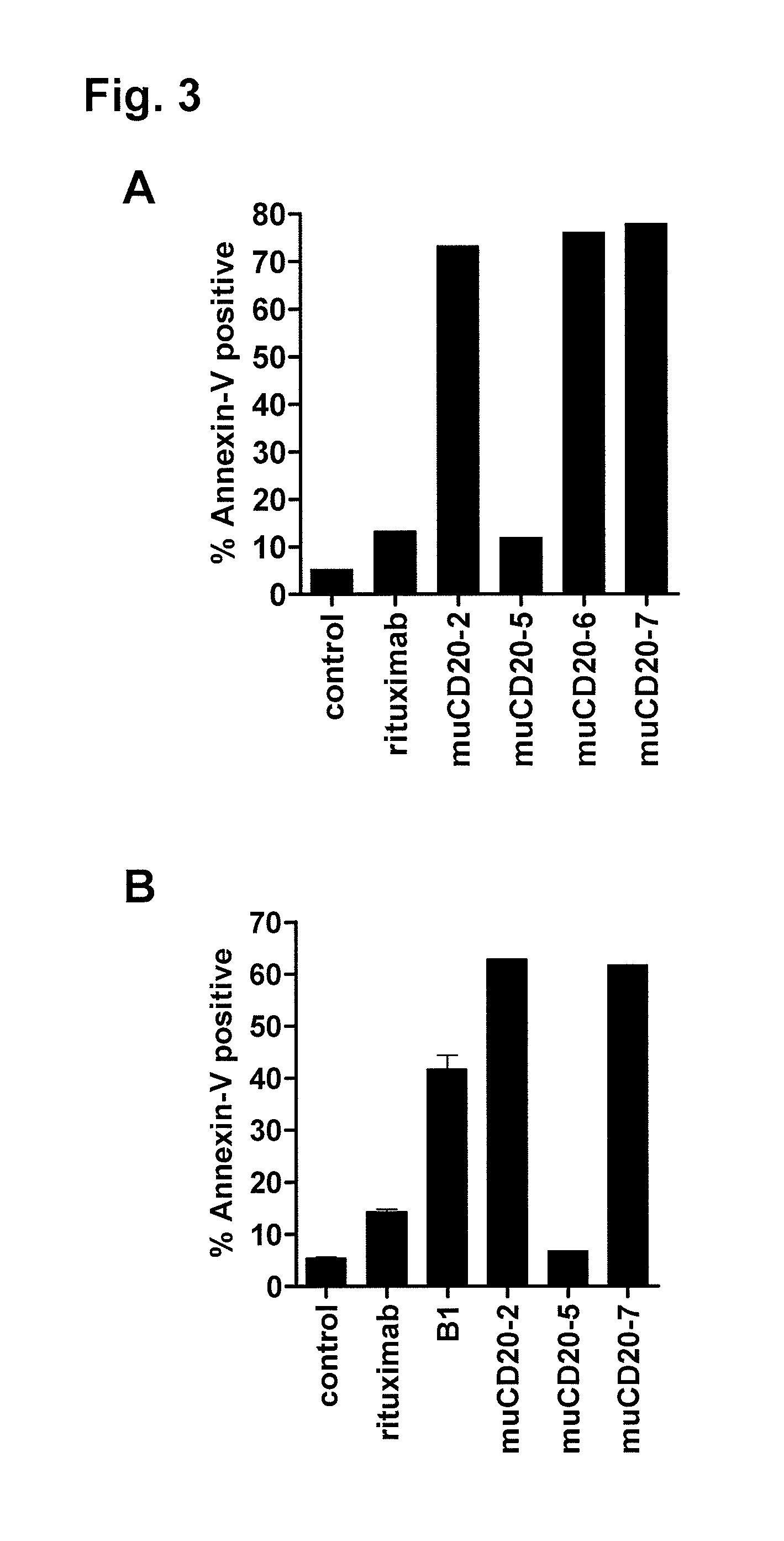
[0512]The anti-CD20 antibody muCD20-2, muCD20-6 and muCD20-7 induced apoptosis of lymphoma cell lines. The degree of apoptosis was measured by flow cytometry analysis after staining with FITC conjugates of Annexin-V (Invitrogen) and with TO-PROS-3 (Invitrogen). In healthy, normal cells, phosphatidylserine is on the inside of the membrane bilayer, and the transition of phosphatidylserine from the inner to the outer leaflet of the plasma membrane is one of the earliest detectable signals of apoptosis. Annexin V binds phosphatidylserine on the outside but not on the inside of the cell membrane bilayer of intact cells. The degree of Annexin V binding is therefore an indicator of the induction of apoptosis. TO-PROS-3 is a monomeric cyanine nucleic acid stain that can only penetrate the plasma membrane when the membrane integrity is breached, as occurs in the later stages of apoptosis. Three populations of cells are distinguishable in t...


PUM

PropertyMeasurementUnit
concentrationaaaaaaaaaa
pHaaaaaaaaaa
pHaaaaaaaaaa

Abstract

CD20 is a transmembrane protein of the tetra-spanin family expressed on the surface of B-cells from peripheral blood as well as lymphoid tissues. CD20 expression persists from the early pre-B cell stage until the plasma cell differentiation stage. In addition to expression in normal B-cells, CD20 is expressed in B-cell derived malignancies such as non-Hodgkin's lymphoma (NHL) and B-cell chronic lymphocytic leukemia (CLL). The present invention includes anti-CD20 antibodies and antigen-binding fragments thereof comprising a light chain variable region and a heavy chain variable region, wherein the CDR-L1, CDR-L2, and CDR-L3 of said light chain variable region comprise the amino acid sequences of SEQ ID NOs: 17-19, respectively, and wherein the CDR-H1, CDR-H2, and CDR-H3 of said heavy chain variable region comprise the amino acid sequences of SEQ ID NOs: 20-22, respectively.

Description

REFERENCE TO SEQUENCE LISTING SUBMITTED ELECTRONICALLY VIA EFS-WEB[0001]The content of the electronically submitted Sequence Listing (Name: SubstituteSequenceListing.ascii.txt; Size: 43,347 bytes; and Date of Creation: Dec. 3, 2012) is herein incorporated by reference in its entirety.BACKGROUND OF THE INVENTION[0002]CD20 and CD20 isoforms (“CD20”) are generally appreciated to be transmembrane proteins of the tetra-spanin family that are expressed on the surface of B-cells (Valentine et al., J. Biol. Chem., 264(19):11282-11287 (1989); Einfeld et al., EMBO J., 7(3):711-717 (1988)). CD20 is expressed by the vast majority of peripheral blood B-cells as well as B cells from various lymphoid tissues. CD20 expression generally persists from the early pre-B cell stage of development until the plasma cell differentiation stage of development (Tedder et al., J. Immunol., 135(2):973-979 (1985)). CD20 is not generally expressed by hematopoietic stem cells, pro-B cells, differentiated plasma cel...

Claims


Application Information

Patent Timeline
Patent Type & Authority Patents(United States)
IPC IPC(8): A61K39/395C07K16/28C07K16/30A61K51/10G01N33/574A61K47/48A61K39/00
CPCA61K51/1027A61K47/48438C07K16/2887G01N33/57426A61K39/39541A61K47/48446A61K47/48561A61K2039/505C07K2317/24C07K2317/30C07K2317/565C07K2317/73C07K2317/732C07K2317/734C07K2317/92C07K2319/55C12N2501/48A61K47/6817A61K47/6819A61K47/6849A61P1/00A61P1/04A61P1/16A61P11/00A61P11/06A61P13/12A61P17/00A61P17/06A61P19/02A61P21/00A61P21/04A61P25/00A61P27/02A61P29/00A61P31/18A61P31/22A61P35/00A61P35/02A61P37/00A61P37/06A61P37/08A61P43/00A61P5/14A61P7/00A61P7/02A61P7/04A61P7/06A61P9/00A61P9/10A61P3/10C07K16/30C07K2317/21C07K2317/76
Inventor DECKERT, JUTTARUI, LINGYUNPARK, PETER U.
Owner IMMUNOGEN INC
Learn More