
Tube lens with long front tube length within optical system for digital pathology

a technology of optical system and tube lens, applied in the field of digital pathology, can solve the problems of increasing the cost of both materials, increasing the cost of processing, and poor spatial resolution of human vision at blue wavelength, and achieves the effects of increasing the demand for lateral color, increasing the cost of manufacturing, and increasing the cost of processing

Active Publication Date: 2016-01-12
L A OMEGA
View PDF21 Cites 5 Cited by
  • Summary
  • Abstract
  • Description
  • Claims
  • Application Information

AI Technical Summary

Benefits of technology

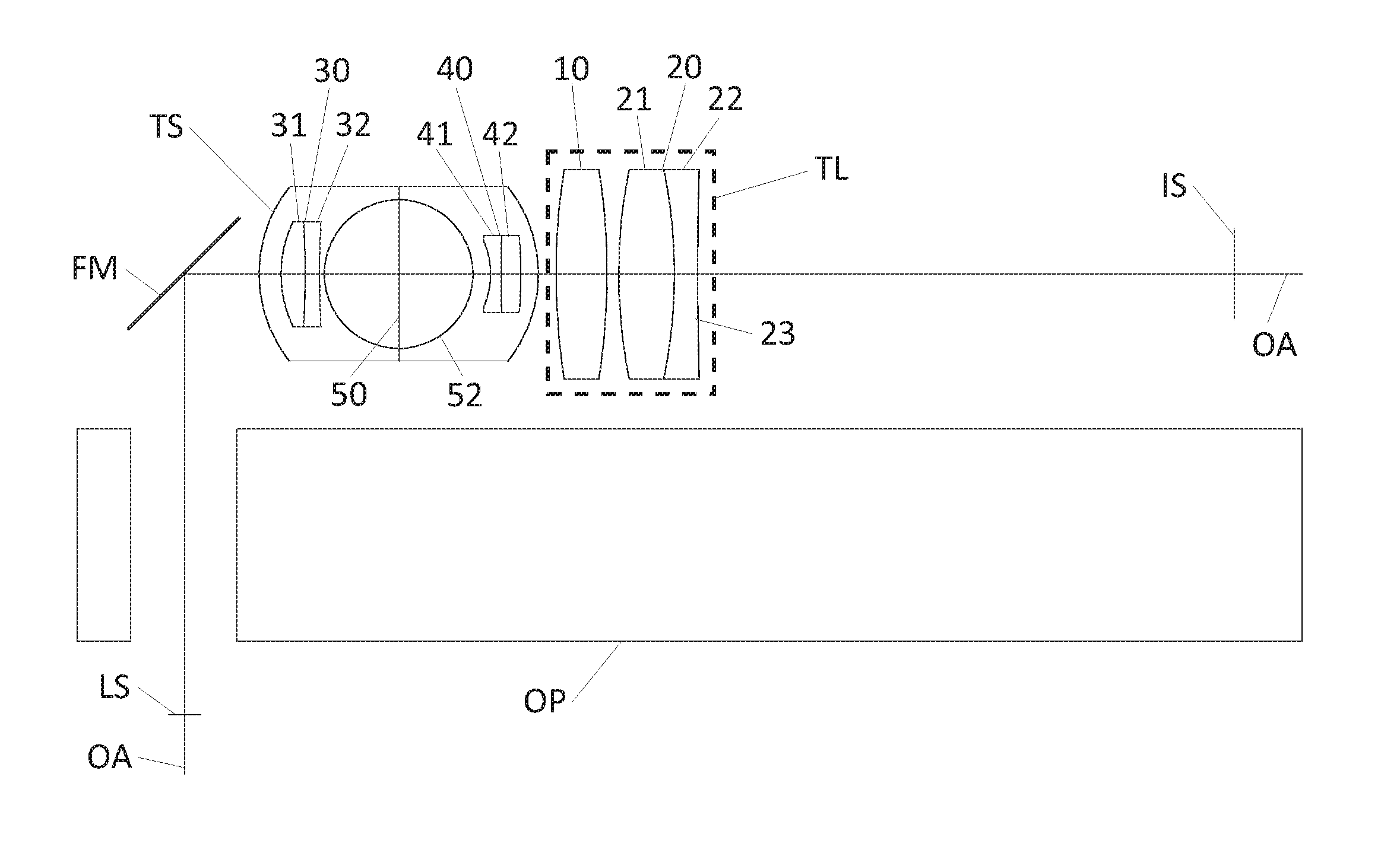
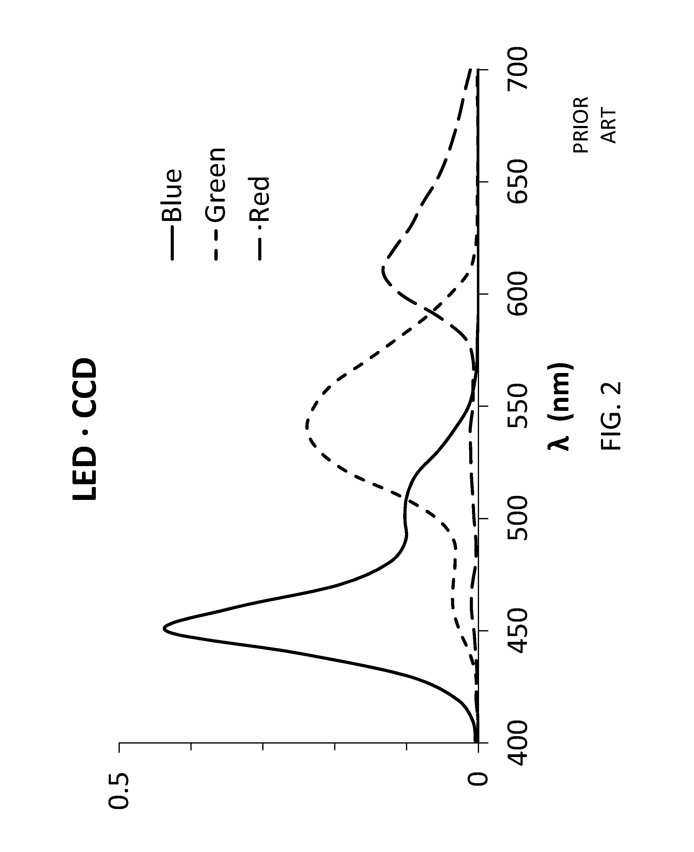
The present invention is about improving optical systems for pathology laboratories. The invention uses a folded optical path to minimize the height of the instrument. The invention also includes a tube lens with a long front tube length and color correction to achieve excellent resolution of tissue stained with heamatoxolin and eosin. The optical system incorporates a bistable telescope and a folding mirror, which provide a rapid shift between magnifications of 20× and 40× and a suitable format for a bench top. The use of novel glass types in the tube lens allows for a long front tube length with sufficient correction of lateral color for the spectra of a white LED and color CCD. The telescope provides additional magnification toward sampling the image without aliasing. Overall, the invention improves the performance of optical systems for digital pathology laboratories.

Problems solved by technology

Human vision has poor spatial resolution at blue wavelengths.
The g (436 nm) line is frequently ignored due to lack of blue sensitivity and blue resolution in human vision.
Consistent orientation of the images to the CCDs requires precise alignment of optical hardware, which is difficult and expensive.
Both materials are more expensive and more difficult to process than crown glass.
However, the large angular size of the KAI-08050 detector occupies a compromised portion of the field of tube lens in the prior art.
Thus, implementation of a larger CCD with 25 mm diagonal has been limited by the quality of the tube lens.
In particular, the lateral color of tube lens is compromised at field margin.
Consequently, numerous authors erroneously indicate “critical illumination” as a source image located at the object.
However, the lens stop of the objective lens is frequently not located at the back focal point of the objective.
It is a high quality lens, but it lacks sufficient tube length for an optical platform, a folding mirror, and a bistable telescope.
It too is a high quality lens, but it lacks sufficient front tube length for an optical platform, a folding mirror, and a bistable telescope.
However this solution can create undesirable lateral color.

Method used


Image

  • Tube lens with long front tube length within optical system for digital pathology
  • Tube lens with long front tube length within optical system for digital pathology
  • Tube lens with long front tube length within optical system for digital pathology

Examples


first embodiment

[0112]The front tube length of the first embodiment is approximately 1.3 times the effective focal length of the tube lens TL. The front tube length is deemed a long front tube length at values greater than 1.0 the effective focal length. Above 1.5 times the effective focal length, a long front tube length creates lateral color beyond the correction of this embodiment. Thus, a long front tube length is defined as 1.0 to 1.5 times the effective focal length for this embodiment.

[0113]The image field of the embodiment of FIG. 6A is 4.0°. The image field is defined by a 25 mm diagonal of the image sensor IS and an effective focal length at 180 mm. At 4.0° in field angle, long crown CAF2 is required for correction of lateral color. The Abbe number of CAF2 is 95.0. At 2.7° in field angle, the long crown N-FK5 is acceptable. The Abbe number of N-FK5 is 70.4. At 2.0° in field angle, the crown silica is not acceptable for correction of lateral color. The Abbe number of silica is 67.8. Thus, ...

third embodiment

[0122]FIGS. 6C, 6D and 6E display the maximum lateral color, the field curvature, and distortion of the tube lens TL of FIG. 6A. The maximum lateral color of the tube lens TL is 2.6 um. The field curvature is less 210 um. At 20× in lateral magnification, the longitudinal magnification at 400× transforms the 210 um of Petzval curvature at the Image sensor IS into 0.5 um at the specimen. The distortion of the third embodiment is 0.48%.

[0123]The prescription of the tube lens TL of FIG. 6A is specified in Table 2 above. The lens stop LS diameter is 9.0 mm. The effective focal length of the tube lens TL is 180 mm. The front tube length is 232.5 mm. The ratio of the front tube length to the effective focal length is 1.29. The front singlet 10 and the front positive element 21 of the back doublet 20 are made of a long crown of crystalline calcium fluoride CAF2. The back negative element 22 of the back doublet 20 is a short flint of amorphous Zr-doped tantalum silicate N-KZFS11. The high Ab...

fourth embodiment

[0146]In a fourth embodiment which is neither claimed nor illustrated, an optical system for digital pathology comprises a Kohler illumination system, an objective lens with lens stop LS, a tube lens TL and a color CCD image sensor IS. The illumination source of the Kohler illumination system is a white LED. The optical system for digital pathology requires a specific combination of glass types, an optimum NA of the objective lens, and a sufficiently large image field. The tube lens includes a long crown and a short flint for accommodation of the white LED and a color CCD sensor. The specific glass types of the objective lens include a long crown and a short flint as essential glass types of an apochromatic or semi-apochromatic objective lens. The NA of the objective lens at 0.50 to 0.55 accommodates the excess cover strata of the tissue specimen. A sufficiently large image field at 2.0° to 4.0° accommodates a typical 20× objective lens. These conditions provide the color correction...


PUM


Abstract

A tube lens includes a singlet and a doublet with a long front tube length between a lens stop and the tube lens. The front tube length is 1.0 to 1.5 times an effective focal length of the tube lens. The front tube length provides sufficient space for an optical platform, a folding mirror, and a telescope. The telescope rotates between a 2× mode with front and back doublets and a 1× mode with a clear aperture. The field angle of the tube lens is 4.0 degrees. Specific glass types correct the lateral color in a system for digital pathology with a white LED and a color CCD sensor. The system for digital pathology further includes an objective lens with a numerical aperture at 0.50-0.55, and a Kohler illumination system with a numerical aperture of critical illumination at 0.75-0.85 times the numerical aperture of the objective lens.

Description

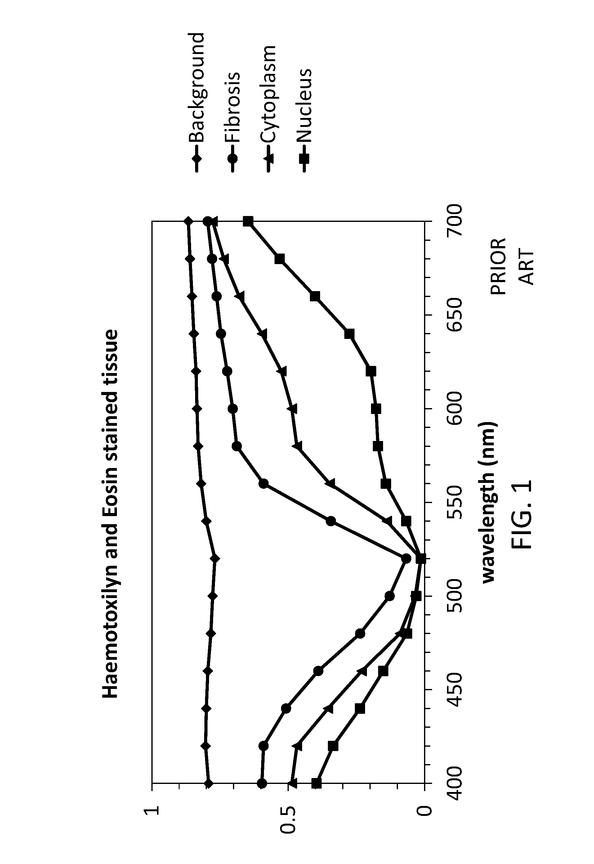
FIELD OF THE INVENTION[0001]The invention relates generally to digital pathology. Tissue stained with haemotoxilyn and eosin display prominent features in both red and blue spectra. Consequently, correction of color from red through blue is essential. Digital images must be optically correct throughout the spatial extent of the image. Thus, lateral color must be corrected throughout the entire image area. Sampling of the image by a pixel array should not create overlap of replicated Fourier spectra. Consequently, the magnification must be properly matched to the pixel size, magnification, and objective NA. The instrument should operate on a bench top. Thus, a folded optical path is beneficial.[0002]Other applications of the tube lens are numerous, including, by way of example, cytology and hematology. Moreover, the tube lens has application beyond clinical tests, as it can be applied to any color digital imaging system wherein an objective lens requires a tube lens with a long front...

Claims


Application Information

Patent Timeline
IPC IPC(8): G02B21/00G02B23/00G02B9/12
CPCG02B9/12G02B21/00G02B23/00G02B21/02G02B21/361
Inventor SEWARD, GEORGE, H.
Owner L A OMEGA
Learn More