Ultrasound based method and apparatus to determine the size of kidney stone fragments before removal via ureteroscopy

a technology of ultrasonic and ultrasonic imaging, applied in the direction of ultrasonic/sonic/infrasonic image/data processing, application, catheter, etc., can solve the problems of inability to measure the size of fragments, too large fragments to be removed,

Active Publication Date: 2016-08-16
THE UNIV OF BRITISH COLUMBIA +1
View PDF16 Cites 2 Cited by
  • Summary
  • Abstract
  • Description
  • Claims
  • Application Information

AI Technical Summary

Problems solved by technology

Urologists occasionally attempt to remove such stone fragments only to find that the fragment is too large to remove through the ureter, since endoscopic stone fragment size estimates are subjective and can be in error.
Currently, no intra-operative tools are available to measure fragment size before such removal is attempted.

Method used


Image

  • Ultrasound based method and apparatus to determine the size of kidney stone fragments before removal via ureteroscopy
  • Ultrasound based method and apparatus to determine the size of kidney stone fragments before removal via ureteroscopy
  • Ultrasound based method and apparatus to determine the size of kidney stone fragments before removal via ureteroscopy

Examples


Embodiment Construction

Figures and Disclosed Embodiments are not Limiting

[0026]Exemplary embodiments are illustrated in referenced Figures of the drawings. It is intended that the embodiments and Figures disclosed herein are to be considered illustrative rather than restrictive. No limitation on the scope of the technology and of the claims that follow is to be imputed to the examples shown in the drawings and discussed herein.

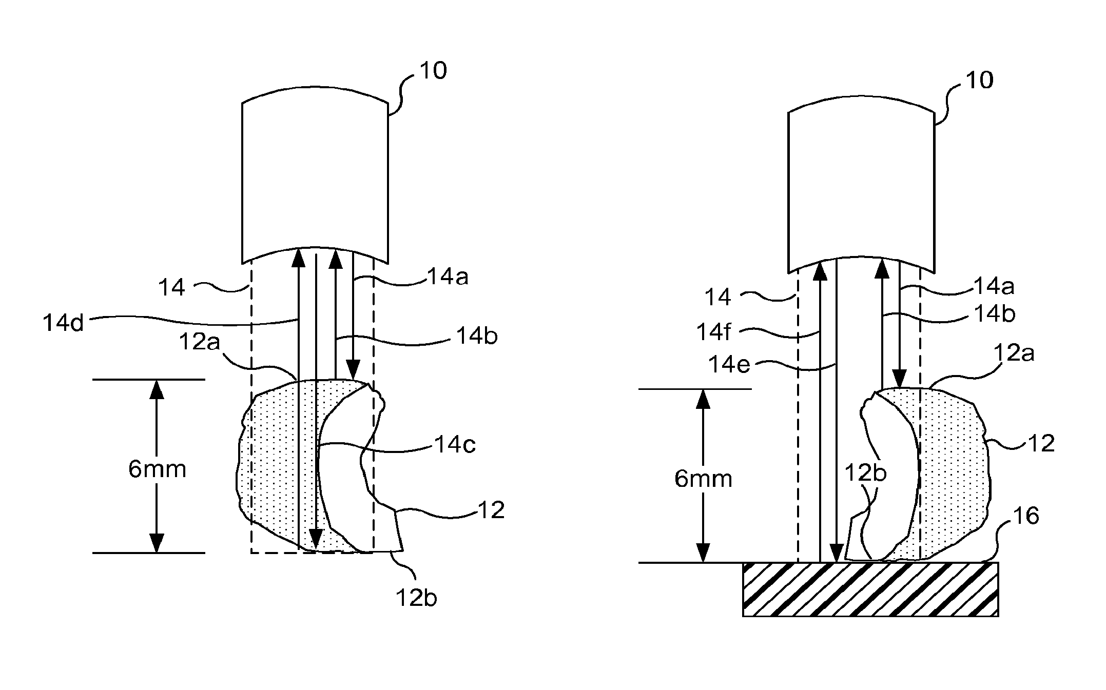
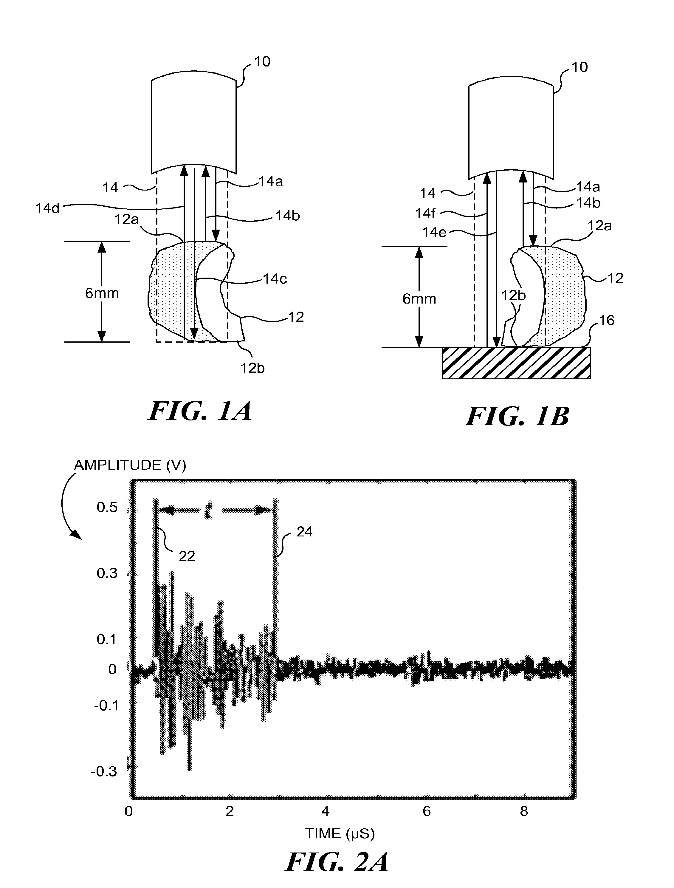
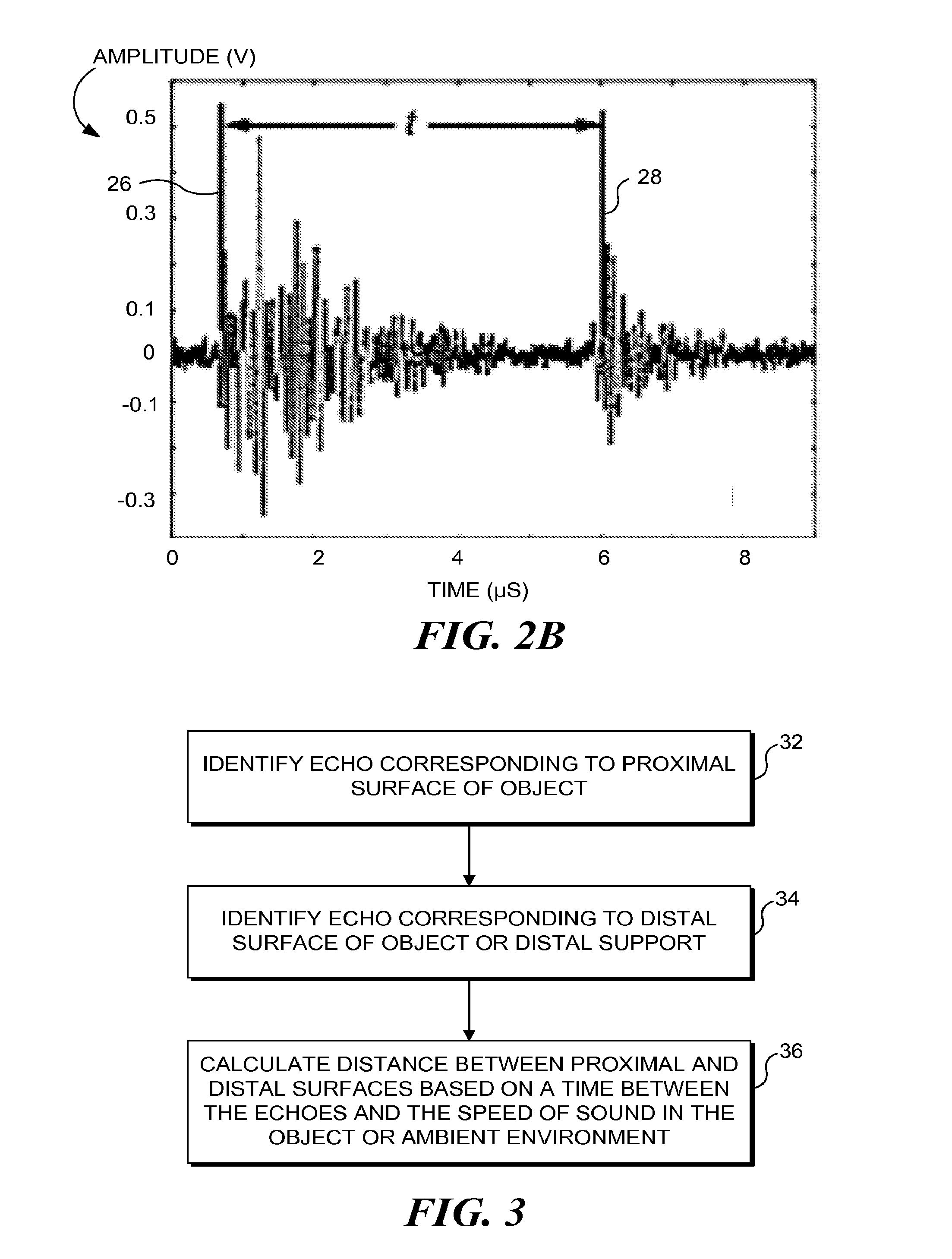
[0027]As noted above, the concepts disclosed herein employ ultrasound to enable a medical tool to estimate a size of an in vivo object. In an exemplary, but non-limiting embodiment, the object is a kidney stone or fragment thereof, and the inter-operative tool is a ureteroscope. Kidney stones or stone fragments are often removed through narrow tubes during ureteroscopy. Thus, one aspect of the concepts described herein is a device to measure stone size before attempting to remove a stone or stone fragment that is too large to fit through an available lumen. Attempting to extract a s...


PUM


Abstract

A transducer is used to send an ultrasound pulse toward a stone and to receive ultrasound reflections from the stone. The recorded time between a pulse that is reflected from the proximal surface and a pulse that is reflected either from the distal surface of the stone or from a surface supporting the stone is used to calculate the stone size. The size of the stone is a function of the time between the two pulses and the speed of sound through the stone (or through the surrounding fluid if the second pulse was reflected by the surface supporting the stone). This technique is equally applicable to measure the size of other in vivo objects, including soft tissue masses, cysts, uterine fibroids, tumors, and polyps.

Description

CROSS-REFERENCES TO RELATED APPLICATIONS[0001]The present application is a Continuation of U.S. Ser. No. 12 / 469,086 filed May 20, 2009 (Allowed), which application claims the benefit of U.S. Provisional Appln. No. 61 / 054,640 filed May 20, 2008 and is also a continuation-in-part of U.S. Ser. No. 11 / 872,668 filed Oct. 15, 2007 (now U.S. Pat. No. 8,535,250). The full disclosures, each of which are incorporated herein by reference in their entirety, for all purposes.GOVERNMENT RIGHTS[0002]This invention was made with government support under grant number DK43881 awarded by National Institutes of Health (NIH) and grant number SMS00402 awarded by National Space Biomedical Research Institute (NSBRI). The government has certain rights in the invention.BACKGROUND OF THE INVENTION[0003]Ureteroscopy often requires extraction of stone fragments by grasping them in a basket and pulling them out of the body along the ureteral tract. Urologists occasionally attempt to remove such stone fragments o...

Claims


Application Information

Patent Timeline
Patent Type & Authority Patents(United States)
IPC IPC(8): A61B8/08A61B5/107A61B17/225A61B8/12A61B17/22A61B1/307
CPCA61B5/1076A61B8/08A61B8/12A61B17/2256A61B8/5215A61B1/307A61B17/22012A61B2090/061
Inventor BAILEY, MICHAELTEICHMAN, JOELSORENSEN, MATHEW
Owner THE UNIV OF BRITISH COLUMBIA
Why Eureka
Try Eureka