
Assays for fungal infection

a technology for fungal infection and assays, applied in the field of fungal infection assays, can solve the problems of morbidity and mortality, fungi are able to cause superficial and often fatal disseminated infections in immunocompromised patients, and the impact of fungal infection is often exacerbated, so as to achieve rapid and reliable detection

Active Publication Date: 2017-05-23
RUTGERS THE STATE UNIV
View PDF12 Cites 2 Cited by
  • Summary
  • Abstract
  • Description
  • Claims
  • Application Information

AI Technical Summary

Benefits of technology

[0008]The inventors have shown that the presence or absence of any fungus in a sample can be rapidly and reliably determined using DNA analysis. This is referred to herein as panfungal detection. In particular, the inventors have shown for the first time that panfungal detection can be carried out on non-biological samples. The inventors have also identified a novel region of fungal DNA that can be used for panfungal detection.

Problems solved by technology

Fungal infections are a significant cause of morbidity and mortality in a variety of severely ill patients.
For instance, fungi are able to cause superficial and often fatal disseminated infections in immunocompromised patients.
The impact of a fungal infection is often exacerbated by a failure to rapidly diagnose and effectively treat the infection.
Numerous studies have shown that a delay in appropriate therapy is associated with increased morbidity and mortality.
At present, clinical methods of detecting the presence of a fungus in a patient are unreliable and time consuming.
For instance, the detection of fungal infections is usually carried out by blood culture, which takes up to 1 to 2 days to perform and often provides false negative results.
In addition, some fungi, such as members of the Pneumocystis genus, cannot be cultured easily.
The lack of rapid diagnostic tests for particular fungal species is one of the major impediments to successful management of infected patients.
The identification of the specific fungal species causing an infection can be even more time consuming that simply detecting the presence of a fungus and this can often further delay effective treatment of the infection.
However, there is currently a lack of rapid tests for the presence of a fungus or for particular fungal genera or species in non-biological samples.

Method used


Image

  • Assays for fungal infection
  • Assays for fungal infection
  • Assays for fungal infection

Examples


example 1

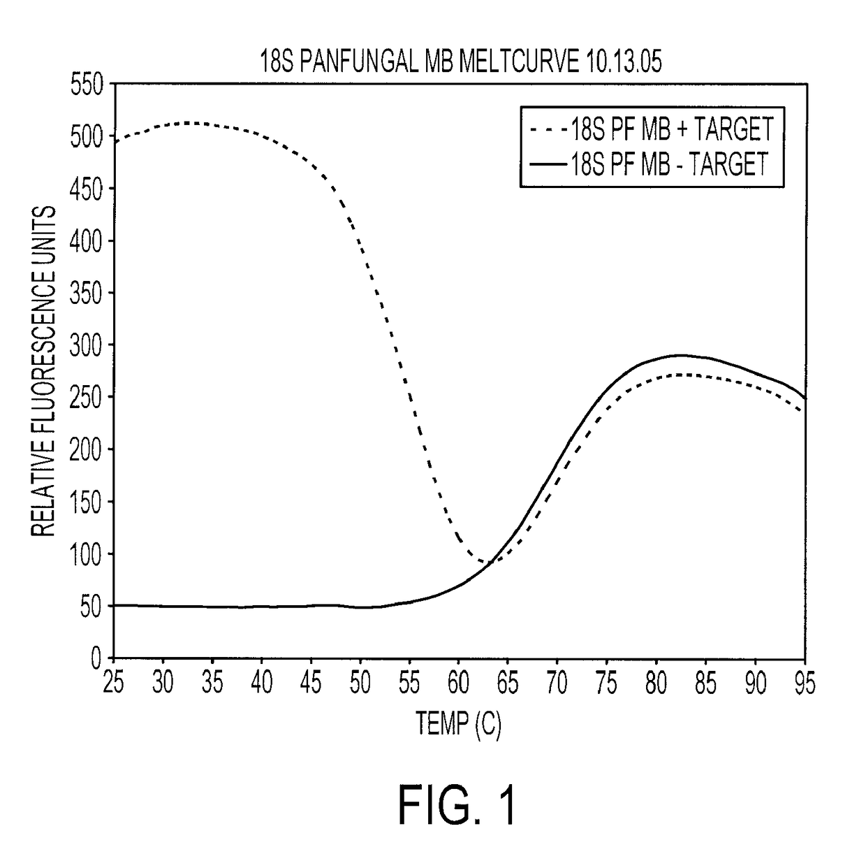
Melting Curve for Panfungal Molecular Beacon Probes

[0190]Reported sequences for the design of primers and molecular beacon probes were used. Molecular beacon probes and DNA primers (Table 1, I) were designed using Beacon Designer 3.0 software (PREMIER Biosoft, Palo Alto, Calif.). The default software parameters were applied for all molecular beacon probe and primer construction. Molecular beacon probes were labeled with fluorophores 5-carboxyfluorescein (FAM) at the 5′ end and with dabcyl at the 3′ end. The molecular beacon probes and primers were purchased from Biosearch Technologies (Biosearch Technologies, Novato, Calif.). The hybridization properties of the molecular beacon probes were tested for the full temperature range, 25° C.-95° C., with single-stranded target oligonucleotides. Molecular beacon probe-target hybridization was performed with the Stratagene MX4000 Multiplex Quantitative PCR system (Stratagene, La Jolla, Calif.). The “Molecular Beacon Melting Curve” experiment...

example 2

Detection of Any Fungus (Panfungal Detection)

[0192]A real-time amplification assay was carried out using the primers and probes described in Table I (SEQ ID NOs: 2 to 4). The assay included DNA amplification by the polymerase chain reaction (PCR) with real-time detection utilizing molecular beacon probes. DNA from multiple fungal species were tested, together with negative controls consisting of DNA extracted from a Gram positive and Gram negative bacteria. Species tested were Aspergillus flavus, Aspergillus fumigatus, Aspergillus japonicus, Aspergillus nidulans, Aspergillus niger, Aspergillus terreus, Candida albicans, Candida dubliniensis, Candida glabrata, Candida keyfr, Candida krusei, Candida lusitaniae, Candida lipolytica, Candida parapsilosis, Candida rugosa, Candida tropicalis, Cryptococcus neoformans, Saccharomyces cerevisiae, Pneumocystis carinii, Pneumocystis jirovecii, Escherichia coli and Staphylococcus aureus. With the exception of the Pneumocystis samples, which were ...

example 3

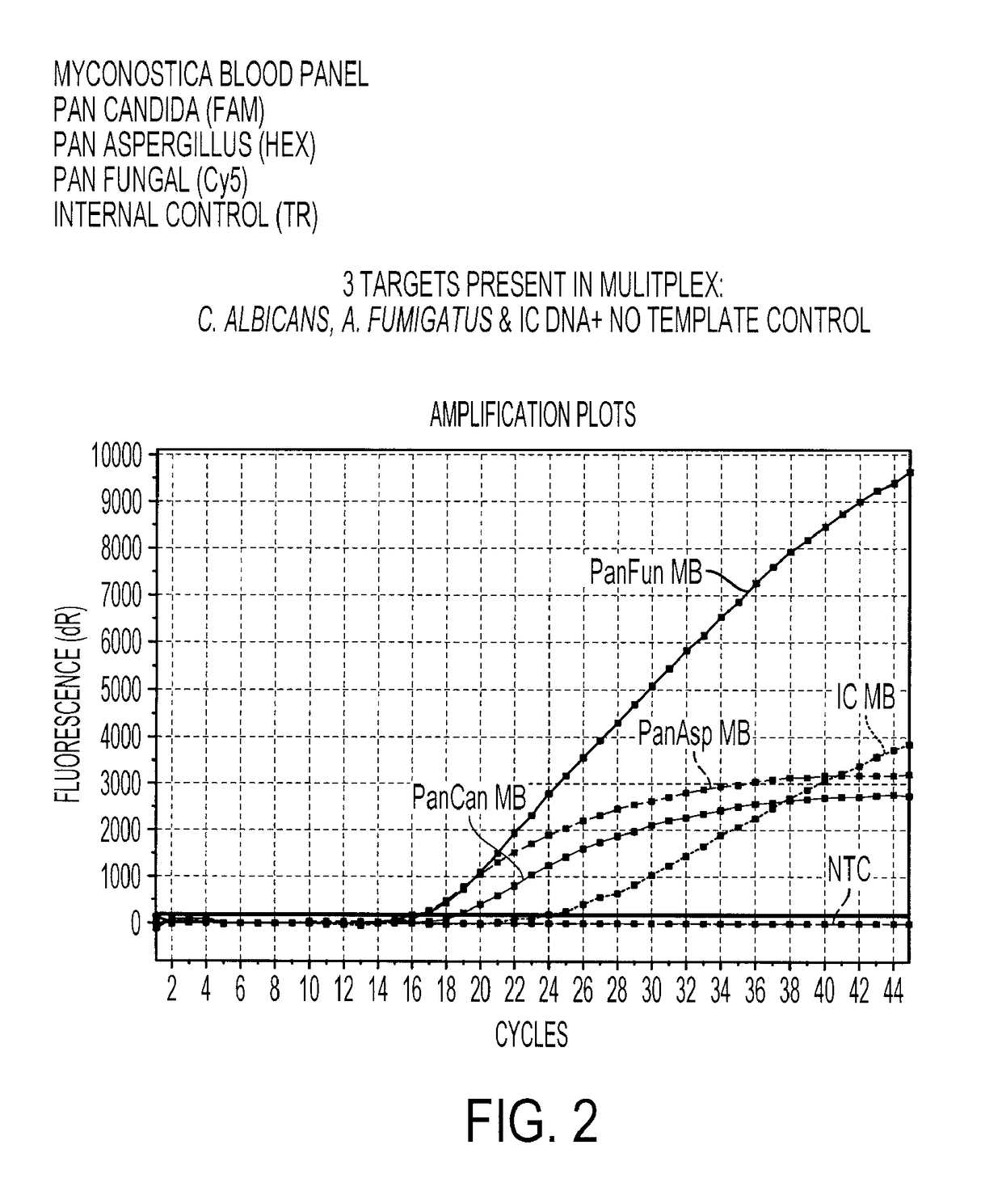
Multiplex Detection of Aspergillus and Candida DNA

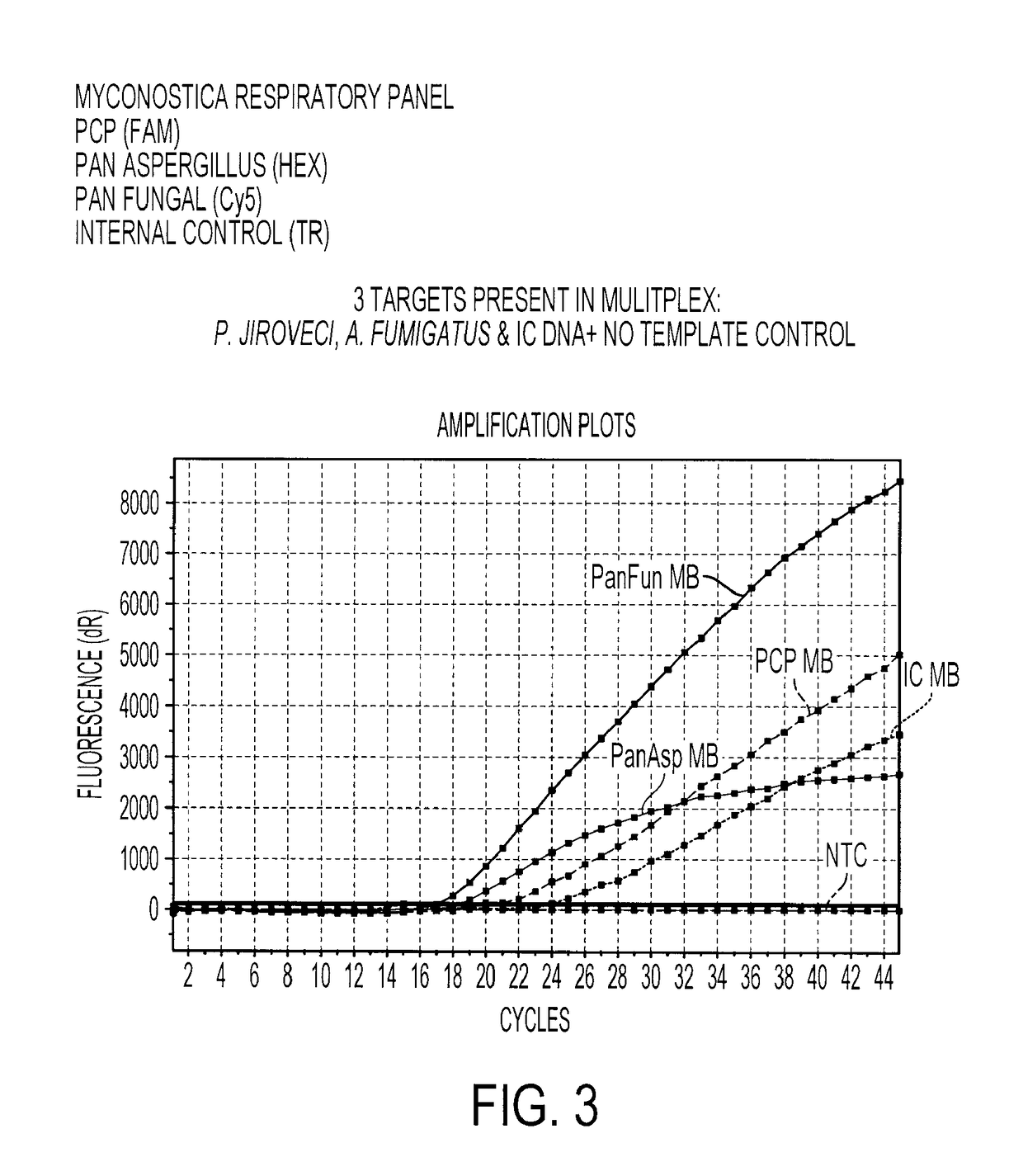
[0196]A real-time amplification assay was carried out for the detection of Aspergillus, Candida and panfungal (presence of any fungal) DNA using the primers and probes described in Table 1, I (SEQ ID NOs: 2 to 4), II (SEQ ID NOs: 6 to 7) and IV (SEQ ID NOs: 15 to 17), together with an assay for the presence of the Internal Control DNA using the primers and probes described in Table 1, III (SEQ ID NOs: 11 to 13). Fluorophores were conjugated to the beacon probes to allow detection. The fluorophores used are shown in Table 3.

[0197]

TABLE 3The fluorophores used to label each molecular beacon probeMolecular BeaconProbeFluorophoreAspergillusHEX (hexachlorofluorescein phosphoramidite)CandidaFAM (carboxyfluorescein)PanfungalCy ®5 (a registered trade mark of GELifesciences. FIG. 11 shows the structure of Cy5mono NHS ester)Internal ControlTexas Red ® (registered trade mark ofMolecular Probes, Inc. FIG. 12 shows the structureof Texas Red sulpho...


PUM

PropertyMeasurementUnit
temperatureaaaaaaaaaa

Abstract

Methods and kits are described for testing for the presence or absence of any fungus in a sample. Examples of fungi that can be detected include, but are not limited to, those belonging to the genera Candida, Aspergillus and Pneumocystis. The methods include obtaining a sample suspected of containing fungal nucleic acid, including at least one universal region of fungal nucleic acid, and testing for the presence or absence in the sample of the at least one universal region of fungal nucleic acid. Samples may be biological or non-biological.

Description

INFORMATION ON RELATED APPLICATIONS[0001]This application claims priority to U.S. Provisional Application No. 60 / 968,413, filed Aug. 28, 2007, and to U.S. Provisional Application No. 60 / 864,146, filed Nov. 2, 2006, and to UK Patent Application No. 0716687.9, filed Aug. 28, 2007, and to UK Patent Application No. 0621864.8, filed Nov. 2, 2006, all of which are hereby incorporated by reference in their entireties.FIELD OF THE INVENTION[0002]The invention relates to detecting fungi in a sample. In particular, the invention relates to the rapid detection of any fungus in a sample and the rapid detection of a particular genus or species of fungus in a sample. The invention also generally relates to fungal identification. Methods and kits are also provided for testing for the presence or absence of a fungus in a sample. Examples of fungi detected include, but are not limited to, those belonging to the genera Candida, Aspergillus and Pneumocystis. BACKGROUND OF THE INVENTION[0003]Fungal inf...

Claims


Application Information

Patent Timeline
Patent Type & Authority Patents(United States)
IPC IPC(8): C12Q1/68C07H21/02
CPCC12Q1/6895C12Q2600/16C12Q2600/166
Inventor PARK, STEVENPERLIN, DAVID S.DENNING, DAVID WEMYSS
Owner RUTGERS THE STATE UNIV
Learn More