Device with simultaneous X-ray and infrared image acquisition and processing system for enhanced breast imaging

a breast imaging and processing system technology, applied in the field of medical diagnostics, can solve the problems of large percentage of patients not attending follow-up procedures, affecting the quality of breast imaging, and most of the women experiencing discomfort during the procedure, so as to reduce the number of false positive results and increase the screening

Active Publication Date: 2017-06-27
TUSCANO EQUIP
View PDF10 Cites 2 Cited by
  • Summary
  • Abstract
  • Description
  • Claims
  • Application Information

AI Technical Summary

Benefits of technology

[0038]According to an embodiment herein, the patient support table is configured to support the patient and exposes only the breast for screening and without making any discomfort to the patient.
[0046]Moreover it increases a screening of a follow up process as there is no pain or compression or any discomfort to the patients during the scan / procedure.

Problems solved by technology

Though the X-Ray based mammography is considered as a gold standard tool available today for breast cancer, most of the women feel discomfort during the procedure.
Each breast is compressed with the help of a compression paddle and this causes an acute discomfort to the patient since breast is considered two to three times more sensitive in women.
Nearly 5%-15% of mammograms require a follow-up testing but a sizeable percentage of patients do not turn up for the follow-up procedures because of the discomfort caused by compression.
Besides a discomfort, another drawback of the mammography is the generation of false negative and false positive results due to a compression of three dimensional breast organ images to a two dimensional film or image, thereby leading to different interpretations.
Ultrasound mammography also has unique advantages but it lacks repeatability & reproducibility as the compression pressure applied by the sonologist during imaging is not tracked and hence it is difficult to reproduce the results.
Secondly it is difficult to position a suspected spot, which is identified in the initial study, in a follow-up procedure.
As a result, the MRI studies may provide more false positives (up to 30%), which may lead to undesirable financial and psychological costs.
The MRI based study requires very expensive MRI scanners and also a coil which is specific for positioning a breast.
Further, an MRI may not be used for screening the patients with a pacemaker or breast reconstruction patients with a tissue expander due to the presence of metal.
As a result very few procedures are performed with the MRI scanners.
A fundamental flaw in such an approach is that the rest of the body also dissipates heat and there is a possibility that this can alter or influence the environment regarding a heat pattern.
Few Thermography techniques do capture the heat patterns of the breast in a patient lying in a prone position but do not capture all the views of the breast in a direct manner.
Each year, millions of women around the world are subjected to the unnecessary breast biopsies because of inadequacies of cancer detection and inability to separate a benign from the cancerous lesions.

Method used


Image

  • Device with simultaneous X-ray and infrared image acquisition and processing system for enhanced breast imaging
  • Device with simultaneous X-ray and infrared image acquisition and processing system for enhanced breast imaging
  • Device with simultaneous X-ray and infrared image acquisition and processing system for enhanced breast imaging

Examples


Embodiment Construction

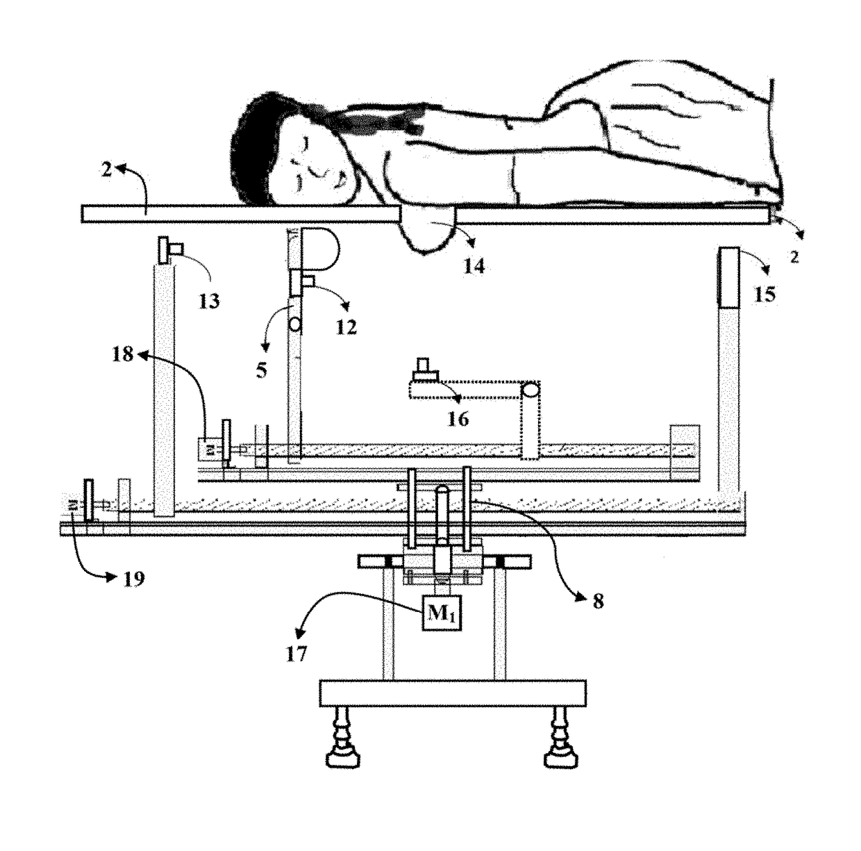
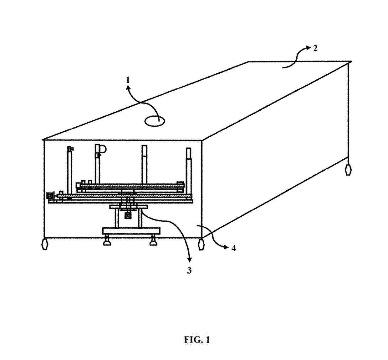
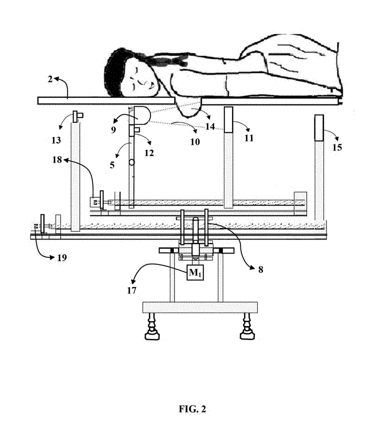
[0016]The primary object of the embodiments herein is to provide an imaging device with a simultaneous digital X-Ray and Infrared red image acquisition and processing systems in conjunction with a positioning apparatus for a minimal radiation.

[0017]Another object of the embodiments herein is to develop an imaging device to provide both the anatomical and physiological views of a breast simultaneously for an early detection of the abnormalities.

[0018]Still another object of the embodiments herein is to develop an imaging device so that each modality is used independently and / or in a concurrent fashion as needed or desired to provide the images of a suspected area.

[0019]Yet another object of the embodiments herein is to develop an imaging device to allow a capturing of time based thermal images of multiple views, including but not limited to cranial, medial, caudal, lateral and frontal views of a breast.

[0020]Yet another object of the embodiments herein is to develop an imaging device...


PUM


Abstract

The embodiments herein discloses a device with simultaneous X-ray and infrared acquisition and processing systems for an enhance breast imaging. The device has a positioning assembly housed inside a closed chamber and provided with the Infrared and X-ray imaging systems to simultaneously capture an infra red image and an X-ray image of the breast under examination. A patient support table with an opening is provided to enable a patient to lie in a prone position without compressing a breast during imaging. The device is used to correlate anatomical and physiological characteristics and post process analysis of a breast tissue thereby reducing a number of to false positive results. Further the device helps in focusing on a suspected area in a follow-up procedure and aids post processing treatment like targeted biopsy and targeted radiation.

Description

CROSS-REFERENCE TO RELATED APPLICATIONS[0001]The present application is a national phase application to the PCT Application entitled, “A Device with Simultaneous X-Ray and Infrared Image Acquisition and Processing System for Enhanced Breast Imaging” with serial number PCT / IN 2012 / 000778, filed at Government of India Patent Office on Nov. 29, 2012, the content of which is incorporated by reference herein.[0002]The present application claims the priority of the Indian Patent application No. 4165 / CHE / 2011 filed on Dec. 1, 2011 with the title, “A Device with Simultaneous X-Ray and Infrared Image Acquisition and Processing System for Enhanced Breast Imaging” and the contents of which are incorporated in entirety by reference herein.BACKGROUND[0003]Technical Field[0004]The embodiments herein generally relate to the field of imaging devices for medical diagnostics and particularly relates to the imaging devices used for detecting a breast cancer. The embodiments herein more particularly re...

Claims


Application Information

Patent Timeline
Patent Type & Authority Patents(United States)
IPC IPC(8): A61B6/00A61B6/04A61B5/00A61B6/03A61B10/02A61B6/12
CPCA61B5/0035A61B5/0091A61B5/708A61B6/032A61B6/0435A61B6/12A61B6/4417A61B6/502A61B6/5247A61B10/02
Inventor NEELAKANTA, KANNANANAND, JAYANTHICHANDRASEKARAN, SUDHANCHERUKANDATH, RAJENDRANVASAN, JAYARAMAN KIRUTHI
Owner TUSCANO EQUIP
Why Eureka
Try Eureka