Treatment of corneal neovascularization

a corneal neovascularization and treatment technology, applied in the direction of sense disorder, pharmaceutical active ingredients, medical preparations, etc., can solve the problems of widespread adverse effects, vision loss, and loss of ocular tissue, and achieve the effect of preventing cnv and treating i

Inactive Publication Date: 2017-10-10
IRBM SCI PARK
View PDF9 Cites 2 Cited by
  • Summary
  • Abstract
  • Description
  • Claims
  • Application Information

AI Technical Summary

Problems solved by technology

As well as having adverse impact on the transparency of the tissue due to the existence of the blood vessels themselves, the infiltrative growth of new blood vessels can disrupt or destroy ocular tissue resulting in widespread adverse effects.
Thus, CNV can lead to corneal scarring, edema (swelling), lipid deposits, and inflammation that may result in vision loss.
In addition, it can lead to loss of immune privilege of the eye which can affect the outcome of corneal transplantation, and worsens the prognosis of penetrating keratoplasty.
Complications also include increased inflammation which is obviously undesirable.
Activation of this compound with low energy laser light generates cytotoxic mediators, which cause selective thrombosis and destruction of newly formed vessels.
Corticosteroids have been a standard treatment for CNV. with variable and limited success.

Method used


Image

  • Treatment of corneal neovascularization
  • Treatment of corneal neovascularization
  • Treatment of corneal neovascularization

Examples


example 1

Inhibition of Corneal Neovascularisation in a Pellet Angiogenesis Model

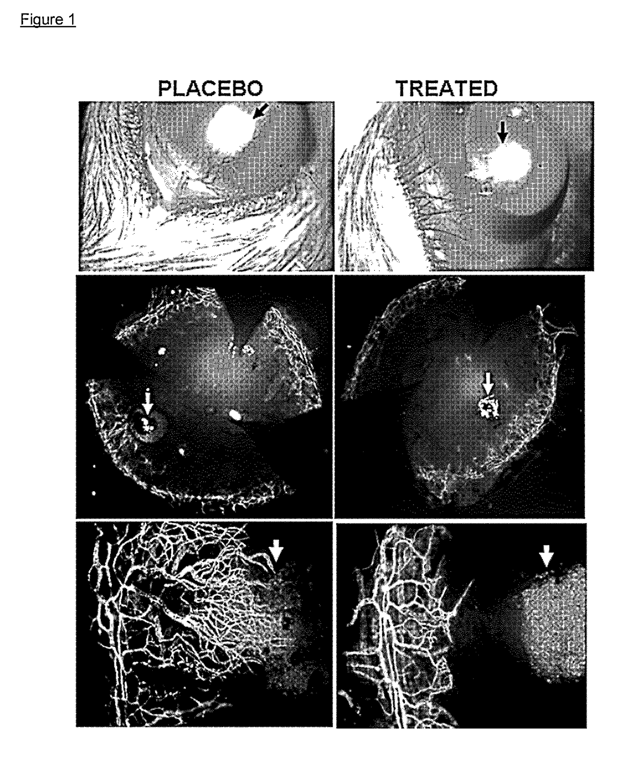
[0592]Male 6- to 8-week-old C57Bl / 6 mice were treated to induce neovascularization using Fibroblast Growth Factor (FGF) 80 ng pellet implantation, a non-inflammatory model of corneal neovascularization (see Experimental Methods).

[0593]Immediately after pellet implantation, on Day 0, each group received one of the following topical treatments: (1) fosaprepitant (purchased from Universal Drug Store, Canada and diluted as per technical sheet instructions with 0.9% NaCl solution to reach a final concentration of 1 mg / ml) at the concentration of 1 mg / ml (one drop, 50 μl, per instillation, for a total amount of 0.05 mg of active principle per instillation) in the conjunctival sac 4 times per day for 1 week; or (2) phosphate buffered saline PBS as placebo. 5 mice per group were used. After taking photographs at the slit lamp, mice were sacrificed. Immunohistochemical staining was performed with FITC-conjugated CD31 / PECA...

example 2

Inhibition of Corneal Neovascularization in a Caustication Neovascularization Model

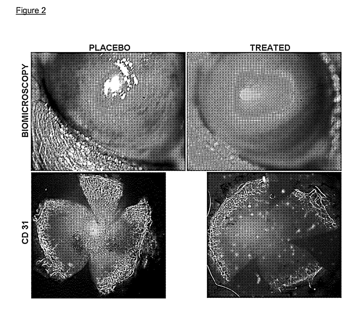
[0596]Male 6- to 8-week-old C57Bl / 6 mice were treated to induce neovascularization using sodium hydroxide caustication (see Experimental Methods)

[0597]Immediately after sodium hydroxide caustication, on Day 0, each group received one of the following topical treatments: (1) fosaprepitant (purchased from Universal Drug Store, Canada and diluted as per technical sheet instructions with 0.9% NaCl solution to reach a final concentration of 1 mg / ml) at the concentration of 1 mg / ml (one drop, 50 μl, per instillation, for a total amount of 0.05 mg of active principle per instillation) in the conjunctival sac 4 times per day for 1 week; or (2) phosphate buffered saline PBS as placebo. 5 mice per group were used. After taking photographs at the slit lamp, mice were sacrificed. Immunohistochemical staining was performed with FITC-conjugated CD31 / PECAM-1 to reveal corneal neovessels.

[0598]Neovascularization was ...

example 3

Toxicity of Fosaprepitant Following Topical Application in Healthy Eyes

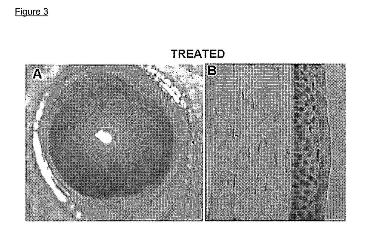
[0600]Fosaprepitant toxicity was tested on the healthy cornea by administering to 3 healthy C57Bl / 6 mice fosaprepitant (purchased from Universal Drug Store, Canada and diluted as per technical sheet instructions with 0.9% NaCl solution to reach a final concentration of 1 mg / ml) at the concentration of 1 mg / ml in the conjunctival sac 4 times per day for 1 week. In vivo corneal biomicroscopy imaging and hematoxylin eosin ocular cross sections were checked as markers for toxicity.

[0601]No alteration of the corneal epithelium, stroma, or endothelium could be found at the slit lamp observation of healthy corneas upon topical fosaprepitant administration for up to 7 days (FIG. 3A). Hematoxylin-eosin staining of eyeball cross-sections appeared normal 7 days after daily administration of fosaprepitant, no anatomical alteration was detected in the epithelium, stroma, or endothelium (FIG. 3B).

[0602]FIG. 3A: slit-lamp exami...


PUM

PropertyMeasurementUnit
pHaaaaaaaaaa
pHaaaaaaaaaa
pHaaaaaaaaaa

Abstract

There is provided inter alia a compound which is an NK-1 receptor antagonist for use in the treatment or prevention of CNV. There is also provided a compound which is an NK-1 antagonist for use in the treatment of chemical burns of the eye particularly alkali burns of the eye. There is also provided a pharmaceutical composition for topical administration to the eye comprising an NK-1 antagonist and an antibiotic agent.

Description

RELATED APPLICATIONS[0001]This Application is a National Stage Application of International Application No. PCT / EP2012 / 063067, filed 4 Jul. 2012; which claims the benefit of Provisional Application Ser. No. US61 / 504,265, filed 4 Jul. 2011; all of which are incorporated herein by reference in their entireties.BACKGROUND TO THE INVENTIONCorneal Neovascularization[0002]The cornea is a transparent avascular tissue of the eye, whose transparency is essential for clarity of vision. Corneal neovascularization (CNV) is a pathology characterized by the excessive growth of blood vessels from the limbus into adjacent corneal tissues in which the new blood vessels can extend into superficial and deep corneal stroma. As well as having adverse impact on the transparency of the tissue due to the existence of the blood vessels themselves, the infiltrative growth of new blood vessels can disrupt or destroy ocular tissue resulting in widespread adverse effects.[0003]Thus, CNV is characterized by angi...

Claims


Application Information

Patent Timeline
Patent Type & Authority Patents(United States)
IPC IPC(8): A61K31/5377A61K31/4545A61K45/06A61K31/453
CPCA61K31/4545A61K31/453A61K31/5377A61K45/06A61K2300/00A61P27/02
Inventor FERRARI, GIULIO
Owner IRBM SCI PARK
Why Eureka
Try Eureka