Method of detecting micrometastasis

A technology for micro-transfer and production testing, applied in biochemical equipment and methods, microbial determination/inspection, organic chemistry, etc., can solve the problems of delayed test results and difficult mRNA processing

Inactive Publication Date: 2007-09-05
TOSOH CORP
View PDF11 Cites 0 Cited by
  • Summary
  • Abstract
  • Description
  • Claims
  • Application Information

AI Technical Summary

Problems solved by technology

In this case mRNAs with different conditions for optimal amplification and detection are practically difficult ...

Method used


Image

  • Method of detecting micrometastasis
  • Method of detecting micrometastasis
  • Method of detecting micrometastasis

Examples


Embodiment 1

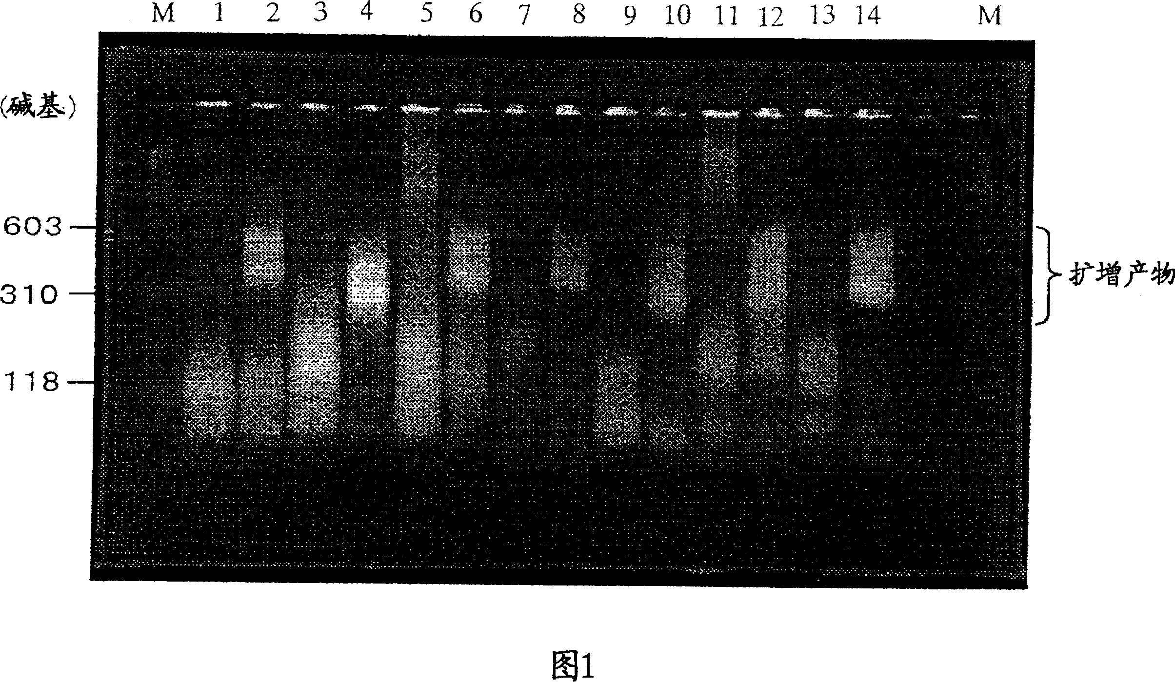
[0041] Carcinoembryonic antigen (CEA) RNA is specifically amplified by using the combination of oligonucleotide primers of the present invention. CEA RNA was prepared and isolated by in vitro transcription of double-stranded DNA containing the nucleotide sequence of the CEA gene (National Center for Biotechnology Information accession number: M29540). In this embodiment, specific sequences refer to the transcripts listed in Table 1 below.

[0042] (1) CEA RNA (2109-mer) as a sample was quantified by measuring the UV absorbance at 260nm, and the RNA diluent (10mM Tris-HCl (pH 8.0), 0.1mM EDTA, 0.5U / μl RNase inhibitor, 5.0mM DTT) diluted to 1.0×10 4 copies / 5 μl. Dilutions were used as controls (negative).

[0043] (2) Add 20.8 μl of reaction solution composed of the following components into a 0.5 ml PCR tube (GeneAmp thin-walled reaction tube, Perkin Elmer), and add 5.0 μl of the above-mentioned CEA RNA sample.

[0044] Composition of reaction solution (final concentration ...

Embodiment 2

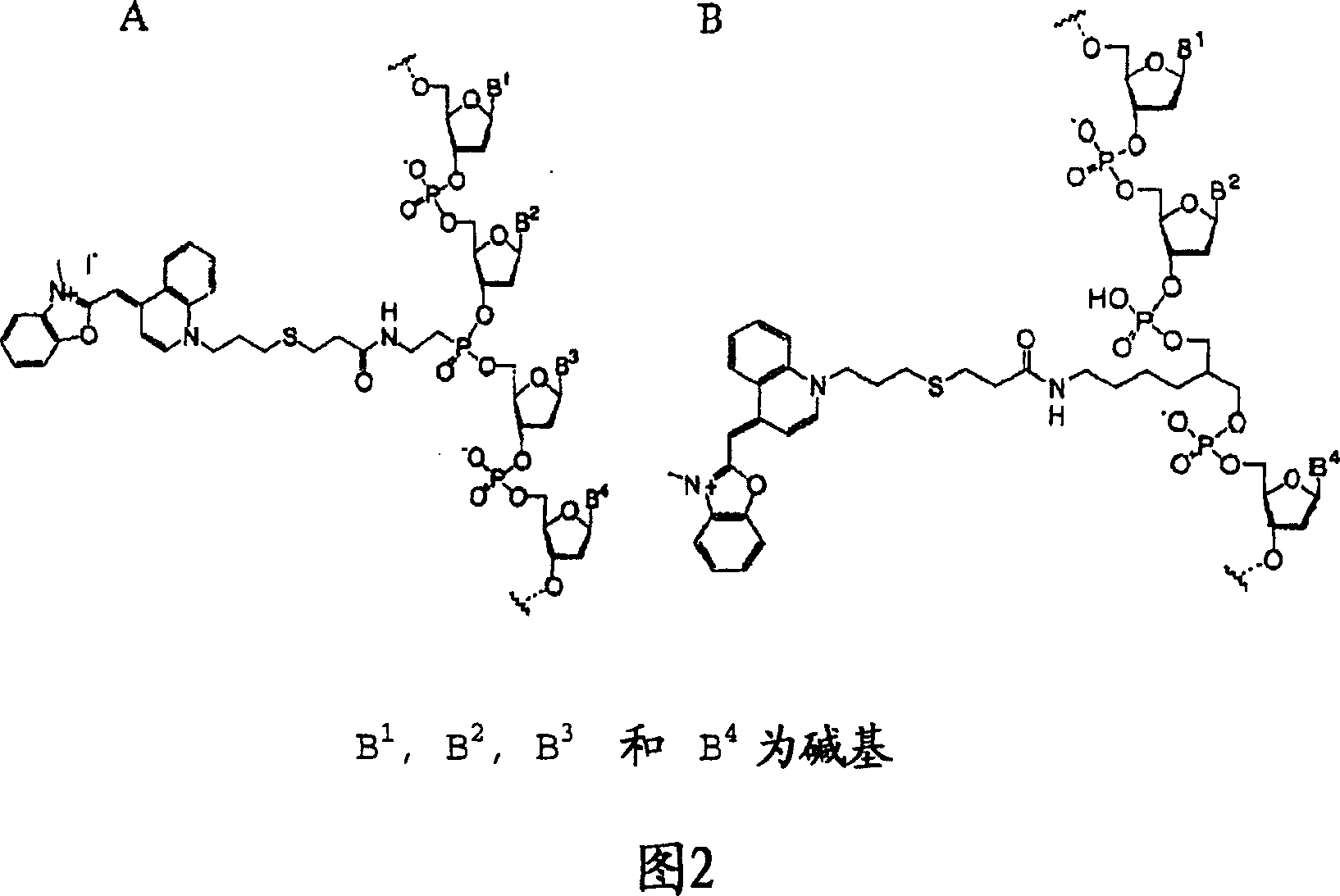
[0071] Preparation of fluorescent intercalating dye-labeled oligonucleotide probes

[0072] The 20-base oligonucleotide having the sequence shown in SEQ ID NO: 19 is modified by C 15 The linker uses Label-ON reagent (Clontech) to connect the amino group between the 5th nucleotide (G) and the 6th nucleotide (A) at its 5' end, and its 3' end is further modified with biotin . Oxazole yellow (oxazole yellow) is a commonly used fluorescent intercalating dye, which can be used as a marker to prepare oxazole yellow-labeled oligonucleotide probes (SEQ ID NO: 19) (Fig. 2B, Ishiguro T (1996) Nucleic Acids Res. 24(24) 4992-4997).

Embodiment 3

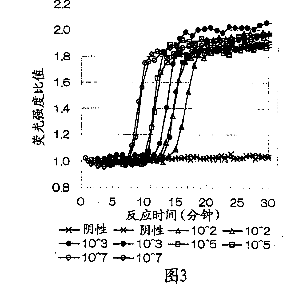
[0074] The combination of oligonucleotide primers in the present invention is used to detect CEA RNA with different initial copy numbers.

[0075] Sample CEA RNA (2109-mer) was quantified by measuring UV absorbance at 260nm, using RNA diluent (10mM Tris-HCl (pH 8.0), 0.1mM EDTA, 0.5U / μl RNase inhibitor, 5.0mM DTT) to Dilute it to 1.0 x 10 7 -10 2 Copies / 5 μl, the dilution was used as a control (negative).

[0076] 20.0 μl of the reaction solution with the following components was added to a 0.5 ml PCR tube (Gene Amp thin-walled reaction tube, Perkin Elmer), and 5.0 μl of the above-mentioned CEA RNA sample was added.

[0077] Composition of the reaction solution (final concentration in the reaction solution after adding the enzyme solution)

[0078] 60.0mM Tris-HCl buffer (pH 8.6)

[0079] 17.0mM magnesium chloride

[0080] 100.0mM potassium chloride

[0081] 1.0mM DTT

[0082] 0.25mM dATP, dCTP, dGTP, dTTP

[0083] 3.0mM ATP, CTP, UTP

[0084] 2.25mM GTP

[0085] 3.6...


PUM


Abstract

A method of detecting micrometastasis of tumor cells in a sample obtained from a region of a subject other than the primary focus of the tumor by using a first primer complementary to part of a specific sequence in the RNA from tumor cells and a second primer homologous to part of the specific sequence (either of which additionally has a promoter sequence for an RNA polymerase at the 5' end), which comprises (1) synthesizing a cDNA by the action of an enzyme having RNA-dependent DNA polymerase activity by using the specific sequence as a template, (2) degrading the RNA strand in the RNA-DNA double strand by an enzyme having a ribonuclease H activity (to give a single-stranded DNA), (3) forming a double-stranded DNA having a promoter sequence which can be transcribed into an RNA homologous or complementary to the specific sequence by using the single-stranded DNA as a template by the action of an enzyme having DNA-dependent DNA polymerase activity, and (4) transcribing the double-stranded DNA into an RNA transcript (which acts as a template in the subsequent cDNA synthesis in the reaction (1)) by the action of an enzyme having RNA polymerase activity and detecting the mRNA.

Description

technical field [0001] The present invention relates to a method for detecting tumor cell micrometastasis, polynucleotides (for detecting carcinoembryonic antigens) suitable for use as primers in the method, and related polynucleotides containing these polynucleotides for tumor cell micrometastasis Detection kits. The method for detecting tumor cell micrometastasis of the present invention is a rapid detection method that enables one-step isothermal amplification and detection of tumor cell RNA, for example, can be performed before surgical removal of tumor cells. Background technique [0002] Cancer cells spread from the primary tumor site through the blood or lymph to develop distant micrometastases. Because successful resection of the primary tumor due to the presence of micrometastases often does not prevent tumor recurrence, surgical removal of the primary tumor and all surrounding lymph nodes is common (lymph node dissection). Although lymphadenectomy can prevent rec...

Claims


Application Information

Patent Timeline
IPC IPC(8): C12Q1/68C07H21/04
CPCC12Q1/6865C12Q2521/101
Inventor 大仲悟林俊典
Owner TOSOH CORP
Why Eureka
Try Eureka