Ultrasound imaging

A technology of ultrasound and noise, applied in the direction of acoustic wave diagnosis, ultrasonic/sonic wave/infrasonic wave diagnosis, infrasonic wave diagnosis, etc. Linear scattering signal, improve the effect of CTR

Active Publication Date: 2007-08-22
比约恩・A・J・安杰尔森 +2
View PDF6 Cites 47 Cited by
  • Summary
  • Abstract
  • Description
  • Claims
  • Application Information

AI Technical Summary

Problems solved by technology

[0016] However, although the last two patents use nonlinear scattering with dual-band pulses to detect contrast agents in tissue, existing methods have limited range and they cannot discriminate the difference be

Method used

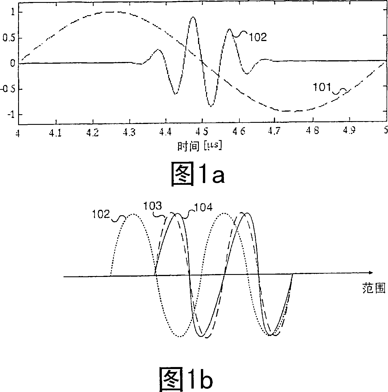
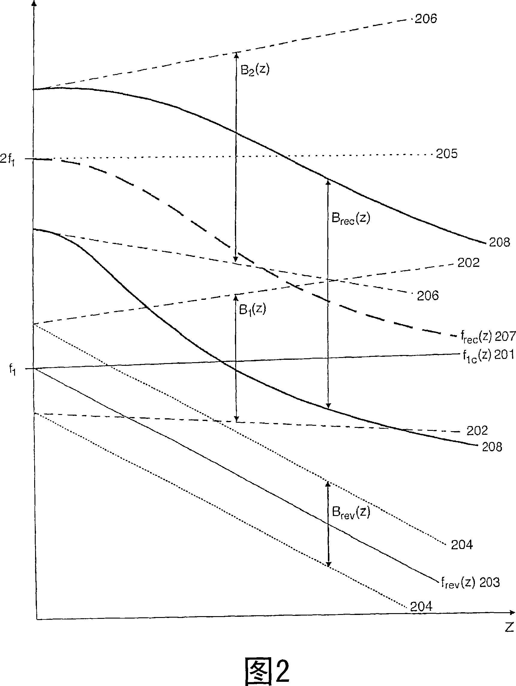
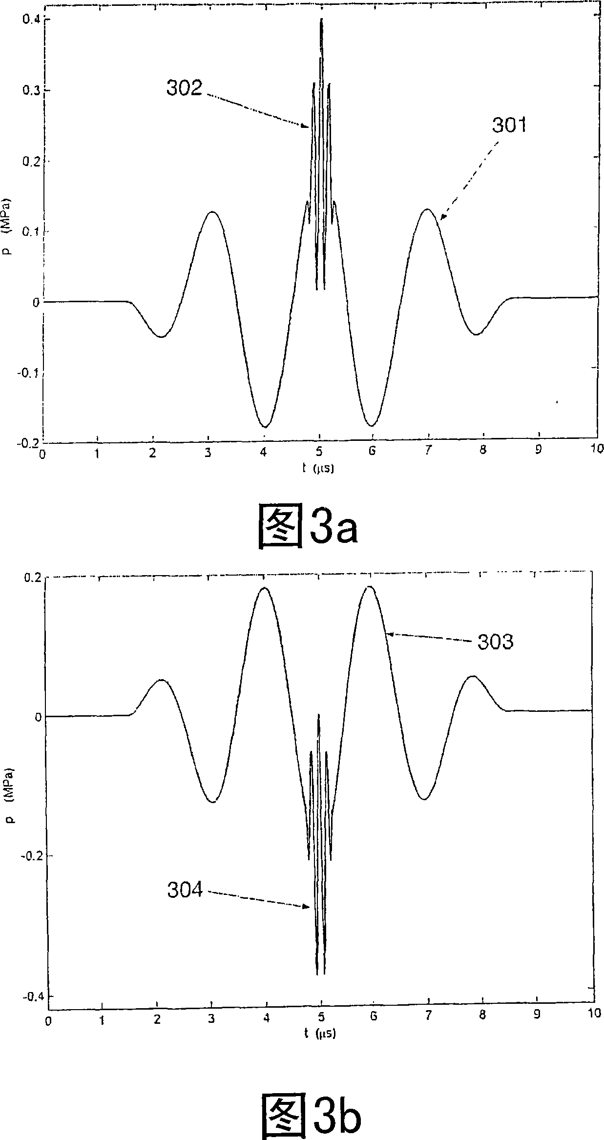

Image

  • Ultrasound imaging
  • Ultrasound imaging
  • Ultrasound imaging

Examples


Embodiment Construction

[0051] Ultrasonic bulk waves in the same kind of matter are in a linear regime determined by the linear wave formula, where, by the mass density ρ of the same type of propagation medium 0 and bulk compressibility κ 0 to determine the body wave propagation velocity c 0 . The volume compressibility is a linear approximation of the volume elasticity defined by the relative amount of compression of the material as follows

[0052] δV ΔV = - ▿ ψ ‾ = κ 0 p - - - ( 1 )

[0053] where δV is the relative volumetric compression of a small volume ΔV subjected to pressure p, ψ is the particle displacement in matter, so - ψ is the relative volumetric compression.

[0054] In soft tissue, there are spatial fluctuations in compressibility and mass...


PUM


Abstract

New methods of ultrasound imaging are presented that provide images with reduced reverberation noise and images of nonlinear scattering and propagation parameters of the object, and estimation of corrections for wave front aberrations produced by spatial variations in the ultrasound propagation velocity. The methods are based on processing of the received signal from transmitted dual frequency band ultrasound pulse complexes with overlapping high and low frequency pulses. The high frequency pulse is used for the image reconstruction and the low frequency pulse is used to manipulate the nonlinear scattering and/or propagation properties of the high frequency pulse. A 1st method uses the scattered signal from a single dual band pulse complex for filtering in the fast time (depth time) to provide a signal with suppression of reverberation noise and with 1st harmonic sensitivity and increased spatial resolution. In other methods two or more dual band pulse complexes are transmitted where the frequency and/or the phase and/or the amplitude of the low frequency pulse vary for each transmitted pulse complex. Through filtering in the pulse number coordinate and corrections of nonlinear propagation delays and optionally also amplitudes, a linear back scattering signal with suppressed pulse reverberation noise, a nonlinear back scattering signal, and quantitative nonlinear scattering and forward propagation parameters are extracted. The reverberation suppressed signals are further useful for estimation of corrections of wave front aberrations, and especially useful with broad transmit beams for multiple parallel receive beams. Approximate estimates of aberration corrections are given. The nonlinear signal is useful for imaging of differences in tissue properties, such as micro-calcifications, in-growth of fibrous tissue or foam cells, or micro gas bubbles as found with decompression or injected as ultrasound contrast agent.

Description

technical field [0001] The present invention relates to methods and systems for imaging spatial variations of ultrasound parameters of objects, especially microbubbles in objects, with particular emphasis being placed on biological tissues and fluids. Background technique [0002] In many patients, image quality obtained using current ultrasound imaging methods is limited by pulse reverberation noise (multiple scattering) and wavefront aberrations. In addition, various types of tissue diseases (eg aneurysm and atherosclerosis of the arterial wall) exhibit little variation in image contrast for proper diagnosis, as well as little variation in diseased tissue. The reason for these problems is that the image construction methods themselves do not fully take into account the physical properties of soft tissues. [0003] Spatial variations in the linear acoustic properties of tissue (mass density and compressibility) are the basis for ultrasound imaging of soft tissues. However...

Claims


Application Information

Patent Timeline
IPC IPC(8): G01S7/52G01S15/89A61B8/15A61B8/14
CPCA61B8/485A61B8/0825A61B8/15A61B8/481A61B8/488G01S7/52022G01S7/52038G01S7/52042G01S7/52049G01S7/52077G01S7/52095G01S15/8925G01S15/8927G01S15/8952
Inventor 比约恩·A·J·安杰尔森鲁内·汉森厄于温·斯坦达
Owner 比约恩・A・J・安杰尔森
Why Eureka
Try Eureka