

Immune fluorescent marking method for naked plant pollen tube microtubule skeleton and its use
A technique of gymnosperm and immunofluorescence, which is applied in the field of immunofluorescence labeling of the microtubule skeleton of gymnosperm pollen tubes, can solve the problems of lagging biological research, achieve good labeling effect, omit the sealing step, and have high application value
- Summary
- Abstract
- Description
- Claims
- Application Information
AI Technical Summary
Problems solved by technology
Method used
Image

Examples
Embodiment 1
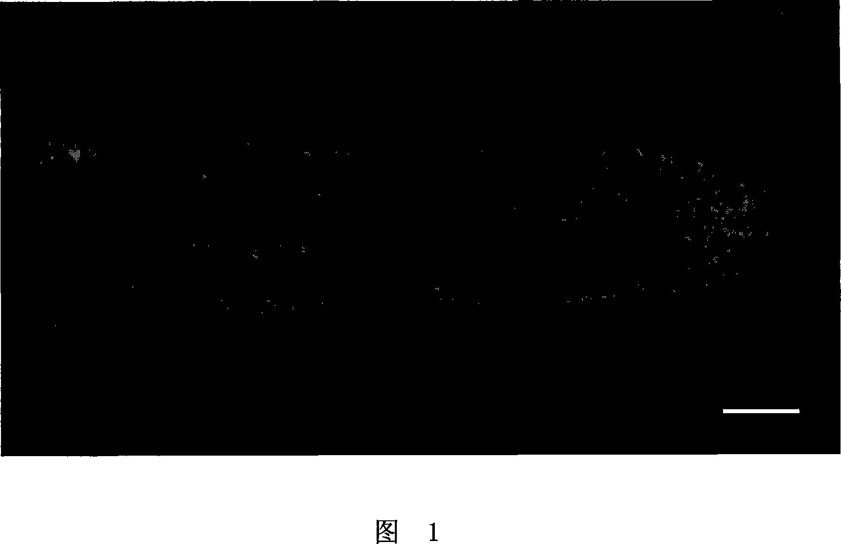
[0032] Example 1. Immunofluorescent labeling of the microtubule skeleton of white rod pollen tubes and its microscopic observation
[0033] Using the method of the present invention to carry out immunofluorescent labeling on the microtubule skeleton of the white rod pollen tube, comprising the following steps:
[0034] 1) collect the white rod pollen tube sample to be marked, in freshly prepared 50mMPipes damping solution containing 4% (g / ml) paraformaldehyde (every liter of water contains 50mM PIPES, 2mM MgCl 2 , 5mM EGTA, pH 6.9) for 60min by fast pumping, the amount of buffer is based on the soaked pollen tube sample;
[0035] 2) wash with 50mM Pipes buffer 3 times, each time for 10min, to remove residual paraformaldehyde;
[0036] 3) Put the pollen tube in 1g / 100ml of pectinase (purchased from Yakult Honsha Co.Ltd, 3000U / g) and cellulase (purchased from Yakult Honsha Co.Ltd, 3000U / g) solution at 25°C Enzymolysis for 35 minutes, wherein the weight-number ratio of pectinas...
Embodiment 2
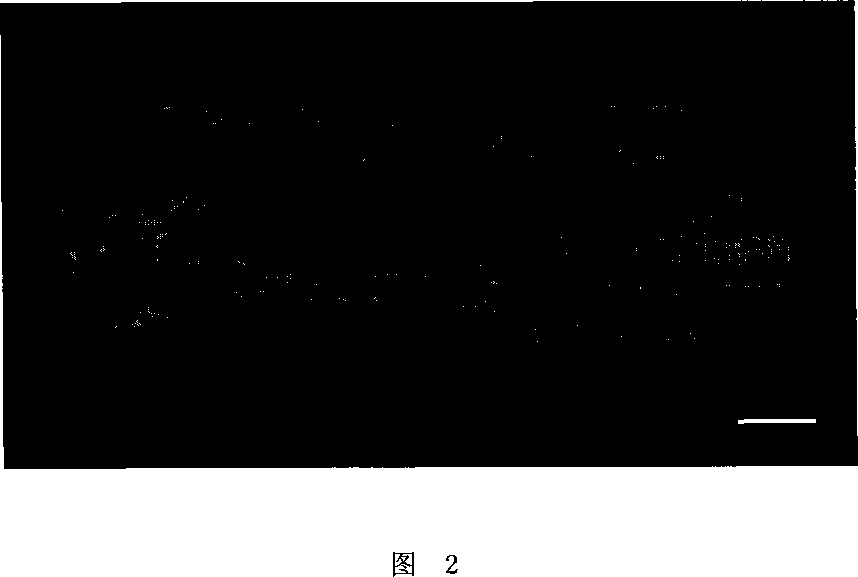
[0044] Example 2. Fluorescence labeling of the microtubule skeleton of the pollen tubes of Pythias lanceolata and its microscopic observation
[0045]Using the method of the present invention to carry out immunofluorescent labeling on the microtubule skeleton of the blue pole pollen tube, comprising the following steps:
[0046] 1) collect the blue stem pollen tube sample to be marked, in freshly prepared 60mMPipes buffer containing 3% (g / ml) paraformaldehyde (every liter of water contains 60mM PIPES, 2mM MgCl 2 , 5mM EGTA, pH 6.9) for 60min by fast pumping, the amount of buffer is based on the soaked pollen tube sample;
[0047] 2) wash with 40mM Pipes buffer 4 times, each time for 5min, to remove residual paraformaldehyde;
[0048] 3) Place the pollen tube in 1g / 100ml of pectinase (purchased from Yakult Honsha Co.Ltd, 3000U / g) and cellulase (purchased from Yakult Honsha Co.Ltd, 3000U / g) solution at 30°C Enzymolysis for 30 minutes, wherein the weight-number ratio of pectina...
Embodiment 3
[0056] Example 3. Fluorescent labeling of the microtubule skeleton of Pinus tabulaeformis and its microscopic observation
[0057] Using the method of the present invention to carry out immunofluorescent labeling on the microtubule skeleton of Pinus tabulaeformis tubes, comprising the following steps:
[0058] 1) collect the pine pollen tube sample to be marked, in freshly prepared 40mMPipes damping solution containing 5% (g / ml) paraformaldehyde (every liter of water contains 40mM PIPES, 2mM MgCl 2 , 5mM EGTA, pH 6.9) and quickly pumped and fixed for 50min, the amount of buffer was based on the soaked pollen tube sample;
[0059] 2) wash with 60mM Pipes buffer twice, each time for 15min, to remove residual paraformaldehyde;
[0060] 3) Put the pollen tube in 0.5% (W / W) pectinase (purchased from Yakult Honsha Co.Ltd, 3000U / g) and cellulase (purchased from Yakult Honsha Co.Ltd, 3000U / g) solution enzymatic hydrolysis at 25°C for 40 minutes, wherein the weight-number ratio of pe...
PUM
 Abstract
Description
Claims
Application Information