Micro-wound percutaneous nephroscope dilating draining instrument

A nephroscopic and instrument technology, applied in the field of minimally invasive percutaneous nephroscopic dilation and drainage surgical instruments, can solve the problems of unsuitability for long-term carrying, poor histocompatibility, and high incidence of infection, so as to reduce blood transfusion and super-selective arterial embolization high probability, good histocompatibility, and the effect of simplifying the surgical procedure

Inactive Publication Date: 2008-01-02
THE AFFILIATED DRUM TOWER HOSPITAL MEDICAL SCHOOL OF NANJING UNIV
View PDF0 Cites 7 Cited by
  • Summary
  • Abstract
  • Description
  • Claims
  • Application Information

AI Technical Summary

Problems solved by technology

[0003] Before the present invention, the drainage tube of the minimally invasive percutaneous nephroscopic dilation and drainage surgical instrument used the traditional latex drainage tube, which could not be fixed by itself. The reaction is severe, the incidence of accompanying infection is high, and it is not suitable for long-term carrying, which brings pain to some patients, especially those who need to undergo two-stage and three-stage surgery; There is no effective hemostasis method for severe bleeding, and medical means are usually used to stop the bleeding. For patients with severe bleeding, superselective renal artery embolization is required to stop the bleeding, which not only costs a lot, but also leads to irreversible loss of part of the renal function.

Method used


Image

  • Micro-wound percutaneous nephroscope dilating draining  instrument

Examples


Embodiment Construction

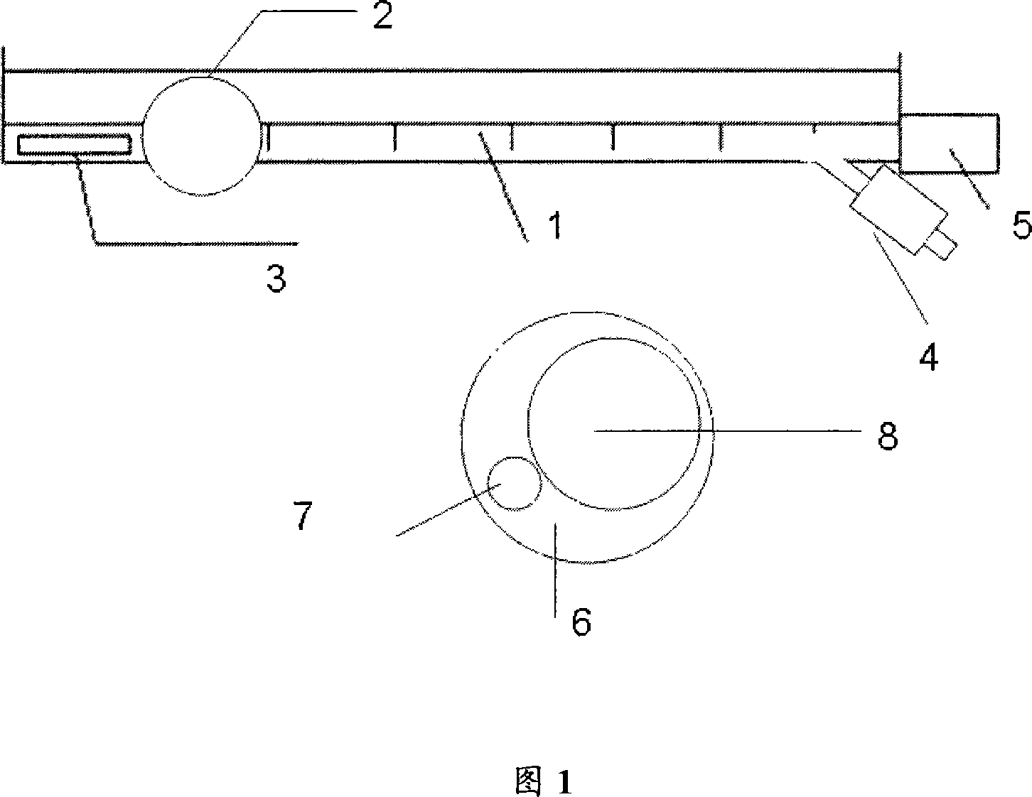
[0011] As shown in Figure 1, a balloon 2 is provided at the front end of the drainage tube body 1, and the balloon 2 is located behind the side hole 3 of the drainage tube. The water injection port 4 at the rear end of the body 1 is connected; behind the water injection port 4 is the drainage tube joint 5; as can be seen from the cut surface 6 of the drainage tube, the water injection tube 7 is arranged in the body 1 of the drainage tube, and is separated from the lumen 8 of the drainage tube. The tube body 1 of the drainage tube is made of silicone material, so that it has good tissue compatibility.

[0012] During the operation, the balloon 2 is located behind the side hole 3 of the drainage port, and the balloon 2 is located in the calices or renal pelvis. After water injection, the drainage tube body 1 can be effectively fixed in the kidney, so that the drainage tube body 1 can be firmly fixed. ; When hemostasis is required, inject water from the water injection port 4 at ...


PUM


Abstract

The invention relates to a kind of micro-injure drainage surgical apparatus, which is characterized in that the front end of drainage tube is equipped with one saccule, the saccule is connected with drainage tube and water injection end. It solves problems that the current traditional latex drainage tube can not be fixed by itself but needs being fixed on skin, the tissue compatibility is bad, inflammation in part area is severe, high infection occurrence rate, inefficient styptic method for bleeding-out in pelvis and kidney calices, and it needs to carry out selective renal artery gland haemostasis, which will cost much money and cause non-reversible kidney function loss. The invention is characterized by simple structure, reasonable design, convenient usage, good tissue compatibility, and no apparent toxic and side effect.

Description

technical field [0001] The invention relates to an operation auxiliary device, in particular to a minimally invasive percutaneous nephroscopic expansion and drainage operation device. Background technique [0002] Minimally invasive percutaneous nephroscopy is an emerging stone treatment technology developed on the basis of the original classic percutaneous nephroscopy. It narrows the expansion channel of renal puncture and uses new technologies such as ureteroscope-assisted pneumatic lithotripsy and holmium laser. The technology carries out the treatment of kidney calculus, has the advantages such as high safety, less bleeding, less trauma, and has been popularized rapidly since its invention. [0003] Before the present invention, the drainage tube of the minimally invasive percutaneous nephroscopic dilation and drainage surgical instrument used the traditional latex drainage tube, which could not be fixed by itself. The reaction is severe, the incidence of accompanying i...

Claims


Application Information

Patent Timeline
Patent Type & Authority Applications(China)
IPC IPC(8): A61B17/00A61B17/12A61B19/00A61M27/00A61M1/00A61M25/10
Inventor 郭宏骞甘卫东李笑弓
Owner THE AFFILIATED DRUM TOWER HOSPITAL MEDICAL SCHOOL OF NANJING UNIV
Why Eureka
Try Eureka