Use of vascular endothelial growth factor receptor 1+ cells in treating and monitoring cancer and in screening for chemotherapeutics
A growth factor receptor, vascular endothelium technology, applied in the application of vascular endothelial growth factor receptor 1+ cells in the treatment and monitoring of cancer and in the screening of chemotherapy drugs, can solve the problem of unclear precise role of tumor metastasis and other issues
- Summary
- Abstract
- Description
- Claims
- Application Information
AI Technical Summary
Problems solved by technology
Method used
Image
Examples
preparation example Construction
[0054] Methods for preparing polyclonal antibodies are also well known. Usually, vascular endothelial growth factor receptor 1 can be + Subcutaneous administration of New Zealand white rabbits (which had previously been bled to obtain preimmune serum) produced such antibodies. Antigen was injected at 6 different sites with a total volume of 100 μl per site. Each injected material contained pluronic polyol, a synthetic surfactant adjuvant, or ground acrylamide gel containing protein or polypeptide after SDS polyacrylamide gel electrophoresis. Rabbits were bled 2 weeks after the first injection, and then boosted 3 times with the same antigen regularly every 6 weeks. Serum samples were collected 10 days after each booster immunization. Polyclonal antibodies are then recovered from the serum by affinity chromatography using the corresponding antigen-capture antibodies. Finally, rabbits were anesthetized with 150 mg / Kg IV pentobarbital. The above and other methods for producin...
Embodiment 1
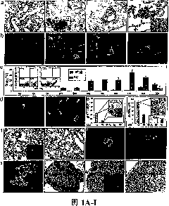
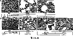
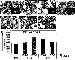
[0087] Wild-type C57B1 / 6 mice were lethally irradiated (950 rads) and then transplanted with 1×10 6 β-galactosidase + or GFP + BM cells (Rosa-26 mice) (Lyden et al., "Impaired Recruitment of Bone-marrow-derived Endothelial and Hematopoietic Precursor Cells Blocks Tumor Angiogenesis and Growth" and Growth), "Nat. Med. 7: 1194-1201 (2001), which is hereby incorporated by reference in its entirety). After 4 weeks, mice were intradermally injected with 2×10 6 LLC (ATCC) or B-16 cells (ATCC).
Embodiment 2
[0088] Tissues and femurs were fixed in 4% paraformaldehyde (PFA) for 4 hours. Samples were stained in X-gal solution at 37°C for 36 hours, see Tam et al., "The Allocation of Epiblast Cells to the Embryonic Heart and other Mesodermal Lineages: The Role of Ingression and Tissue Movement during Gastrulation" Other mesoderm lineages: role of inward migration and tissue motility during gastrulation)," Development. 124: 1631-1642 (1999), which is hereby incorporated by reference in its entirety. X-gal stained tumors and BM were embedded as described in: Lyden et al., "Impaired Recruitment of Bone-marrow-derived Endothelial and Hematopoietic Precursor Cells Blocks TumorAngiogenesis and Growth can block tumor angiogenesis and growth)," Nat. Med. 7: 1194-1201 (2001), which is hereby incorporated by reference in its entirety.
PUM
 Abstract
Description
Claims
Application Information
 - R&D
- Intellectual Property
- Life Sciences
- Materials
- Tech Scout
- Unparalleled Data Quality
- Higher Quality Content
- 60% Fewer Hallucinations
Browse by: Latest US Patents, China's latest patents, Technical Efficacy Thesaurus, Application Domain, Technology Topic, Popular Technical Reports.
© 2025 PatSnap. All rights reserved.Legal|Privacy policy|Modern Slavery Act Transparency Statement|Sitemap|About US| Contact US: help@patsnap.com


