

Four dimensional rebuilding method of coronary artery vessels axis
A coronary artery and blood vessel technology, applied in the field of medical detection, can solve problems such as poor continuity of reconstruction results, high calculation costs, and difficulty in achieving accuracy, and achieve the effects of avoiding point-by-point matching, enhancing visual effects, and improving reconstruction accuracy
Inactive Publication Date: 2008-10-15
NORTH CHINA ELECTRIC POWER UNIV (BAODING)
View PDF0 Cites 34 Cited by
- Summary
- Abstract
- Description
- Claims
- Application Information
AI Technical Summary
Problems solved by technology
For discrete two-dimensional axes, the epipolar constraints in stereo imaging are generally used for point-by-point matching. This method is difficult to achieve high accuracy, the continuity of reconstruction results is not good, and the amount of calculation is also large.
In addition, since the above steps need to be repeated for the images at each moment in the sequence, the calculation cost required to complete the blood vessel tracking of the entire sequence is too high, and a large amount of manual participation of the operator is required, so the practical value is low
Method used
View moreImage
Smart ImageViewing Examples


Examples
Embodiment Construction
Login to View More PUM
 Abstract
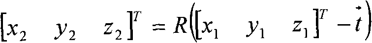
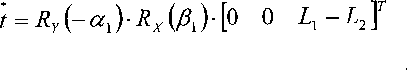
A 4D reconstruction method of the vascular axis of coronary artery, which belongs to the field of medical detection technology, comprises following steps: constructing the projection models of an X-ray angiography system at two angles according to X-ray coronary artery angiogram sequences at two angles, deducing the geometric transformation relationship between the two angle images, 3D reconstructing the sampled points of interested blood vessels selected by hand in the image of the first moment, connecting the reconstructed points to obtain a broken line, acquiring the 3D axis of the blood vessel at the first moment by snake transformation with respect to the broken line as the initial position, and acquiring 3D vascular axis in each subsequent moment in the image sequence by snake transformation with respect to the 3D vascular axis of the previous moment as the initial position, thus completing the 4D reconstruction of the vascular axis in the entire sequence. The method can greatly reduce the uncertainty and the error due to the operation of operators, thus improving the repeatability of the result and achieving convenient operation and high efficiency.
Description
A Four-dimensional Reconstruction Method of Coronary Artery Axis technical field The invention relates to a method for four-dimensional (three-dimensional + time) reconstruction of a blood vessel axis according to a digital X-ray coronary angiography image sequence covering one or more cardiac cycles, and belongs to the technical field of medical detection. Background technique X-ray coronary angiography (X-raycoronary angiography, CAG) is an imaging method widely used in clinical diagnosis and treatment of coronary heart disease. The biggest feature of CAG diagnosis is visibility, that is, to judge the location, nature and degree of abnormal cardiovascular anatomy by observing the filling and disappearance of contrast agent statically or dynamically. X-ray contrast imaging is to superimpose the spatial vascular structure on the two-dimensional imaging plane, that is, projection imaging, thus losing most of the three-dimensional spatial information required in clinical dia...
Claims
Login to View More Application Information
Patent Timeline
 Patent Type & AuthorityApplications(China)
IPC IPC(8): A61B6/00G06T15/00
Inventor孙正
OwnerNORTH CHINA ELECTRIC POWER UNIV (BAODING)