Variable visual field scanning microscope and the method based on fixed light path system

A scanning microscopy, fixed light technology, applied in the field of fluorescence microscopy, can solve the problems of limited application, less than 5% utilization of light energy, single field of view and resolution, and achieve the effect of flexible conversion

Inactive Publication Date: 2009-01-07
SHENZHEN UNIV
View PDF0 Cites 37 Cited by
  • Summary
  • Abstract
  • Description
  • Claims
  • Application Information

AI Technical Summary

Problems solved by technology

In fact, limited by the nonlinear damage of the sample, the optical power required by the traditional single-point scanning multi-photon excitation fluorescence microscope to achieve sufficient photon number density is about 3-10mW
Therefore, the utilization rate o...

Method used


Image

  • Variable visual field scanning microscope and the method based on fixed light path system

Examples


Embodiment Construction

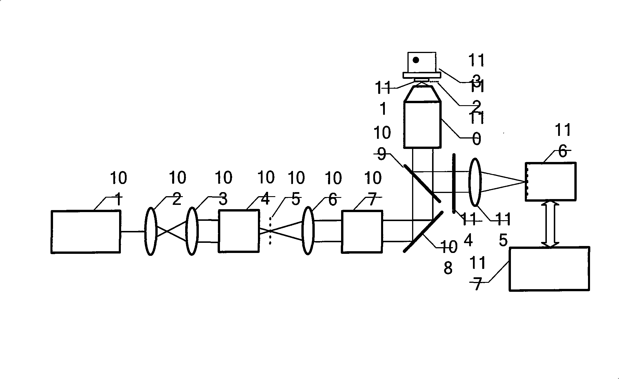
[0031] A kind of variable field of view scanning microscopy method based on the fixed optical path system of the present invention comprises the following steps:

[0032] (1) Use a Ti:Sapphire mode-locked femtosecond laser to generate an ultrashort pulse laser beam. The wavelength of the laser beam is allowed to vary from 700-1000nm, and the repetition rate varies from 50MHz to 500MHz, but the operating frequency remains stable for each operation.

[0033] (2) Use a pair of beam expander lenses to uniformly expand the high repetition rate ultrashort pulse laser beam output by the laser to form a uniform laser beam.

[0034] (3) Using a two-dimensional space discrete system composed of a microlens array to perform two-dimensional space discretization processing on a uniform laser beam to form M×N sub-beams, the M×N sub-beams are two-dimensional space discrete distribution, where M>1 , N>1.

[0035] (4) Focus the M×N sub-beams on the sample placed on the sample platform after s...


PUM

PropertyMeasurementUnit
Wavelengthaaaaaaaaaa

Abstract

The invention discloses a variable-field scanning microscopic method based on a fixed optical path system, and a device thereof. At first, a beam expanding system is used for uniformly expanding laser beams which are transmitted from a laser so as to form uniform laser beams; then a two-dimensional space discrete system is used for the two-dimensional space discretization of the uniform laser beams so as to form a sub-beam of M multiplied by N; the sub-beam of M multiplied by N orderly passes through a collimating lens, a scanning galvanometer, a reflector, a dichroic mirror and a fluorescence microscope, and then focuses on the sample on a sample platform; thus the excited fluorescence can be formed on the sample; the fluorescence microscope is used for collecting the excited fluorescence, which is transmitted into an imaging system after being reflected by the dichroic mirror and focused by a focusing lens. The scanning of the galvanometer and the crossing movement of the sample platform are combined: the scanning step length and the scanning step number of the galvanometer are changed; the area of the unit viewing field is used as the step length to move the sample platform; thus the scanned viewing field can be changed. The scanning microscopic method realizes the flexible switching between the low-resolution large viewing field and the high-resolution small viewing field, and satisfies the requirements in different biomedical applications.

Description

technical field [0001] The invention relates to fluorescence microscopy technology, in particular to a scanning microscopy method and device for variable field of view based on a fixed optical path system. Background technique [0002] Fluorescence microscopy has become an important tool in life sciences, especially cell biology research. In recent years, with the development of life sciences, higher and higher requirements have been put forward for fluorescence microscopy technology. The continuous development of laser technology, fluorescent probe labeling technology, new fluorescence detection technology and imaging methods has become an important factor to promote the development of life sciences. important driving force. Laser scanning confocal microscopy and multiphoton excitation fluorescence microscopy can be used to achieve high spatial resolution three-dimensional imaging of biological samples. But their common disadvantage is that the sample needs to be scanned ...

Claims


Application Information

Patent Timeline
IPC IPC(8): G01N21/64G02B23/00
Inventor 屈军乐刘立新王磊林子扬牛憨笨
Owner SHENZHEN UNIV
Why Eureka
Try Eureka