Automatic microscopic imager for detecting cast-off cells and detection method

A technique of exfoliated cells and microscopic imaging, applied in the field of automatic analysis of cervical smears, can solve the problems of heavy workload, cumbersome and fatigued, high false negative rate and false positive rate, etc., achieving a high degree of automation and high reliability. , the effect of accurate analysis results

Active Publication Date: 2009-01-07
HEER MEDICAL TECH DEV CO LTD
View PDF0 Cites 39 Cited by
  • Summary
  • Abstract
  • Description
  • Claims
  • Application Information

AI Technical Summary

Problems solved by technology

But at present, the application of cervical smear is still in the stage of manual identification: the cervical smear is stained with Pap staining method, and the cells are manually inspected with the naked eye one by one under the light microscope, and the female reproductive tract is classified according to the five-level classification of Pap smear. diagnosis of malignant tumors
The workload of this method is heavy, ea...

Method used


Image

  • Automatic microscopic imager for detecting cast-off cells and detection method
  • Automatic microscopic imager for detecting cast-off cells and detection method
  • Automatic microscopic imager for detecting cast-off cells and detection method

Examples


Embodiment Construction

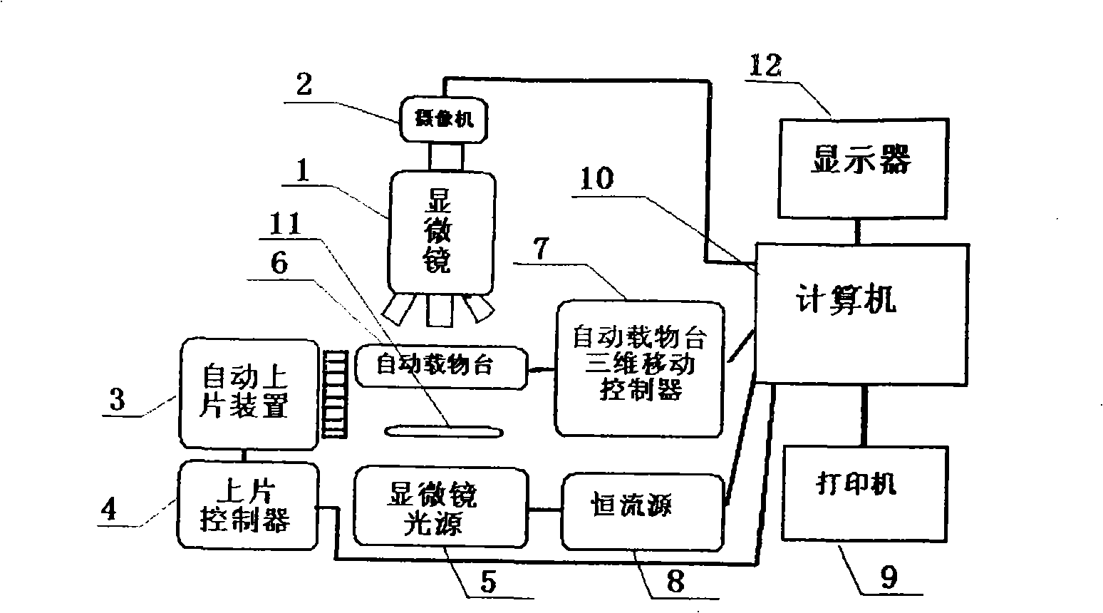
[0018] refer to figure 1 , the digital video camera 2 installed on the microscope 1 of the present invention is connected to the computer 10, the automatic film loading device 3 is connected to the computer 10 through the automatic film loading control system 4, and the automatic stage 6 is connected to the automatic stage three-dimensional movement control system 7 through the automatic stage. The computer 10 is connected, and the computer 10 is respectively connected to the printer and other peripheral devices 9 , the display 12 , the lighting system 5 , and the lighting stabilization system 8 , and there are filters and mirrors 11 under the automatic stage 6 . The computer 10 controls the automatic loading device 3 to load the cell smear on the automatic stage 6 and unload the analyzed cell smear from the automatic stage 6 .

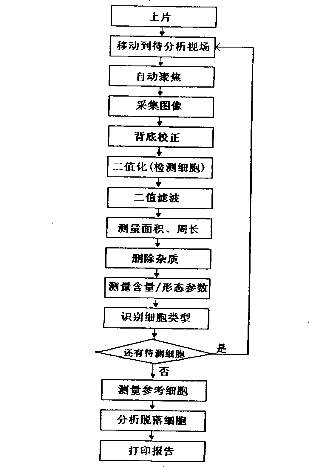
[0019] The present invention is described below by specific embodiment:

[0020] In the present invention, the DNA in cells is selected to be measur...


PUM


Abstract

The invention relates to an automatic micro imaging instrument used for detecting cast-off cells and a detecting method, in particular to an automatic analysis instrument of a uterine neck cell smear and a detecting method. The instrument comprises a microscope, a three dimensional automatic object stage, an automatic film loading device and a control system thereof, a light system, a stable system, a light filter, a computer, a display and a digital camcorder, etc. The detecting method comprises the following steps: the smear is analyzed and colored, then arranged on the automatic object stage which moves up and down to focus automatically, when the automatic object stage moves horizontally, the smear scanning is realized; cell images are amplified by the microscope, picked up and sent to the computer to carry out image processing and analyzing. The detected cells are filtered by two-value filtration to remove the noises, the area and the perimeter are measured, and the impurity is deleted, then the spectrum and other shape parameters of the cells are measured; the measured cells are classified with a pattern recognition algorithm into the cast-off cells, white blood cells, lymphocyte cells and the impurity; after a viewing field is finished, the cells move down automatically, auxiliary diagnostic data is obtained by the computer. The instrument and the detecting method have the advantages of high degree of automation, accurate analysis results and reliability.

Description

technical field [0001] The invention relates to an instrument and a detection method for automatic analysis of cervical cell smears. Background technique [0002] The incidence of cervical cancer ranks first in gynecological malignant tumors, and the incidence of cervical cancer in my country ranks second in the world. Pap smear is an important part of the early diagnosis of cervical cancer. As a successful screening method for cervical cancer, it can provide a lot of useful information on whether there is cancer, the type and severity of inflammation. Cervical cancer mortality has decreased significantly. However, the current application of Pap smear is still in the stage of manual identification: that is, the Pap smear is stained with Pap staining, and the cells are manually examined under a light microscope. diagnosis of malignant tumors. This method has a large workload, and each film has an average of hundreds of thousands of cells. It is very cumbersome and tiring t...

Claims


Application Information

Patent Timeline
IPC IPC(8): G01N33/48G01N21/84
Inventor 曾立波
Owner HEER MEDICAL TECH DEV CO LTD
Why Eureka
Try Eureka