Eureka AIR delivers breakthrough ideas for toughest innovation challenges, trusted by R&D personnel around the world.

Method for supporting, regulating and controlling amplification of hematopoietic stem/progenitor cell in vitro with microencapsulation osteoblast in hypoxia condition

A technology for osteoblast and in vitro expansion, applied in biochemical equipment and methods, bone/connective tissue cells, animal cells, etc., can solve problems such as unsatisfactory cell expansion

Inactive Publication Date: 2009-08-19
DALIAN UNIV OF TECH
View PDF0 Cites 17 Cited by
  • Summary
  • Abstract
  • Description
  • Claims
  • Application Information

AI Technical Summary

Problems solved by technology

At present, there are very few studies on using osteoblasts as stromal cells to support the expansion of hematopoietic stem and progenitor cells in vitro. Only Huang Xiaobing et al. Induced osteoblast-like cells for hematopoietic support studies, but CD34 + The expansion of cells is not ideal, after two weeks of culture, CD34 + Cell content drops to 34%-63% of initial concentration

Method used


Image

  • Method for supporting, regulating and controlling amplification of hematopoietic stem/progenitor cell in vitro with microencapsulation osteoblast in hypoxia condition
  • Method for supporting, regulating and controlling amplification of hematopoietic stem/progenitor cell in vitro with microencapsulation osteoblast in hypoxia condition
  • Method for supporting, regulating and controlling amplification of hematopoietic stem/progenitor cell in vitro with microencapsulation osteoblast in hypoxia condition

Examples


Embodiment 1

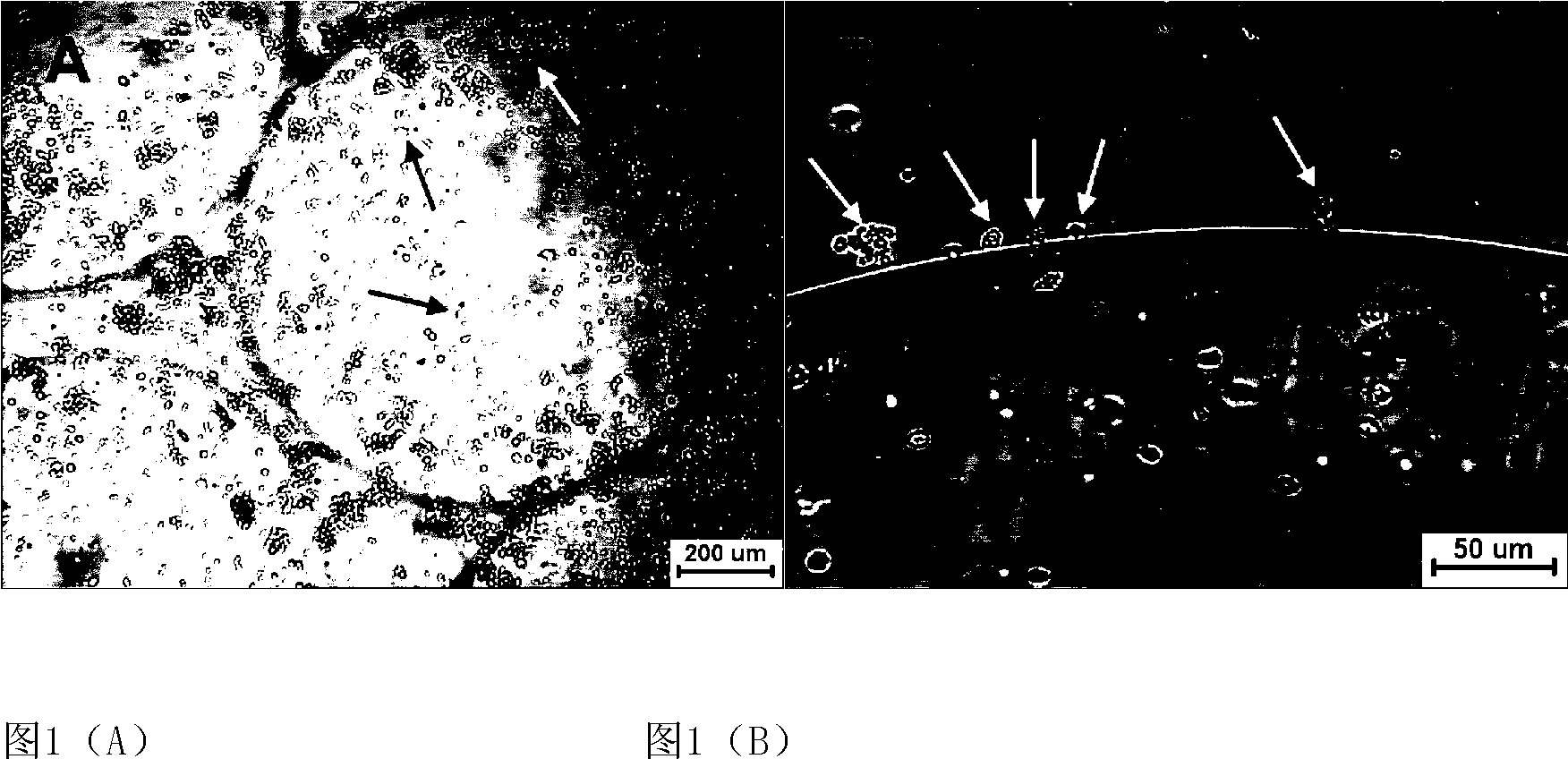
[0043] Example 1 Co-cultivation of microencapsulated osteoblasts and umbilical cord blood HSPCs under normoxia

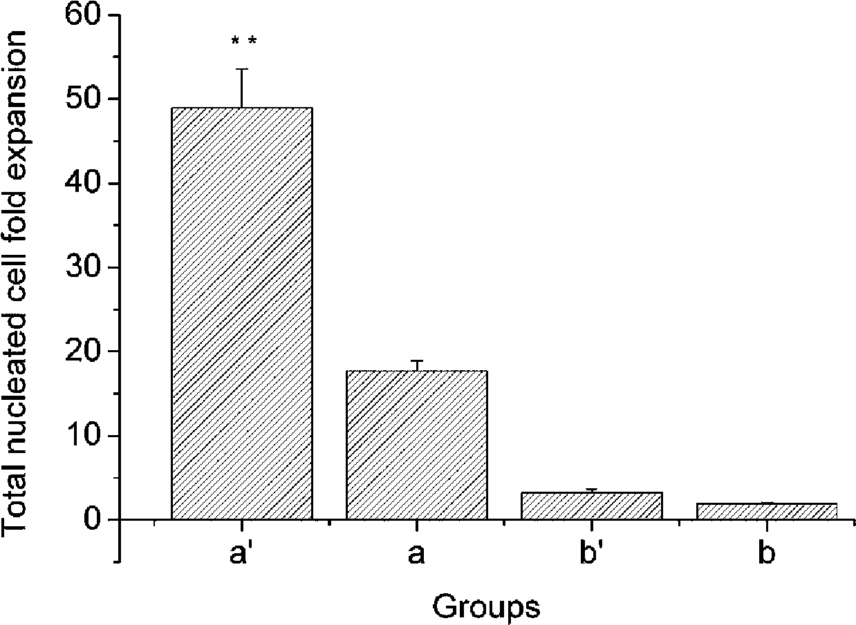
[0044] The embedding density of osteoblasts was 8×10 5 cells / mL, 1mL was added to each well, and three wells were paralleled in each group, a total of 2 groups. One group was used for co-culture with hematopoietic cells, and the other group was used as a control without hematopoietic cells. The diameter of the microbeads is 0.5 mm. The co-culture group with osteoblasts in a normoxic environment is marked as a, and the single culture of cord blood mononuclear cells is marked as b. Adjust the freshly isolated umbilical cord blood total nucleated cell density to 2×10 5 cells / mL, inoculated in a 6-well plate containing human osteoblast microbeads, added 2 mL to each well, and paralleled 3 wells in each group. Serum-free IMDM was used as the medium, and a small amount of growth factors were added, including 2.4ng / mLIL-3, 20ng / mLSCF, 10ng / mLFL, 2.0ng / mLGM-SCF and 3.2n...

Embodiment 2

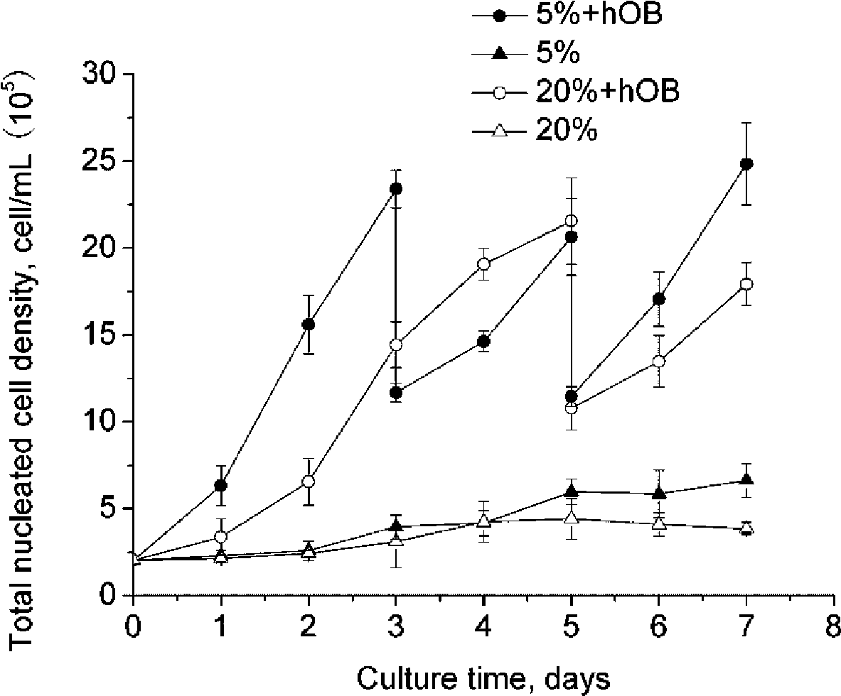
[0047] Example 2 Co-cultivation of osteoblast microgel beads with different diameters and umbilical cord blood HSPCs

[0048] Adjust the cord blood mononuclear cell density to 2×10 5 cells / mL, seeded in a 6-well plate, and added 3 mL of cell suspension to each well. Then add 1 mL of osteoblasts to each well at a density of 5 × 10 5 cells / mL microgel beads. The ratio of osteoblasts to umbilical cord blood hematopoietic cells was 5:6. placed in a hypoxic incubator (5% O 2 , 20% CO 2 , 37°C). The 0.5mm microgel bead group is marked as A, and the 2mm microbead group is marked as B. Serum-free IMDM was used as the co-culture medium, and a small amount of growth factors were added according to the standard in Example 1. Co-cultivate for 7 days, gently blow off the cells before sampling every day, so that the signal factors and hematopoietic growth factors secreted by osteoblasts are evenly dispersed, take 0.2mL cell suspension from each well for various tests, and add the sam...

Embodiment 3

[0051] Example 3 Co-cultivation of microencapsulated osteoblasts and umbilical cord blood HSPCs under optimized conditions

[0052] The embedding density of osteoblasts was 8×10 5 cells / mL, 1mL was added to each well, and three wells were paralleled in each group, a total of 2 groups. One group was used for co-culture with hematopoietic cells, and the other group was used as a control without hematopoietic cells. The diameter of the microbeads is 0.5 mm. The group co-cultured with human osteoblasts in a hypoxic environment was marked as a', and the group cultured with umbilical cord blood mononuclear cells alone was marked as b'. Adjust the total nucleated cell density of freshly isolated umbilical cord blood to 2×10 5 cells / mL, inoculated in a 6-well plate containing human osteoblast microbeads, added 2 mL to each well, and paralleled 3 wells in each group. Serum-free IMDM was used as the medium, and a small amount of growth factors were added according to the standard in...


PUM

PropertyMeasurementUnit
Diameteraaaaaaaaaa
Diameteraaaaaaaaaa

Abstract

The invention discloses a method for encapsulated osteoblasts to support and regulate the in-vitro amplification of cord blood hematopoieticstem / progenitorcells (He) in the hypoxia condition, belonging to the fields of biotechnology and tissue TSPCS engineering. The method is characterized in that the human osteoblasts are embedded by gelatin-alginate-chitosan (GAC) microcapsule as matrix feeder cells and co-cultured with the cord blood HSPCs in the 5% hypoxia environment to obtain the HSPCs. The invention adopts GAC microcapsule to separate the human osteoblasts from the cord blood HSPCs to avoid the cell contamination and immunological rejection. Furthermore, the pore passages of the microcapsule are capable of causing hemopoietic growth factors secreted by the osteoblasts in the microcapsule to expand to the habitat of the HSPCs and play a role of stimulating the amplification of the hematopoietic stem / progenitor cells. Moreover, the material of the microcapsule has the slow release function to the expansion of the growth factors to increase the concentration gradient of the growth factors on the surface of the microcapsule, which is helpful for the long-term function of the growth factor. In addition, the microencapsulated osteoblasts form the 3D environment, which not only causes the culture system to contain more osteoblasts, but also causes the culture system to have bigger HSPCs-OBs action area compared with the co-culture pattern of the osteoblasts cultured by adherence. Besides, in the 5% hypoxia environment, the co-culture system is capable of better simulating the hematopoietic microenvironment iche of human bone marrow the osteoblasts have significant effects on the characteristic maintenance and number amplification of stem cells of the HSPCs.

Description

technical field [0001] The invention belongs to the field of biotechnology and tissue engineering, and relates to a kind of microencapsulated osteoblasts supporting and regulating umbilical cord blood hematopoietic stem / progenitor cells (Hematopoieticstem / progenitor cells, HSPC) under hypoxic conditions. S ) method of in vitro amplification. Background technique [0002] Rare hematopoietic stem / progenitor cells are the only source of various mature blood cells in the body, and they mainly exist in umbilical cord blood, bone marrow and peripheral blood (Weissman, Cell, 2000, 100(1): 157-168). Umbilical cord blood is currently an important source of hematopoietic stem / progenitor cells in clinical practice. It is rich in earlier hematopoietic stem / progenitor cells, has weak immunogenicity, produces fewer antibodies against heterologous antigens, and has fewer mature T cells. and easy preservation, no tumor cell contamination, no damage to the donor and no side effects, CD34 +...

Claims


Application Information

Patent Timeline
IPC IPC(8): C12N5/08C12N5/077C12N5/0789
Inventor 宋克东刘天庆赵国峰崔占峰马学虎
Owner DALIAN UNIV OF TECH
Why Eureka
Eureka Blog
Learn More