Method for identifying tumors that are responsive to treatment with anti-ErbB2 antibodies

A technology of humanized antibody and its application, which is applied in the direction of antineoplastic drugs, antibodies, chemical instruments and methods, etc.

Inactive Publication Date: 2010-05-26
GENENTECH INC +1
View PDF109 Cites 0 Cited by
  • Summary
  • Abstract
  • Description
  • Claims
  • Application Information

AI Technical Summary

Problems solved by technology

However, the mechanisms by which these receptors aggregate and how they contribute to signaling are not well understood (Brennan, P.J. et al., Oncogene, 19 : 6093-6101 (2000))

Method used


Image

  • Method for identifying tumors that are responsive to treatment with anti-ErbB2 antibodies
  • Method for identifying tumors that are responsive to treatment with anti-ErbB2 antibodies
  • Method for identifying tumors that are responsive to treatment with anti-ErbB2 antibodies

Examples


Embodiment 1

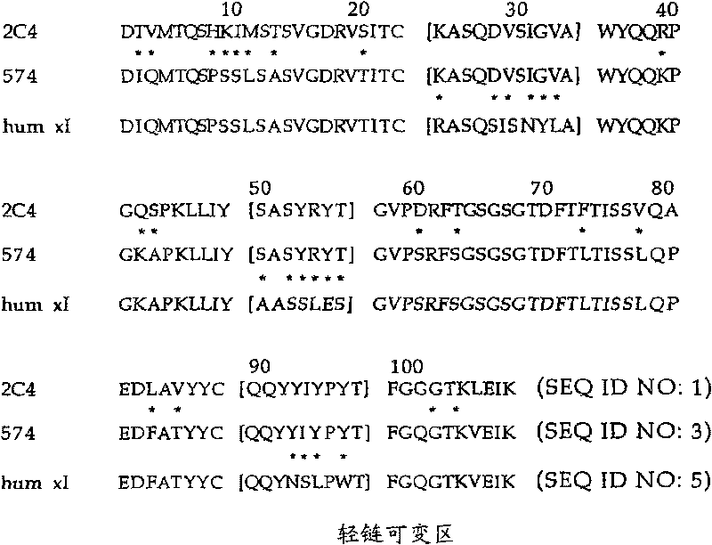
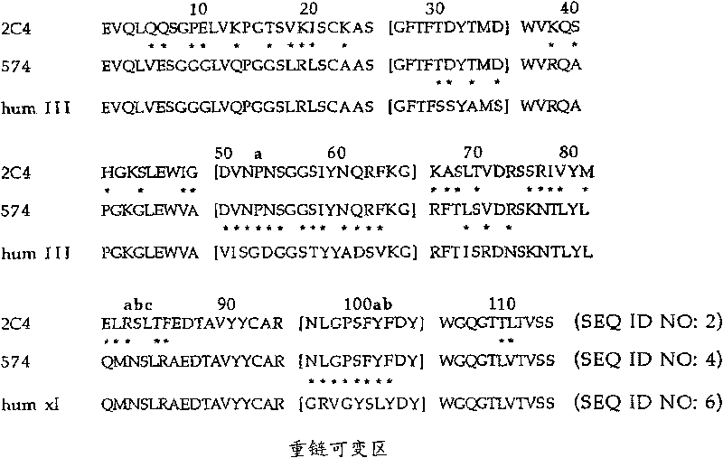
[0508] HRG-dependent binding of ErbB2 to ErbB3 is blocked by monoclonal antibody 2C4

[0509] The murine monoclonal antibody 2C4, which specifically binds the extracellular domain of ErbB2, is described in WO 01 / 89566, the disclosure of which is hereby expressly incorporated by reference in its entirety.

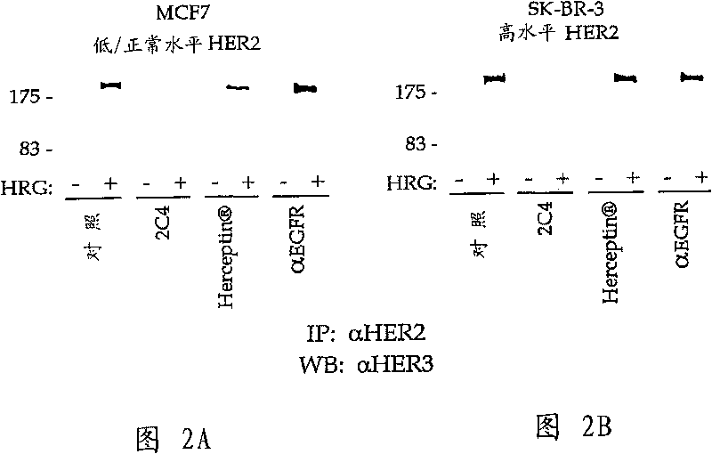
[0510] The ability of ErbB3 to combine with ErbB2 was tested in co-immunoprecipitation experiments. 1.0×10 6 MCF7 or SK-BR-3 cells were seeded into 6-well tissue culture plates containing 10% fetal bovine serum (FBS) and 10 mM HEPES, pH 7.2 in 50:50 DMEM / Ham's F12 medium (growth medium) , and let it attach overnight. Cells were starved for 2 hours in serum-free growth medium before starting the experiment. Cells were briefly washed with phosphate-buffered saline (PBS), then mixed with 100 nM of the indicated antibody diluted in 0.2% w / v bovine serum albumin (BSA), RPMI medium (binding buffer) with 10 mM HEPES, pH 7.2, Or incubate with binding buffer only (control). Af...

Embodiment 2

[0516] Responsiveness of cell lines and human tumor xenograft models to 2C4

[0517] Approximately 40 tumor models were tested for reactivity to 2C4. These models represent major cancers such as breast, lung, prostate, and large intestine. 50-60% of the models responded to 2C4 treatment. Table 1 below lists selected tumor models tested for responsiveness to 2C4. Briefly, human tumor xenograft fragments approximately 3 mm in size were implanted under the skin of athymic nude mice. Alternatively, in vitro grown human tumor cells are isolated from culture dishes, resuspended in phosphate-buffered saline, and injected subcutaneously into the flank of immunocompromised mice. Tumor growth was monitored every 2-3 days using electric calipers.

[0518] When tumors reached approximately 30-100 mm in size, animals were randomly divided into different treatment and control groups. 2C4 was administered by intraperitoneal injection once a week. Control animals received the same vol...

Embodiment 3

[0532] Detection of heterodimers in 2C4-responsive tumors by immunoprecipitation

[0533] Anti-ErbB2 antibody immunoprecipitation was performed on 2C4-responsive and non-responsive tumors to analyze the presence of ErbB2-ErbB3 and EGFR-ErbB2 heterodimers. Unless otherwise stated, the method was performed according to "Maniatis T. et al., Molecular Cloning: A Laboratory Manual, Cold Spring Harbor, New York, USA, Cold Spring Harbor Press, 1982".

[0534] Choose anti-HER2, anti-HER3 and anti-HER1 antibodies that do not cross-react. To determine whether antibodies cross-react, HER1, HER2, HER3, and HER4 receptors were expressed in human embryonic kidney (HEK) 293 cells. Triton containing HEPES buffer (pH 7.5) TM Cells were lysed with X100 (1% w / v). Approximately 20 μg of total cellular protein from control cells and cells expressing HER1, HER2, HER3, and HER4 were separated on SDS gels and transferred to nitrocellulose membranes by semi-dry blotting. Various anti-HER1, anti...


PUM

PropertyMeasurementUnit
diameteraaaaaaaaaa

Abstract

The invention relates to a method for identifying tumors that are responsive to treatment with ANTI-ERBB2 antibodie. Tumors are identified as responsive to treatment with anti-HER2 antibodies by detecting the presence of a HER2 / HER3 and / or HER2 / HER1 protein complex or for HER2 phosphorylation in a sample of tumor cells. Patients suffering from a tumor comprising HER / 2 / HER1 and / or HER2 / HER3 heterodimers and / or HER2 phosphorylation are treated with anti-HER2 antibodies, such as rhuMAb 2C4.

Description

[0001] This application is filed on July 11, 2003, the application number is 03821625.6 (the international application number is PCT / US2003 / 021590), and the invention title is "Method for identifying tumors responding to treatment with anti-ErbB2 antibody". Divisional application. technical field [0002] The present invention relates to methods of identifying tumors that respond to treatment with anti-HER2 antibodies, and methods of treating patients with such tumors. Background technique [0003] The ErbB family of receptor tyrosine kinases are important mediators of cell growth, differentiation and survival. The receptor family includes four distinct members, including epidermal growth factor receptor (EGFR or ErbB1), HER2 (ErbB2 or p185 neu ), HER3 (ErbB3) and HER4 (ErbB4 or tyro2). [0004] EGFR, encoded by the erbB1 gene, has been implicated in human malignancy. In particular, elevated EGFR expression has been observed in breast, bladder, lung, head, neck and gastri...

Claims


Application Information

Patent Timeline
Patent Type & Authority Applications(China)
IPC IPC(8): A61K39/395A61P35/00C07K14/485C07K16/32G01N33/50G01N33/53G01N33/574
CPCC07K16/32G01N2333/485A61K2039/505G01N33/57492G01N2800/52G01N2333/71C07K2317/24A61P35/00A61P35/02A61P43/00
Inventor 汉斯·科尔伯吉特·博森梅尔汉斯-乔基姆·米勒马克·X·斯利科夫斯基斯蒂芬·M·凯尔西
Owner GENENTECH INC
Why Eureka
Try Eureka