Cell morphology microscopic examination device and method based on staining technology

A morphological and cell technology, applied in the field of cell analysis, can solve the problems of complex structure, counting error, easy to get together, etc., and achieve the effect of ensuring statistics and reliability, reliable automatic inspection device, and speeding up staining

Inactive Publication Date: 2010-06-30
孔兵
View PDF0 Cites 4 Cited by
  • Summary
  • Abstract
  • Description
  • Claims
  • Application Information

AI Technical Summary

Problems solved by technology

[0005] The existing automatic slide staining machines mainly have the following problems: complex structure, large volume, and high price; two-dimensional mechanical scanning is required in the process of acquiring multi-frame images, and the acquisition speed is slow; the number of cells in the image is too large, and it is easy to get together Phenomena, it is difficult to use image processing methods for cell identification and segmentation, and even manual counting can easily lead to counting errors; there are also shortcomings such as high failure rate and unstable quality of blood smears

Method used


Image

  • Cell morphology microscopic examination device and method based on staining technology
  • Cell morphology microscopic examination device and method based on staining technology
  • Cell morphology microscopic examination device and method based on staining technology

Examples


Embodiment Construction

[0016] Wright's staining of blood cells is used as an embodiment of the present invention.

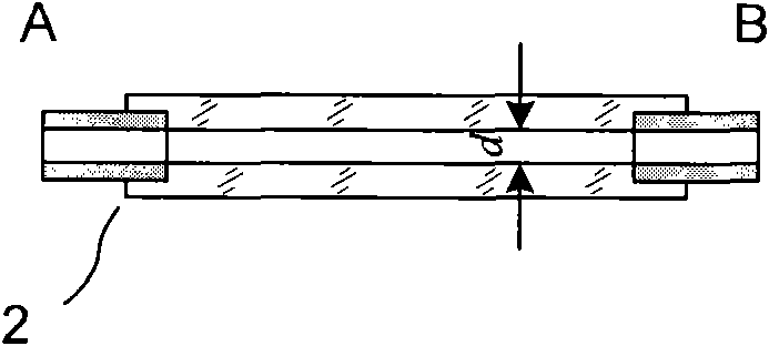
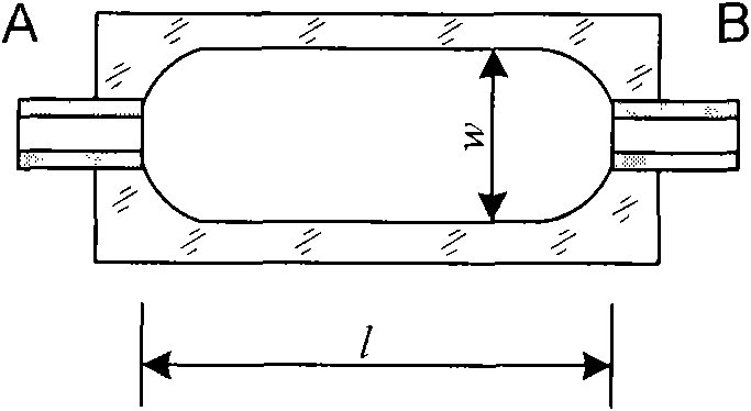
[0017] figure 1 It is the principle diagram of this embodiment, a rapid microscope inspection device for blood cell morphology based on staining technology, including a reaction pool 1 , a measurement chamber 2 , a liquid circuit unit 3 , a microscopic optical system 4 and a control unit 5 . It is characterized in that the liquid circuit unit 3 can inject a precise quantitative blood sample 7 and a staining solution 8 into the reaction pool 1 for staining, inject a quantitative buffer solution 9 to fix the stained sample, and inject a diluent 10 to dilute the fixed sample, Or inject cleaning solution 11 to clean the reaction pool; the measurement chamber 2 is connected to the reaction pool 1 and the liquid path unit 3 respectively, and the measurement chamber 2 is also a component of the micro-optical system 4, and is located on the object surface of the micro-optical system 4 . The ...


PUM

PropertyMeasurementUnit
diameteraaaaaaaaaa

Abstract

A cell morphology microscopic examination device based on the staining technology comprises a reaction bath, a measuring room, a liquid road unit and a micro-optical system. The device is characterized in that the liquid road unit comprises a cell sample interface and at least one reagent interface; the liquid road unit is at least communicated with one of the reaction bath and the measuring room; the reaction bath is communicated with the measuring room; and the measuring room is arranged in the micro-optical system. A cell morphology microscopic examination method based on the staining technology is characterized in that the liquid road unit is used to inject the cell sample and at least one agent in the reaction bath to mix together; after a certain time, the cell sample in the reaction bath is injected in the measuring room; and the flowing cell sample in the measuring room is imaged by the micro-optical system. The invention does not need the complicated mechanical smear device, the micro-optical system is fixed and the stained cell sample fast passes through the micro-optical system in an assembly-line mode, thus the fast cell morphology microscopic examination with high automation and compact structure can be realized.

Description

technical field [0001] The invention relates to the field of cell analysis, in particular to a cell morphology microscope inspection device based on staining technology. Background technique [0002] Existing hematology analyzers can provide rich data, greatly improve the quality and speed of clinical hematology tests, and reduce the workload of testers. However, they can only be used as a screening method in the process of routine blood analysis. When the automatic blood cell counter shows abnormal results or "marks" the results, it must be checked for whole blood smear staining, blood smear staining and microscopic examination It is an important tool to provide differential diagnosis and suggest further necessary examinations, especially in the rapid diagnosis of certain specific infections (such as malaria parasites, kala-azar protozoa and filariasis, etc.), blood smear staining and microscopy play an important role. [0003] The traditional way is to make by hand, first...

Claims


Application Information

Patent Timeline
Patent Type & Authority Applications(China)
IPC IPC(8): G01N21/85G01N1/30
Inventor 孔兵
Owner 孔兵
Why Eureka
Try Eureka