Living body fluorescent endoscopic spectrum imaging device

A technology of spectral imaging and in vivo fluorescence, which is applied in medical science, diagnosis, diagnostic recording/measurement, etc., can solve the problems of low spectral resolution and unfavorable resolution of emission, and achieve high spectral resolution and adjustable spectral resolution

Active Publication Date: 2010-12-08
HUAZHONG UNIV OF SCI & TECH
View PDF4 Cites 25 Cited by
  • Summary
  • Abstract
  • Description
  • Claims
  • Application Information

AI Technical Summary

Problems solved by technology

Although this method is compatible with CCD detection, the spectral resolution is low due to the use of prisms, wi

Method used


Image

  • Living body fluorescent endoscopic spectrum imaging device
  • Living body fluorescent endoscopic spectrum imaging device
  • Living body fluorescent endoscopic spectrum imaging device

Examples


Embodiment Construction

[0018] The present invention will be further described below in conjunction with the embodiments and accompanying drawings, but the protection scope of the present invention should not be limited thereby.

[0019] The present invention can be realized in the following ways:

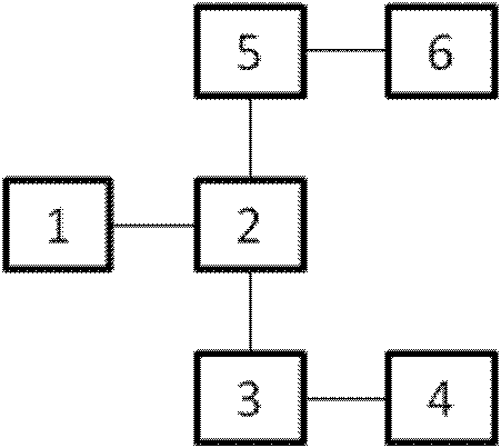
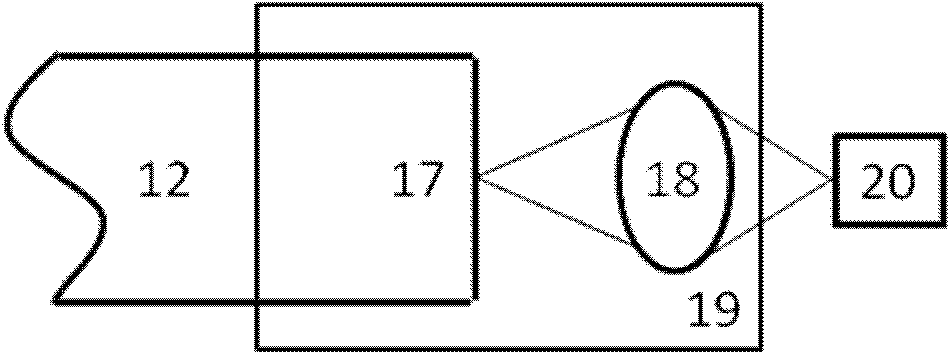
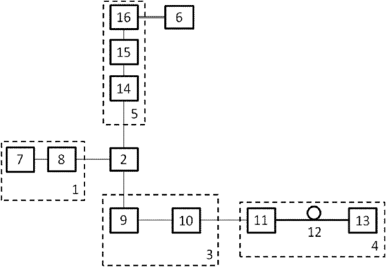
[0020] exist figure 1 , figure 2 Among them, the present invention includes a light source unit 1, a spectroscopic unit 2, a scanning light guide unit 3, an optical fiber bundle endoscopic unit 4, a photoelectric signal detection and acquisition unit 5, and a computer 6; the light source unit 1 is composed of a collimated light source 7 and a belt The light of the collimated light source 7 enters the light-splitting unit 2 after passing through the band-pass filter 8. The light-splitting unit 2 has two ports, and one port of the light-splitting unit 2 is connected with the scanning light guide unit 3, and the scanning light guide unit 3 is connected to the optical fiber bundle endoscopic unit 4 , the o...


PUM


Abstract

The invention relates to a live body fluorescent endoscopic spectrum imaging device, comprising a light source unit, a light split unit, a scanning light guide unit, a fiber bundle endoscopic unit, an electrooptical signal detection and acquisition unit and a computer, wherein the light source unit consists of a collimating unit and a band filter. The imaging device is characterized in that the light of the collimating unit passes through the band filter and then enters the light split unit, the light split unit is provided with two paths of interfaces, one path of the interfaces of the light split unit is connected with the scanning light guide unit, the scanning light guide unit is connected with the fiber bundle endoscopic unit, and the other path of the interfaces of the light split unit is connected with the electrooptical signal detection and acquisition unit which is connected with the computer. The imaging device has the characteristics that: 1, a Fourier transform spectrometer is used to detect a sample excitation spectrum, and has the advantages of high spectral resolution (1nm), adjustable spectral resolution and the like; and 2, the fluorescent live body endoscopic spectrum imaging system can provide not only imaging diagnosis at tissue level but also spectrum diagnosis at molecular level.

Description

technical field [0001] The invention belongs to the technical field of optical imaging, in particular to a living body fluorescent endoscopic spectral imaging device. Background technique [0002] Optical microscopy technology can provide effective information from the molecular level to the cellular level to the tissue level for the research of modern medicine, and help people diagnose, understand, and study various complex diseases and their pathogenic mechanisms at different scales. Since various representations in living organisms are easily affected by complex factors such as the pH, humidity, and temperature of the surrounding environment, it is of great practical significance to vigorously develop in vivo observation methods. Compared with in vitro observation, the data obtained by in vivo observation can more truly reflect the actual nature and state of the observed target, and the detection results are more accurate. In vivo fluorescence endoscopic imaging technolo...

Claims


Application Information

Patent Timeline
IPC IPC(8): A61B5/00
Inventor 袁菁付玲曾绍群骆清铭张红明
Owner HUAZHONG UNIV OF SCI & TECH
Why Eureka
Try Eureka